Jump to content
In this section we have spot diagnoses posted on a daily basis since June 2010, now over 4000! You can review the archived cases and read the suggested diagnoses by users and the final comment by the contributors.
Case are uploaded each week day by 10 am UK time with the correct diagnosis will generally be posted at 8 pm UK time. Why not view the most recent spot diagnosis and proffer a diagnosis?

Case Number : Case 1130 - 22nd October Posted By: Guest

Please read the clinical history and view the images by clicking on them before you proffer your diagnosis.
Submitted Date :
   (0 reviews)

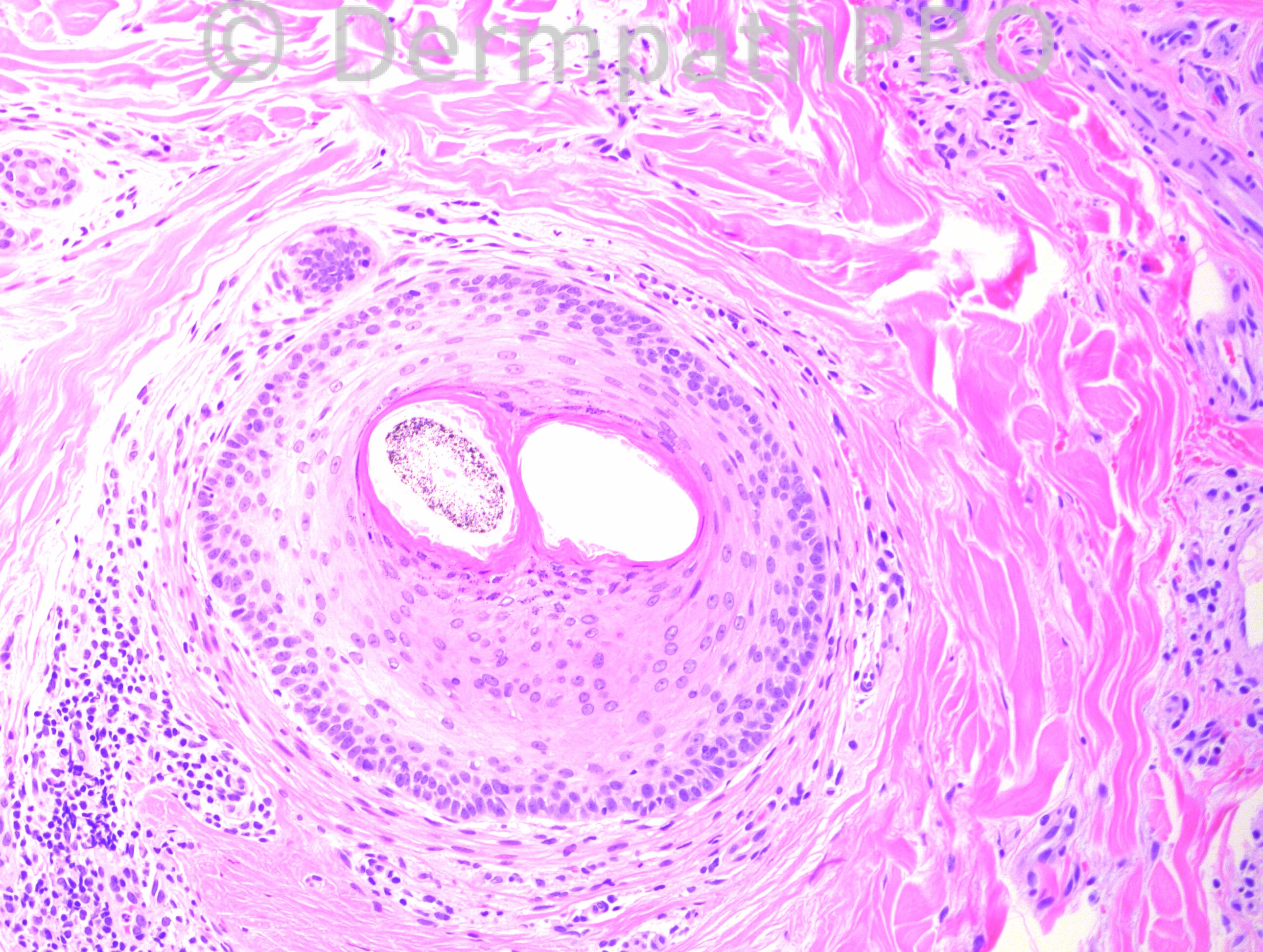
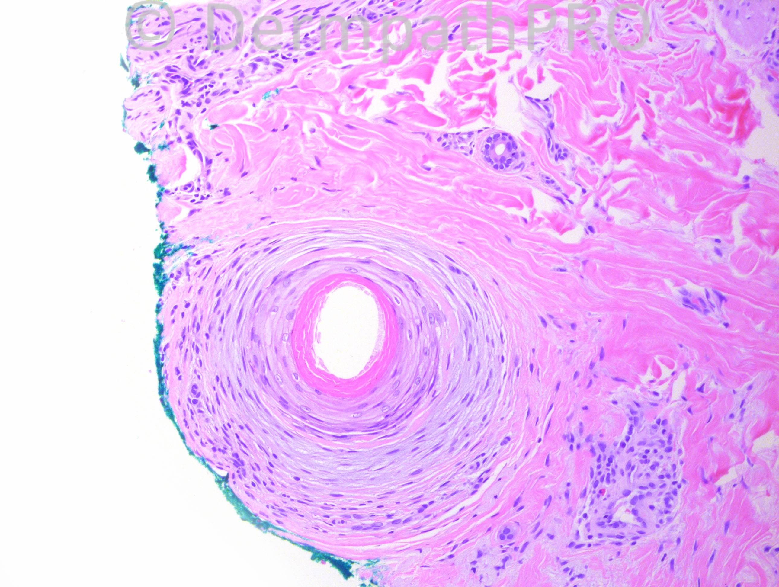
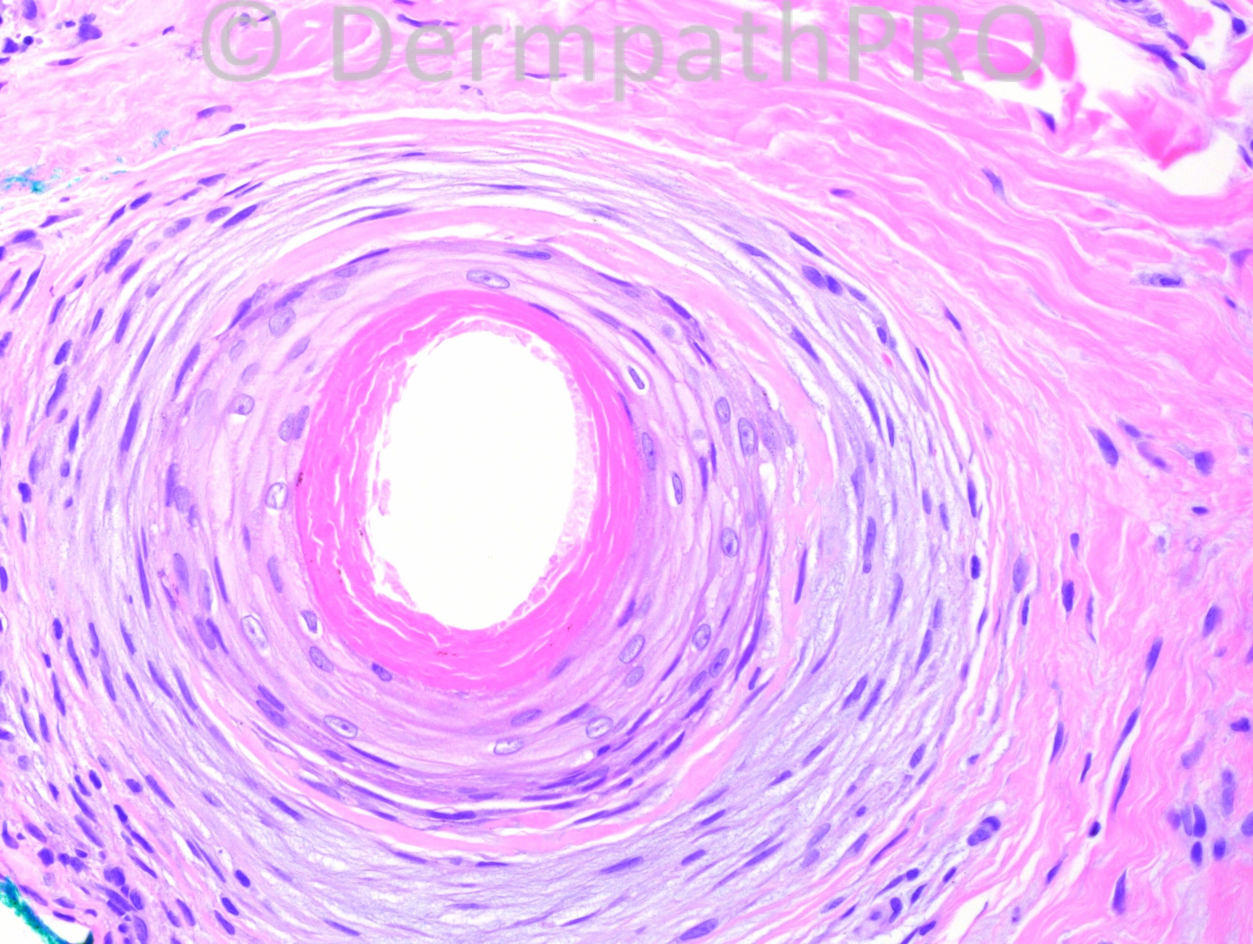
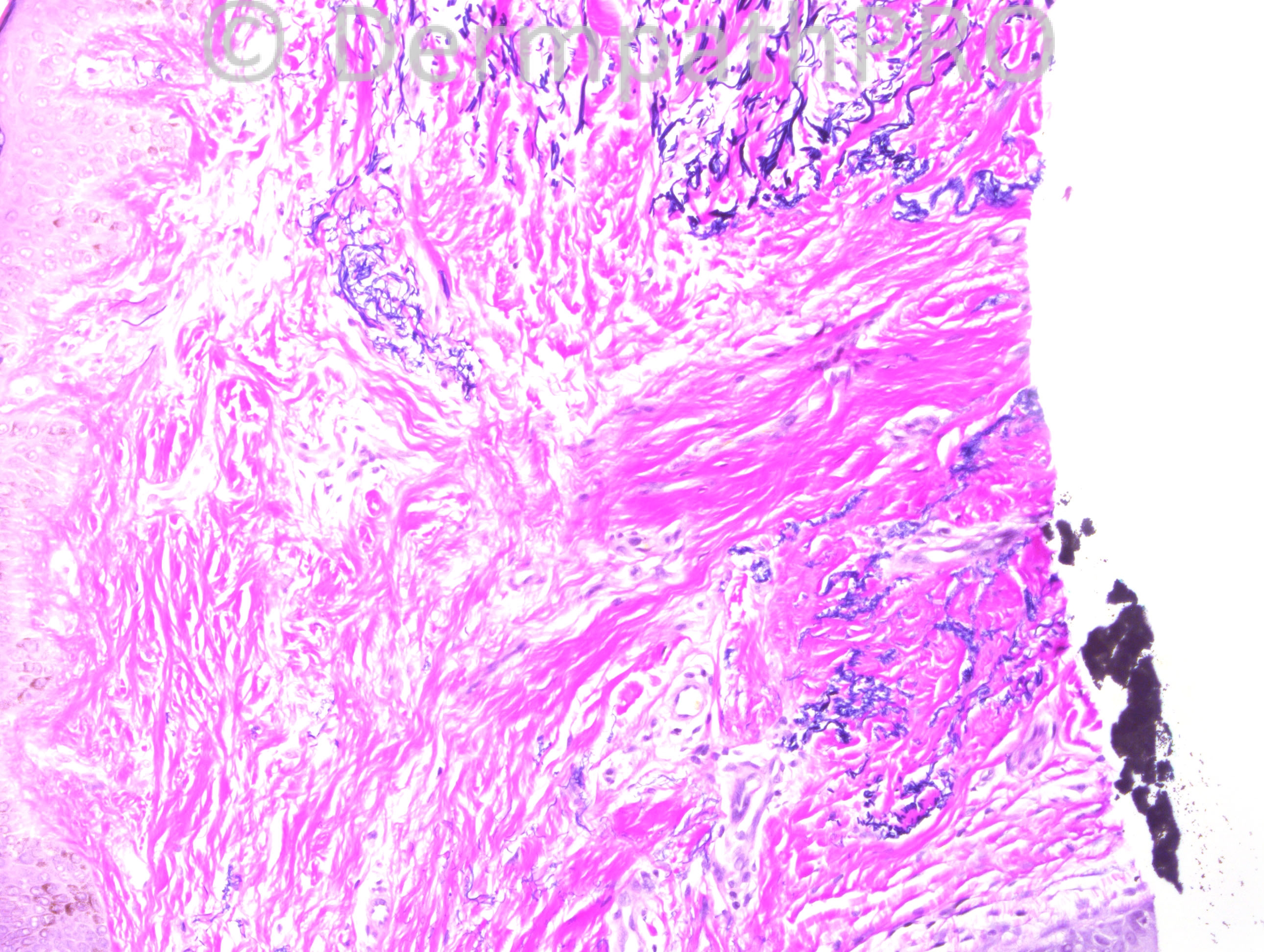
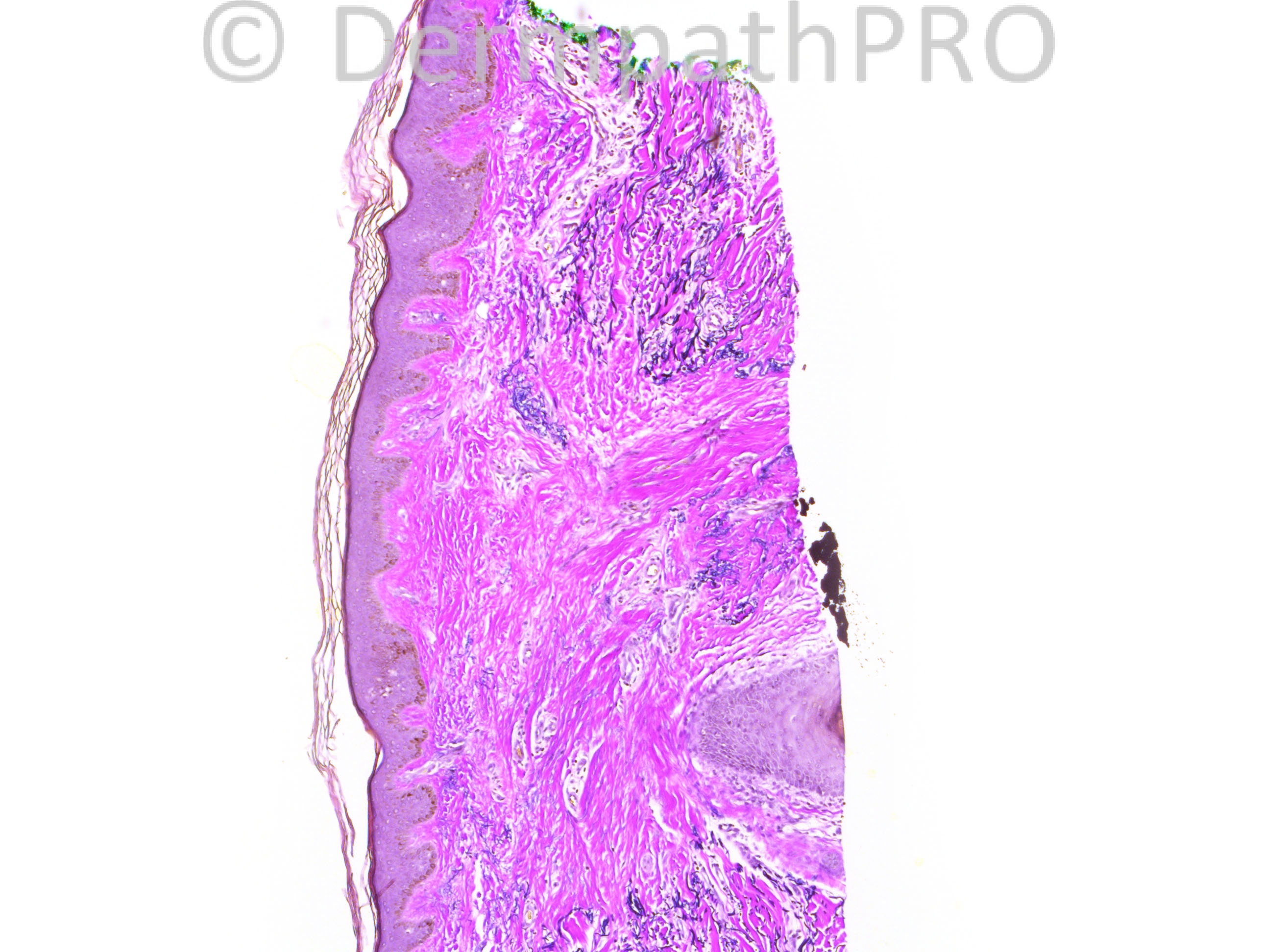
45 year-old African American female with alopecia. This biopsy is from the left parietal scalp.

Case posted by Dr. Hafeez Diwan.

  Report Record

User Feedback


Robledo F. Rocha

Posted

Late-stage lymphocytic scarring alopecia. I favor lichen planopilaris due to lichenoid lymphocytic infiltrate at the outside of perifollicular concentric lamellar fibrosis, squamotization of basal layer, and loss of elastic fibers within the scar.

Share this comment


Link to comment
share_externally

Dr. Mona Abdel-Halim

Posted

I took the clue of desquamation of the inner root sheath as a clue for central centrifugal cicatricial alopecia but the clinical refers to parietal scalp biopsy, not a crown or vertex biopsy. May be it is a wide area of centrifugally expanding alopecia!!

Share this comment


Link to comment
share_externally

Dr. Mona Abdel-Halim

Posted

According to McKee's Pathology of the skin, central centrifugal cicatricial alopecia includes other entities previously described as: Follicular Degeneration Syndrome (hot comb alopecia), Pseudopelade of African American and Central Elleptical Pseudopelade of Caucasians.. All are included now under one name: central centrifugal cicatricial alopecia.

Share this comment


Link to comment
share_externally

Dr. Richard Carr

Posted

Lichen planopilaris is my favoured diagnosis although my experience with CCCA is very limited.

Share this comment


Link to comment
share_externally

Robledo F. Rocha

Posted

There is really desquamation of the inner root sheath, but I don't think it is premature once I didn't find subcutaneous fat or eccrine coils to indicate the horizontal sections were taken at the level of lower isthmus. Also, I'm nor convinced about eccentric atrophy of the follicular epithelium, a sign expected to be found during the late stage of central centrifugal cicatricial alopecia.

Share this comment


Link to comment
share_externally

Dr. Hafeez Diwan

Posted

Central centrifugal cicatricial alopecia. The perifollicular fibrosis is the kind one sees in CCCA. There are fused follicles.

Share this comment


Link to comment
share_externally


Join the conversation

You can post now and register later. If you have an account, sign in now to post with your account.

Guest
Add a comment...

×   Pasted as rich text.   Paste as plain text instead

  Only 75 emoji are allowed.

×   Your link has been automatically embedded.   Display as a link instead

×   Your previous content has been restored.   Clear editor

×   You cannot paste images directly. Upload or insert images from URL.

Loading...

×
×
  • Create New...