Jump to content
In this section we have spot diagnoses posted on a daily basis since June 2010, now over 4000! You can review the archived cases and read the suggested diagnoses by users and the final comment by the contributors.
Case are uploaded each week day by 10 am UK time with the correct diagnosis will generally be posted at 8 pm UK time. Why not view the most recent spot diagnosis and proffer a diagnosis?

Case Number : Case 1608 - 24 August Posted By: Guest

Please read the clinical history and view the images by clicking on them before you proffer your diagnosis.
Submitted Date :
   (0 reviews)

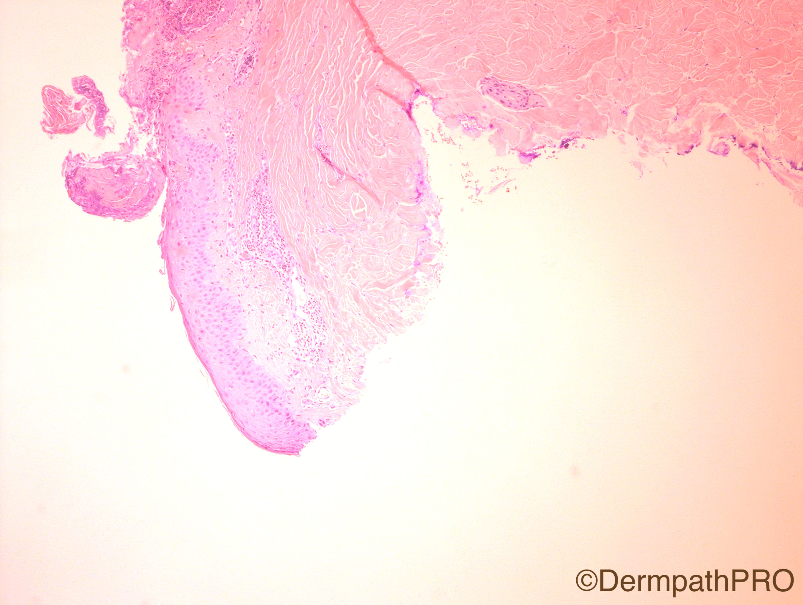
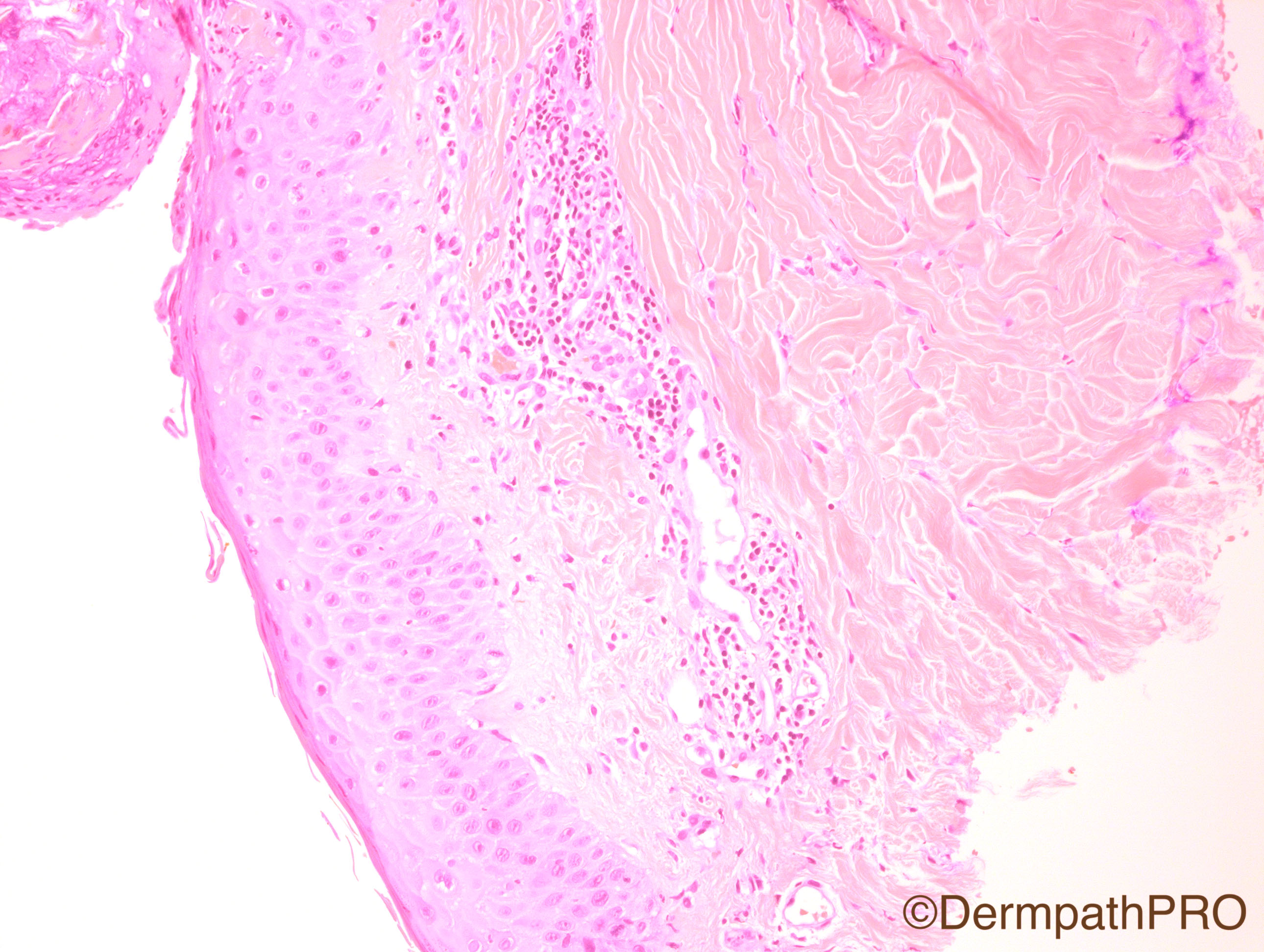
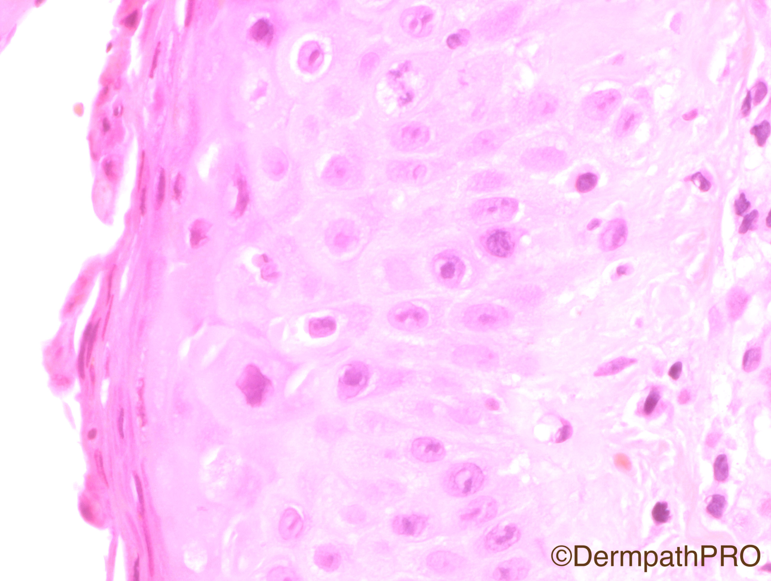
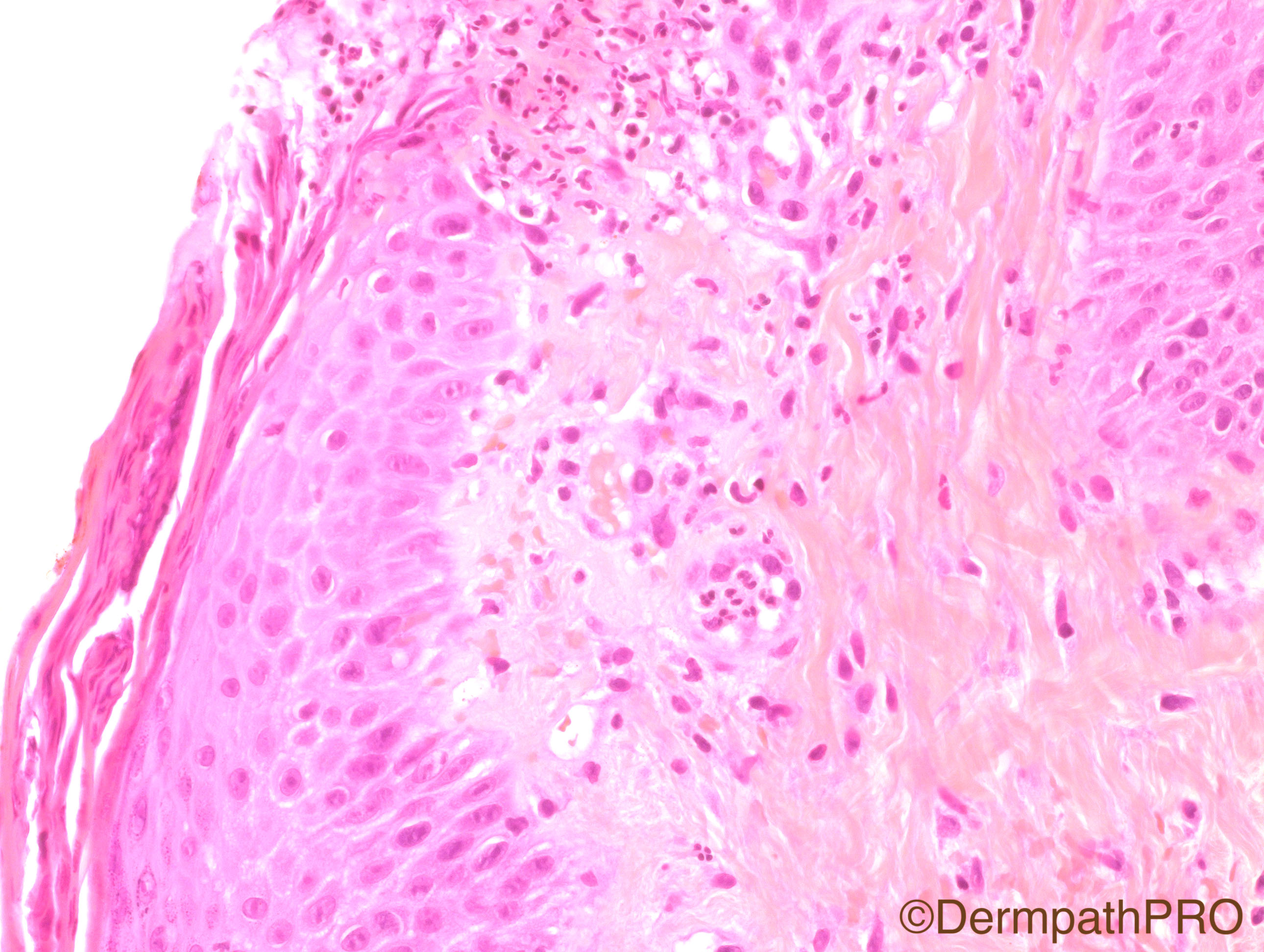
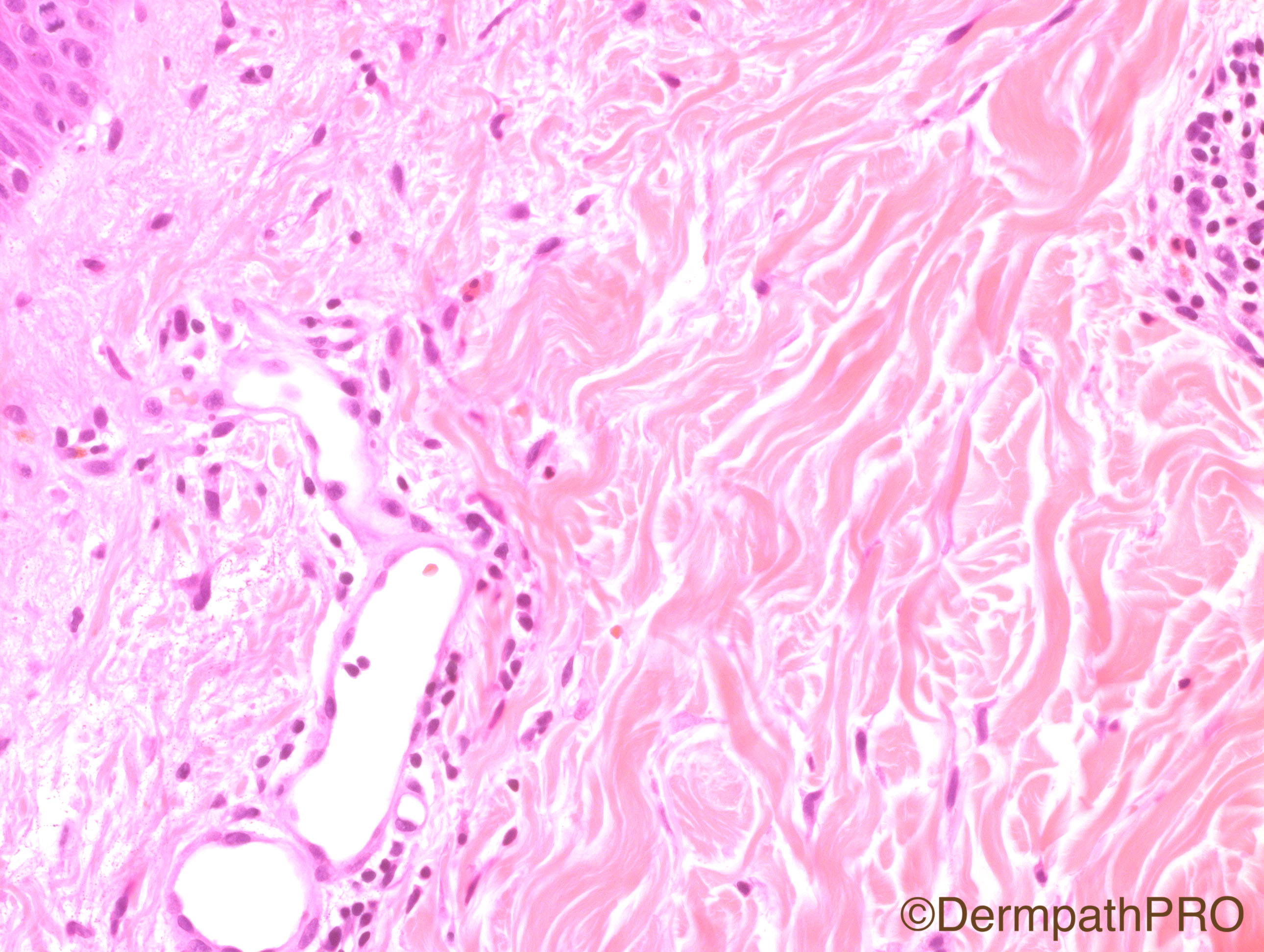
45 year-old male with itchy rash on head, trunk and extremities for several months. Physical examination revealed confluent erythematous, non-scaly plaques on face and erosions with scale on border on chest, abdomen and back. A few scaly macules were present on the thighs and upper extremities. There were erythematous scaly patches on the scalp. This biopsy is from the right upper arm.

Case Posted by Dr Hafeez Diwan


  Report Record

User Feedback


Raul Perret

Posted

I think there are some cells with morphological changes suggestive of viral affection. The clinical description is curious, particularly the long duration of the symptoms. Would perform immunos for herpes family of virus. Other differential could be disseminated dermatophytosis. Psoriasis also crossed my mind because I think there is absent granular layer in the second image and there are dilated blood vessels so  it could also be a consideration if the differentials mentioned above are ruled out.

Share this comment


Link to comment
share_externally

vincenzo polizzi

Posted

What about Reactive Perforating Collagenosis?

Share this comment


Link to comment
share_externally

biswanath behera

Posted

Whether it is a case of Leukemia cutis ??


I am not sure but i am seeing some large cells (?blast cell) in Figure 4 and 5.


In figure 5 , whether thr is a band cell present??

Share this comment


Link to comment
share_externally

Dr. Hafeez Diwan

Posted

Pemphigus foliaceus.  Direct immunofluorescence was positive.  There are dyskeratotic cells, and absent stratum corneum focally, and in the last image I have tried to photograph an eosinophil.

Share this comment


Link to comment
share_externally


Join the conversation

You can post now and register later. If you have an account, sign in now to post with your account.

Guest
Add a comment...

×   Pasted as rich text.   Paste as plain text instead

  Only 75 emoji are allowed.

×   Your link has been automatically embedded.   Display as a link instead

×   Your previous content has been restored.   Clear editor

×   You cannot paste images directly. Upload or insert images from URL.

Loading...

×
×
  • Create New...