Jump to content
In this section we have spot diagnoses posted on a daily basis since June 2010, now over 4000! You can review the archived cases and read the suggested diagnoses by users and the final comment by the contributors.
Case are uploaded each week day by 10 am UK time with the correct diagnosis will generally be posted at 8 pm UK time. Why not view the most recent spot diagnosis and proffer a diagnosis?

Case Number : Case 1488- 08 March Posted By: Guest

Please read the clinical history and view the images by clicking on them before you proffer your diagnosis.
Submitted Date :
   (0 reviews)

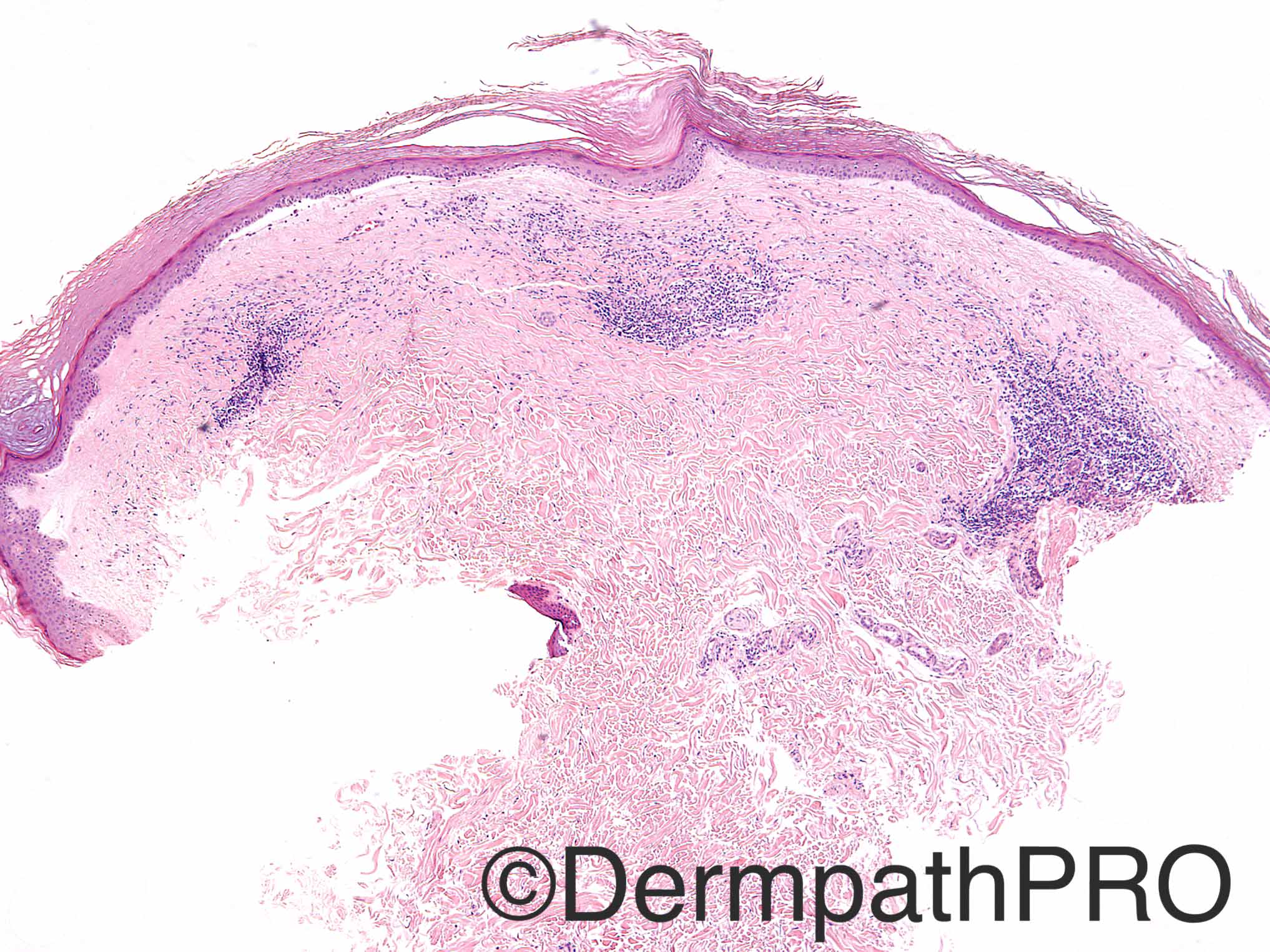
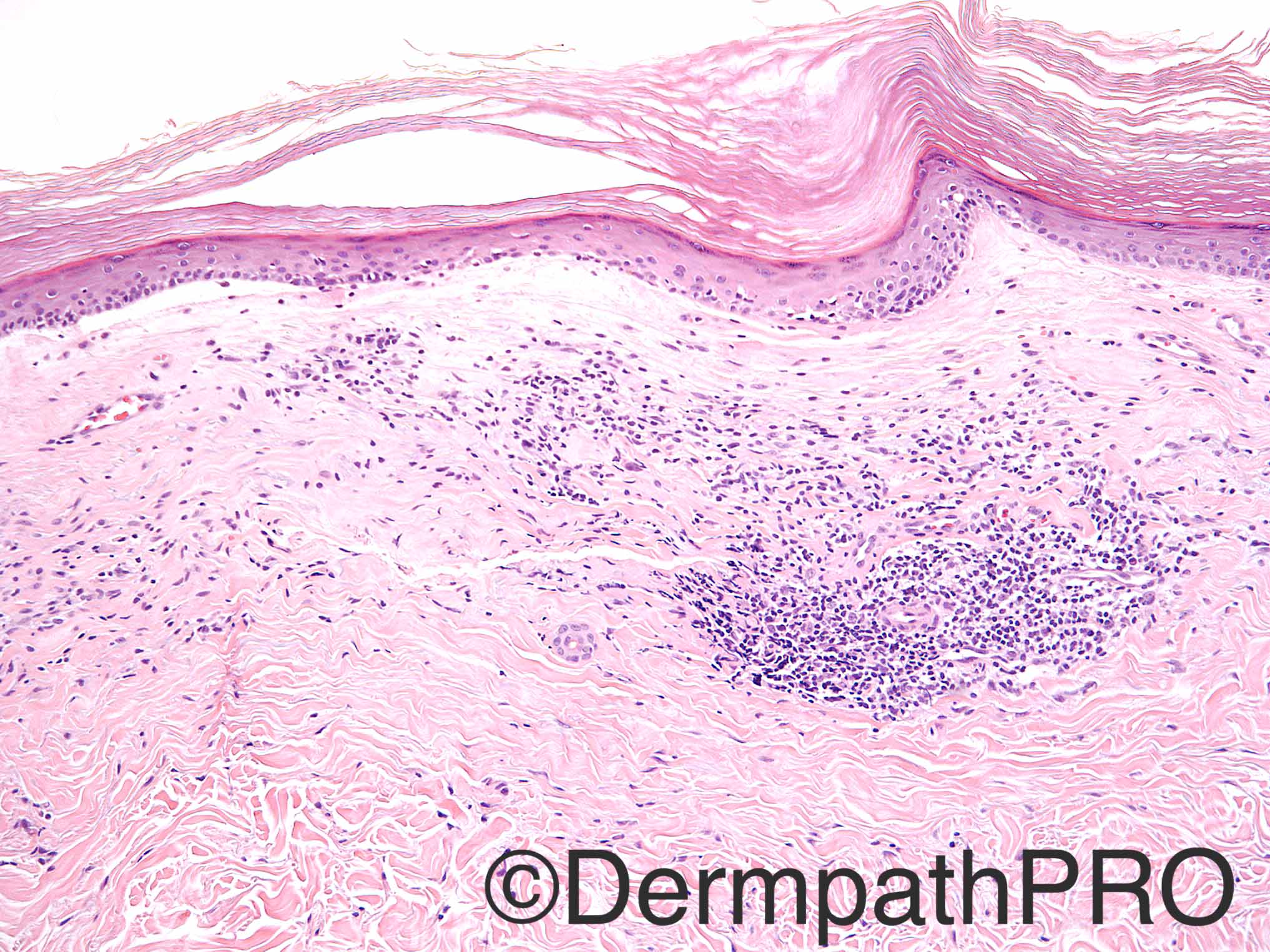
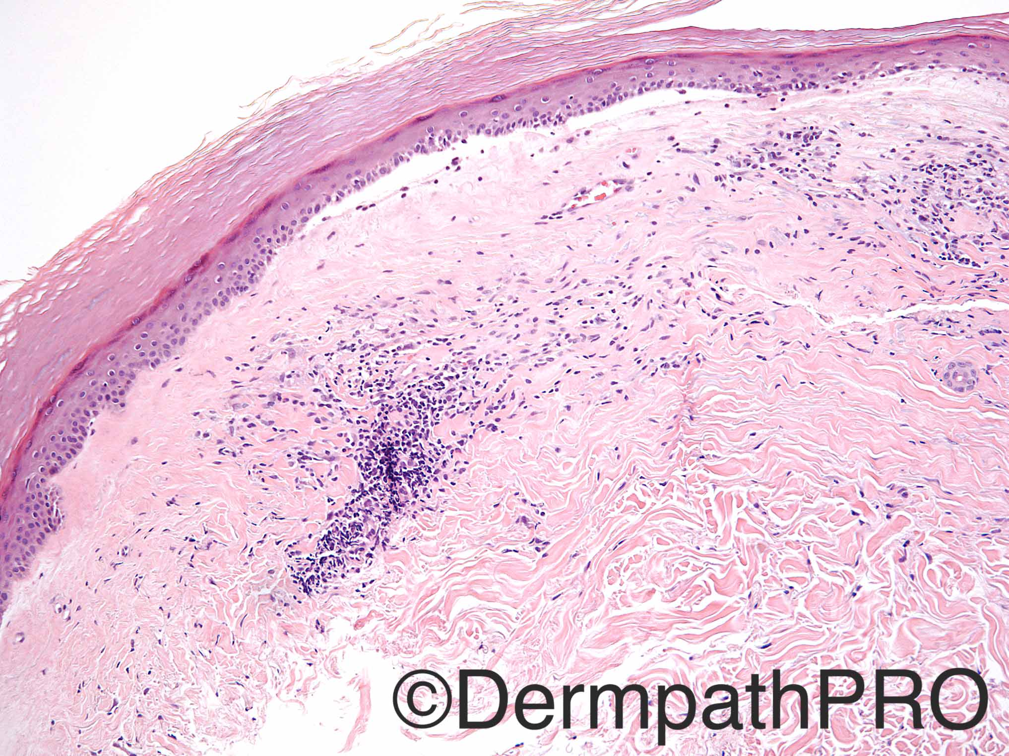
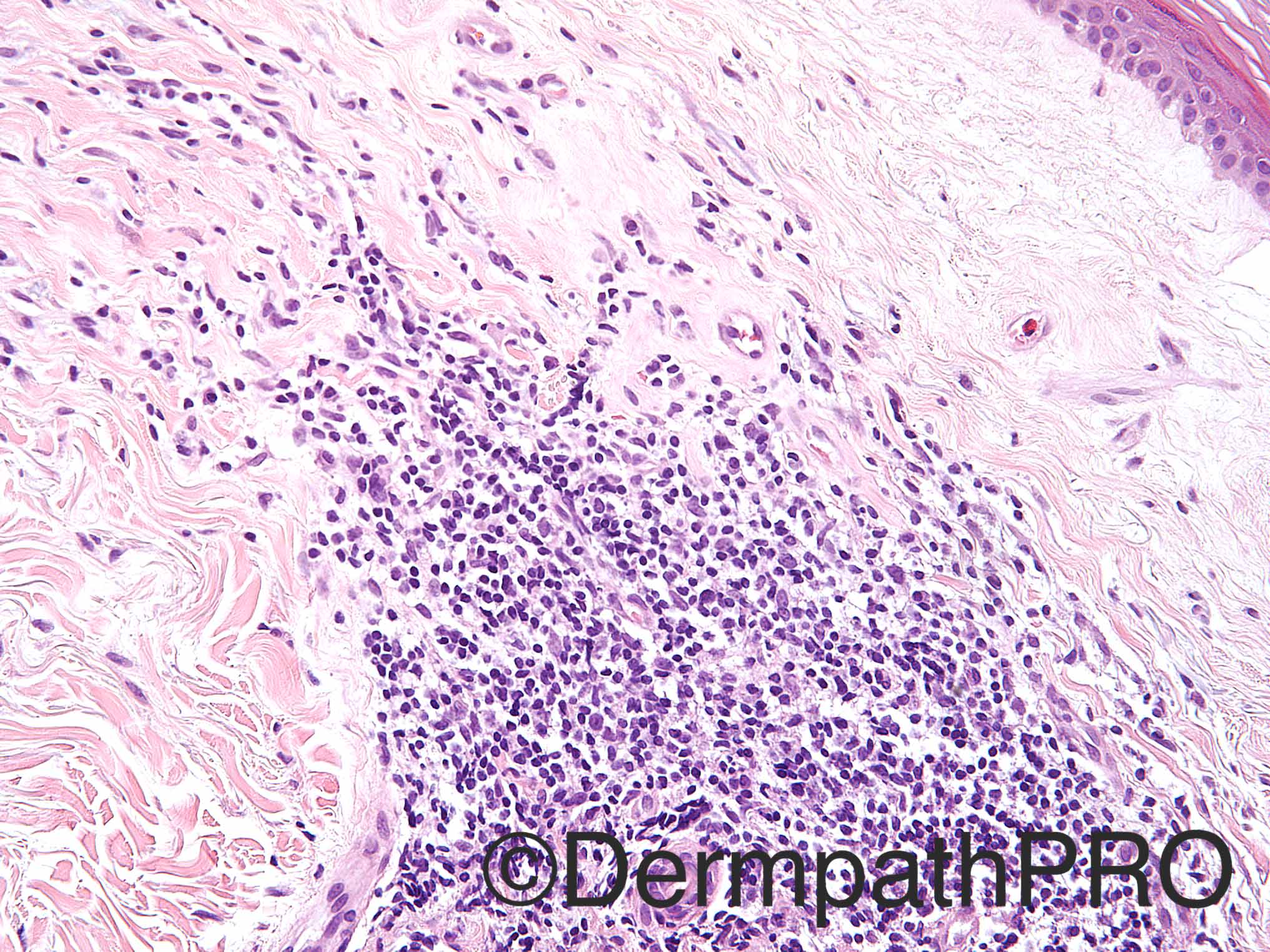
Case History: 55 year old woman with multiple new lesions which the patient thought were spider bites.

Case posted by Dr Uma Sundram


  Report Record

User Feedback


Atrophic epidermis, hyperkeratosis, interface change, superficial sclerosis of the dermis with lymphocytic inflammatory infiltrate under the sclerosis. Lichen sclerosus.

Share this comment


Link to comment
share_externally

Raul Perret

Posted

Let me bring to the differential lupus erythematosus, I think there is a hint of mucin on the dermis

Share this comment


Link to comment
share_externally

vincenzo polizzi

Posted

Hello! Happy Women's Day...Also favor Lichen Sclerosus et Atrophicus

Share this comment


Link to comment
share_externally

Nitin Khirwadkar

Posted

Agree with the differentials. Favour LSet A. Would add morphoea (early phase) to the list.

Share this comment


Link to comment
share_externally

Favor Lichen sclerosis et atrophicus

Share this comment


Link to comment
share_externally

Dr. Mona Abdel-Halim

Posted

Pathologically fits with LS, clinically strange perception as spider bites !!

Share this comment


Link to comment
share_externally

?Tick bites.  ?Acrodermatitis chronica atrophicans.  Like Mona, I think the history does not fit with LS. 

Share this comment


Link to comment
share_externally

vincenzo polizzi

Posted

Yes ...the history doesn't fit! Acrodermatitis chronic atrophicans fit, but pathologically the intradermal inflammatory infiltrate isn't usually under a sclerotic papillary dermis...However like the suggestion of Abdul.

Share this comment


Link to comment
share_externally

Uma Sundram

Posted

Interestingly, when the patient was seen by an experienced dermatologist, she had small pinkish grouped papules that involved the trunk and arms; many were umbilicated and some had depressed centers. Others were the classic ‘porcelain white’ lesions with an erythematous border. The dermatologist was quite concerned about malignant atrophic papulosis (Degos' disease) and we suggested that the histology fit this diagnosis in this clinical context. The patient recently had a full work up and appears to have skin limited disease. 

Share this comment


Link to comment
share_externally

Raul Perret

Posted

Great case. Dr. Sundram was there any evidence of mucin deposition in the biopsy?

Share this comment


Link to comment
share_externally

Dr. Richard Carr

Posted

Histology looks typical / diagnostic of lichen sclerosus - I still prefer LS histologicaly even if this differs from the favoured clinical diagnosis.

Share this comment


Link to comment
share_externally


Join the conversation

You can post now and register later. If you have an account, sign in now to post with your account.

Guest
Add a comment...

×   Pasted as rich text.   Paste as plain text instead

  Only 75 emoji are allowed.

×   Your link has been automatically embedded.   Display as a link instead

×   Your previous content has been restored.   Clear editor

×   You cannot paste images directly. Upload or insert images from URL.

Loading...

×
×
  • Create New...