Jump to content
In this section we have spot diagnoses posted on a daily basis since June 2010, now over 4000! You can review the archived cases and read the suggested diagnoses by users and the final comment by the contributors.
Case are uploaded each week day by 10 am UK time with the correct diagnosis will generally be posted at 8 pm UK time. Why not view the most recent spot diagnosis and proffer a diagnosis?

Case Number : Case 1491- 11 March Posted By: Guest

Please read the clinical history and view the images by clicking on them before you proffer your diagnosis.
Submitted Date :
   (0 reviews)

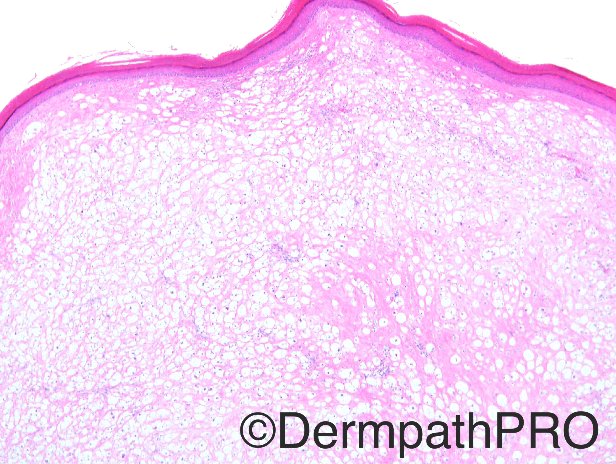
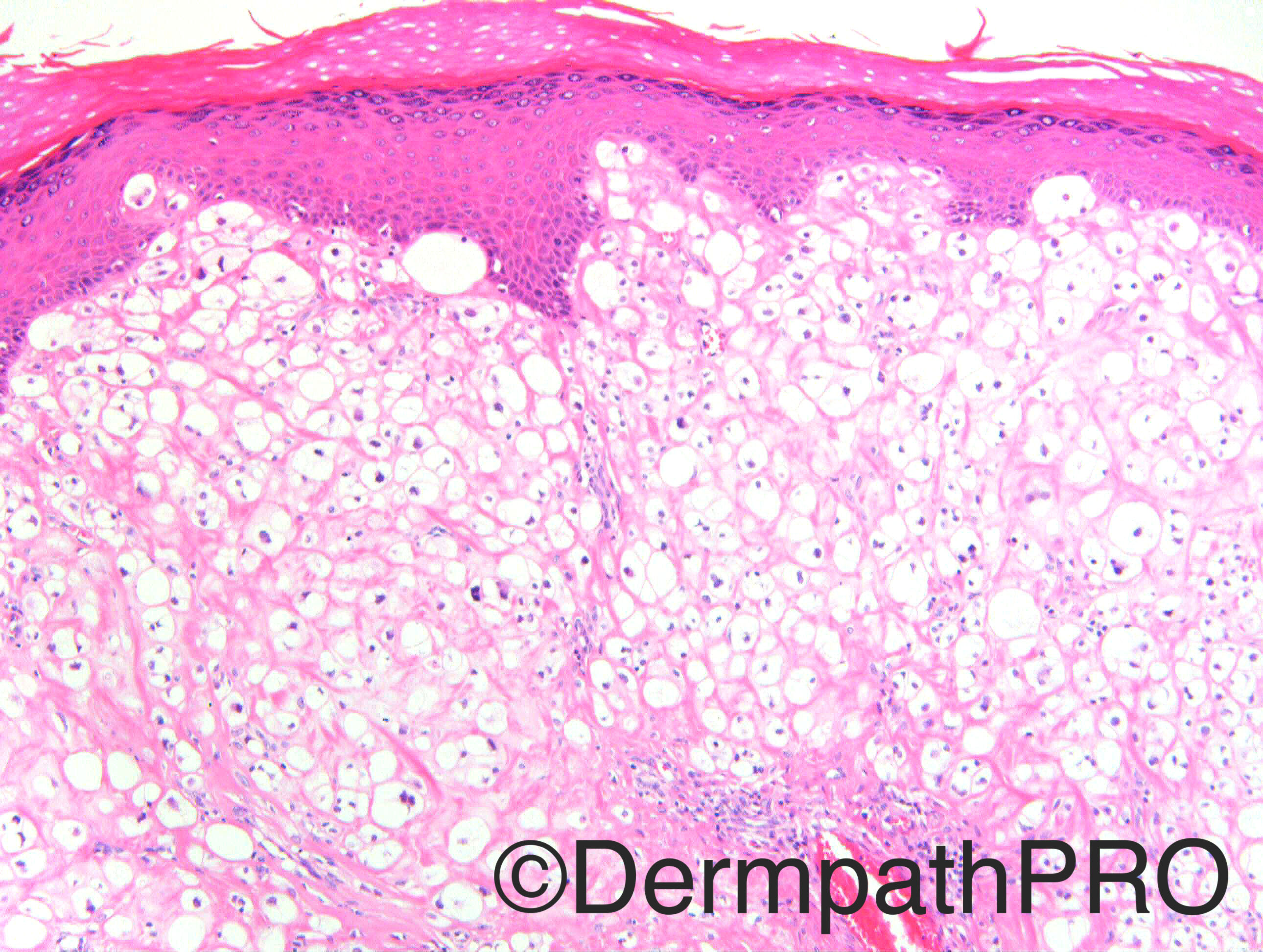
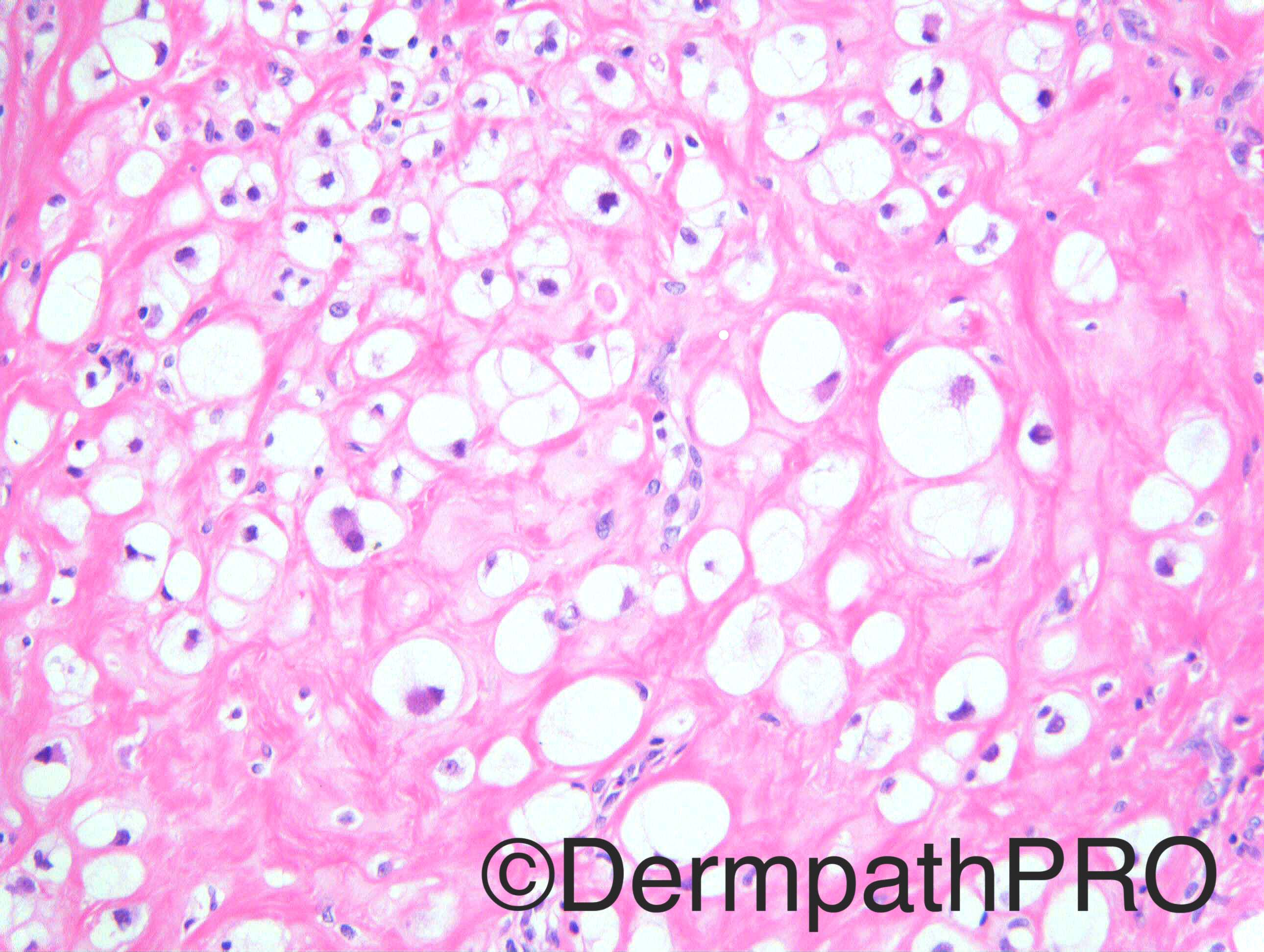
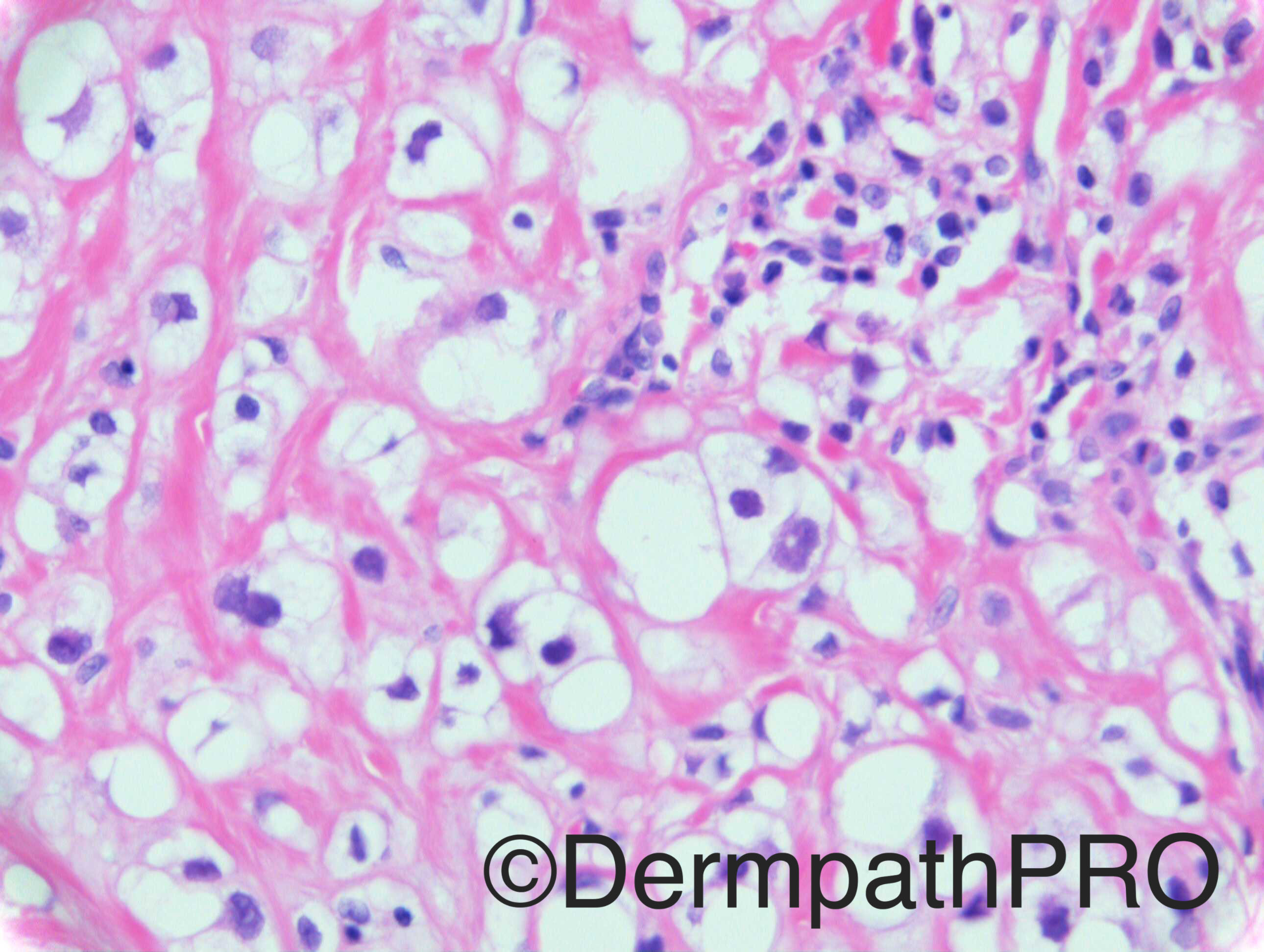
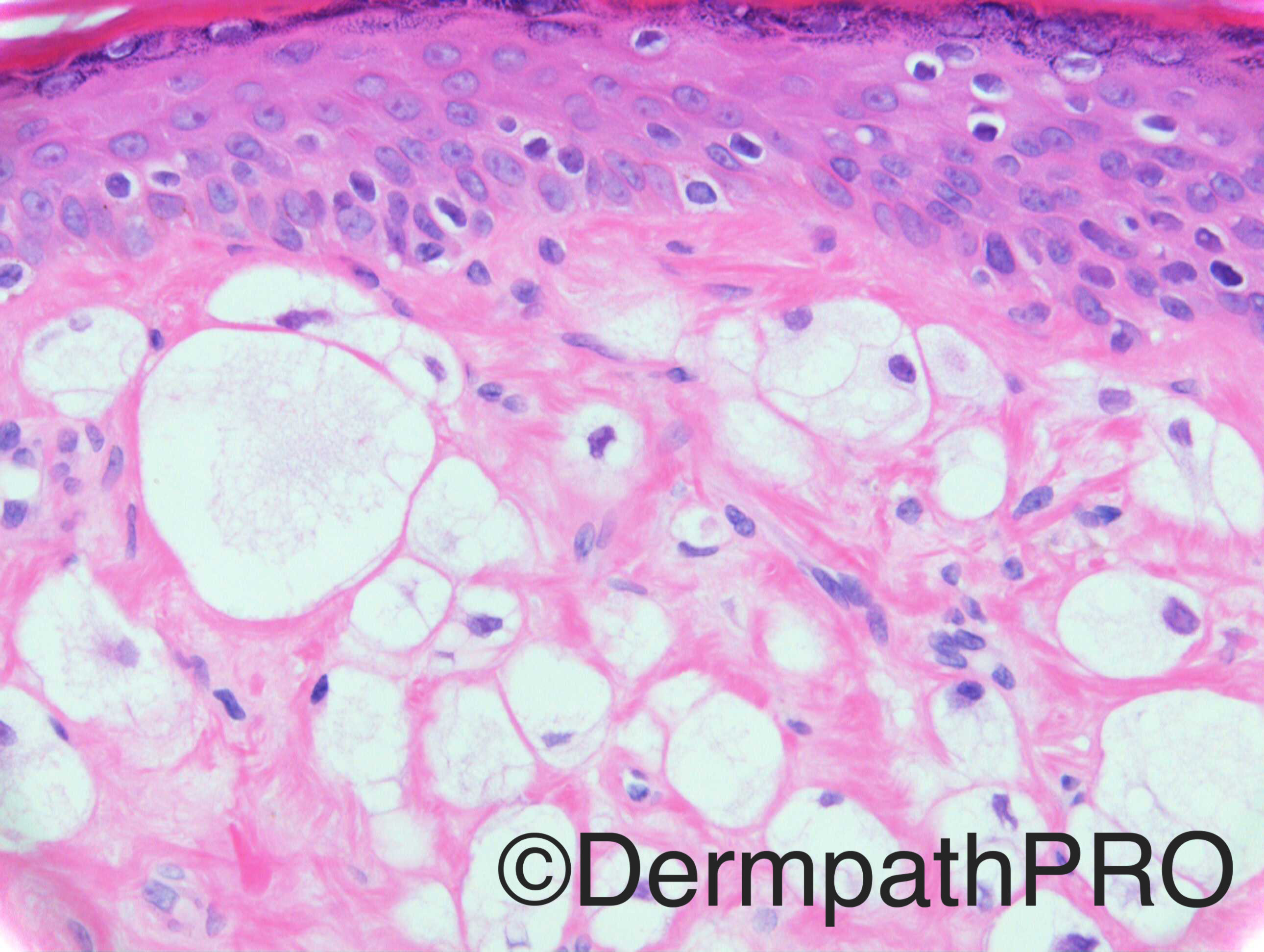
Case History: M55: Papule on face.

Case posted by Dr Richard Carr


  Report Record

User Feedback


vincenzo polizzi

Posted

?Balloon Cell Melanoma ?Fibrous Papule...well circumscribed lesion, with hyalinized stroma, i favour FP.

Share this comment


Link to comment
share_externally

Clear cell fibrous papule.  CD68 and CD63 to confirm.

Share this comment


Link to comment
share_externally

Dr. Mona Abdel-Halim

Posted

Balloon cell nevus vs. Fibrous papule for IHC

Share this comment


Link to comment
share_externally

Guest Romualdo

Posted

More irregular and eccentrically located nuclei favor clear cell fibrous papule over balloon cell nevus. NuCD68 and NKI/C3 positivity would confirm this possibility.

Share this comment


Link to comment
share_externally

Dr. Richard Carr

Posted

My understanding is the chap has had multiple lesions (wide anatomic distribution) excised previously with similar histology.  One was a 2cm lesion on his back.

Share this comment


Link to comment
share_externally

vincenzo polizzi

Posted

Metastasis of malignant clear cell tumor...favor melanoma. The previous back lesion was probably the primary tumor

Share this comment


Link to comment
share_externally

Dr. Mona Abdel-Halim

Posted

Could be than metastasis .. Let wait for the rest of the images

Share this comment


Link to comment
share_externally

Raul Perret

Posted

Sincerely I will wait for the next images but on morphology alone my diagnosis was balloon cell nevus to confirm with IHQ. I have just seen one real case of balloon cell melanoma in my life (appart from pictures on articles and books) and it was clearly evident that was malignant (ulcerated, nuclear atypia and pagetoid extensión to the epidermis). I think the only feature that looks weird in this lesion is that the cells are disposed mainly in sheets (usually balloon cell nevus keeps the architecture of a nevus with theques). However if this is indeed a case of balloon cell melanoma I think I would miss the diagnosis without the clinical information of multiple lesions in different locations that Dr. Carr added.

Share this comment


Link to comment
share_externally

Nitin Khirwadkar

Posted

Agree with the differentials. Clear cell fibrous papule- sclerotic collagen is striking in some of the images, any syndromic association as there are multiple lesions? DD: Balloon cell naevus, metastasis from a RCC, clear cell DF, dermal clear cell mesenchymal neoplasm. Definitely requires a panel of IHC.

Share this comment


Link to comment
share_externally

vincenzo polizzi

Posted

Agree...also i'm waiting for the other imges...but taking into account the CPC (one lesion on back and 2cm and subtle atypia), my first provisional diagnosis is melanoma metastatic. Have a good week end.

Share this comment


Link to comment
share_externally

Raul Perret

Posted

Just to continue adding to the differential and trying also to explain the mutifocality of the tumor there are some reports of eruptive dermal clear cell desmo-plastic mesenchymal tumors with perivascular myoid differentiation (a tumor related to PeComa). Morphology is similar except for the vascular parts that dont seem quite prominent here. Another difference is that in the case reported the lesions were present since infancy

https://iris.unito.it/retrieve/handle/2318/148613/22412/Tomasini%20J%20Cutan%20Pathol%202014%20(1)_1237895.pdf

As everybody agrees a wide panel of immunos is mandatory in this case

Share this comment


Link to comment
share_externally

Arti Bakshi

Posted

Interesting ref by Raul…wasn't aware of this entity. (but they do seem to stress on the close relationship with blood vessels , which is lacking in this case).

I am inclined to agree with Mark's suggestion of Chordoma/parachordoma. The (rather) large vacuolated cells are reminiscent of 'physalipharous cells' and the 2cm tumour on back may be the clue! Of course metastatic melanoma and other clear cell tumours (comprehensively listed by Nitin) still in d/d.

Share this comment


Link to comment
share_externally

Dr. Richard Carr

Posted

Hmm!  Again I think the images I sent originally were subtly different with more cytoplasmic detail (foamy nature) which has been lost.  I have asked dermpathpro team to replace a couple of the high power images and post the macrophage marker (positive).  I am attaching the report my colleague issued on the 2cm nodule (but that was not the first lesion to be excised from this patient, my understanding is that he has had a long history of lesions reported at St Elsewhere as xanthomas).


Well circumscribed nodular dermal lesion, comprising sheets of large cells with foamy to granular cytoplasm. The nuclei are round to oval, with occasional nucleoli and mitotic figures. Residual collange fibres are seen between the large cells. PAS and Oil Red O (frozen post fixation in formalin) were negative.  The cells stain positive for CD68, LCA, vimentin and CD10. Staining for AE1/3, Cam5.2, BerEP4, EMA, RCC, S100, melanA, CD34, Actin, Desmin all negative. Ki67 index very low.


The features favour an excised tuberous xanthoma. The differential diagnosis includes a dermal clear cell mesenchymal neoplasm, however this may be less likely. Please consider checking lipid profile. If other skin lesions are present, biopsy may be of value.


We then received 3 facial lesions as shown here which I concurred with tuberous xanthoma and again enquired regarding lipid profile (cholesterol 6.2, triglyceride 0.8; HDL 1.5; LDL 3.3). Confess my knowledge of lipid chemistry insufficient to make an interpretation. Patient has been referred to a consultant chemical pathologist and I'm awaiting follow-up.

Share this comment


Link to comment
share_externally


Join the conversation

You can post now and register later. If you have an account, sign in now to post with your account.

Guest
Add a comment...

×   Pasted as rich text.   Paste as plain text instead

  Only 75 emoji are allowed.

×   Your link has been automatically embedded.   Display as a link instead

×   Your previous content has been restored.   Clear editor

×   You cannot paste images directly. Upload or insert images from URL.

Loading...

×
×
  • Create New...