Jump to content
In this section we have spot diagnoses posted on a daily basis since June 2010, now over 4000! You can review the archived cases and read the suggested diagnoses by users and the final comment by the contributors.
Case are uploaded each week day by 10 am UK time with the correct diagnosis will generally be posted at 8 pm UK time. Why not view the most recent spot diagnosis and proffer a diagnosis?

Case Number : Case 1492- 14 March Posted By: Guest

Please read the clinical history and view the images by clicking on them before you proffer your diagnosis.
Submitted Date :
   (0 reviews)

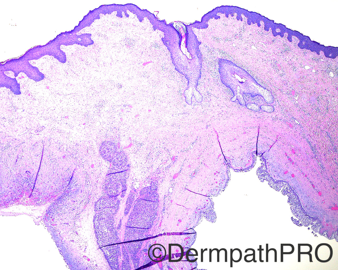
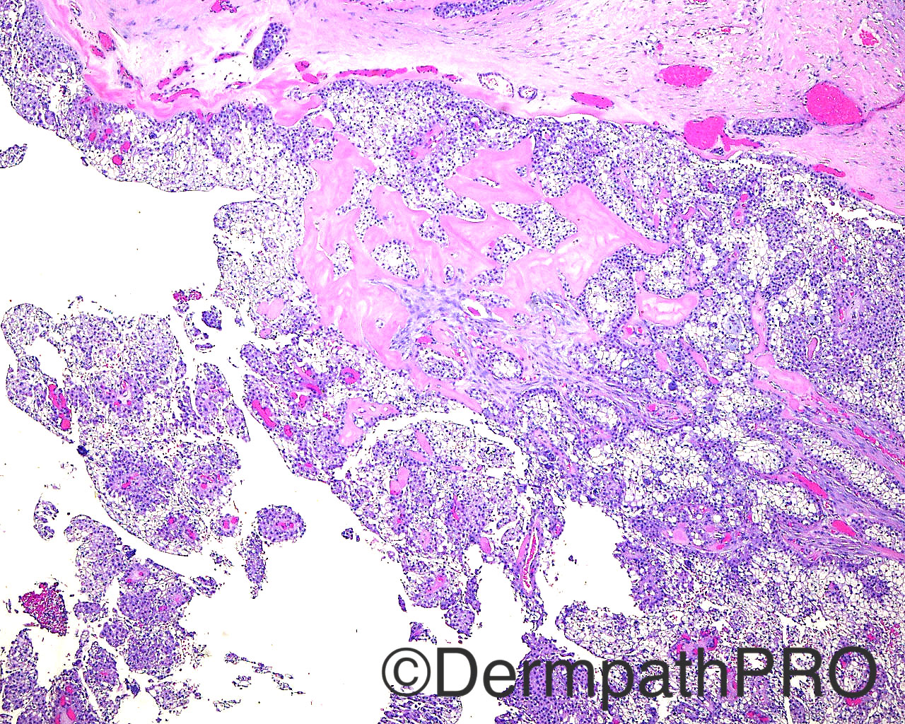
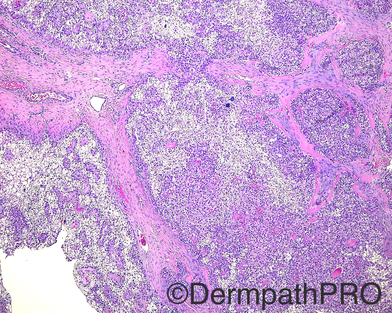
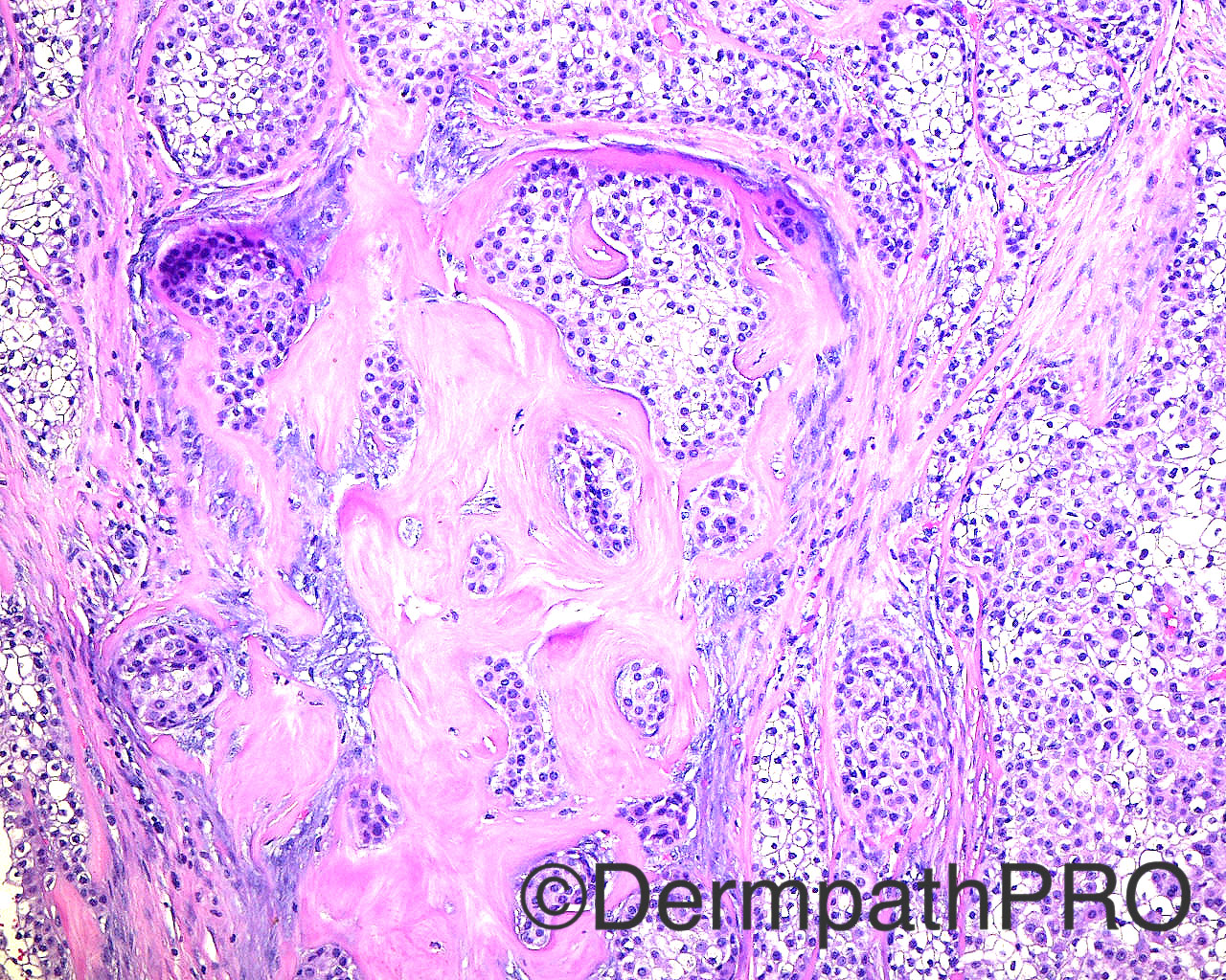
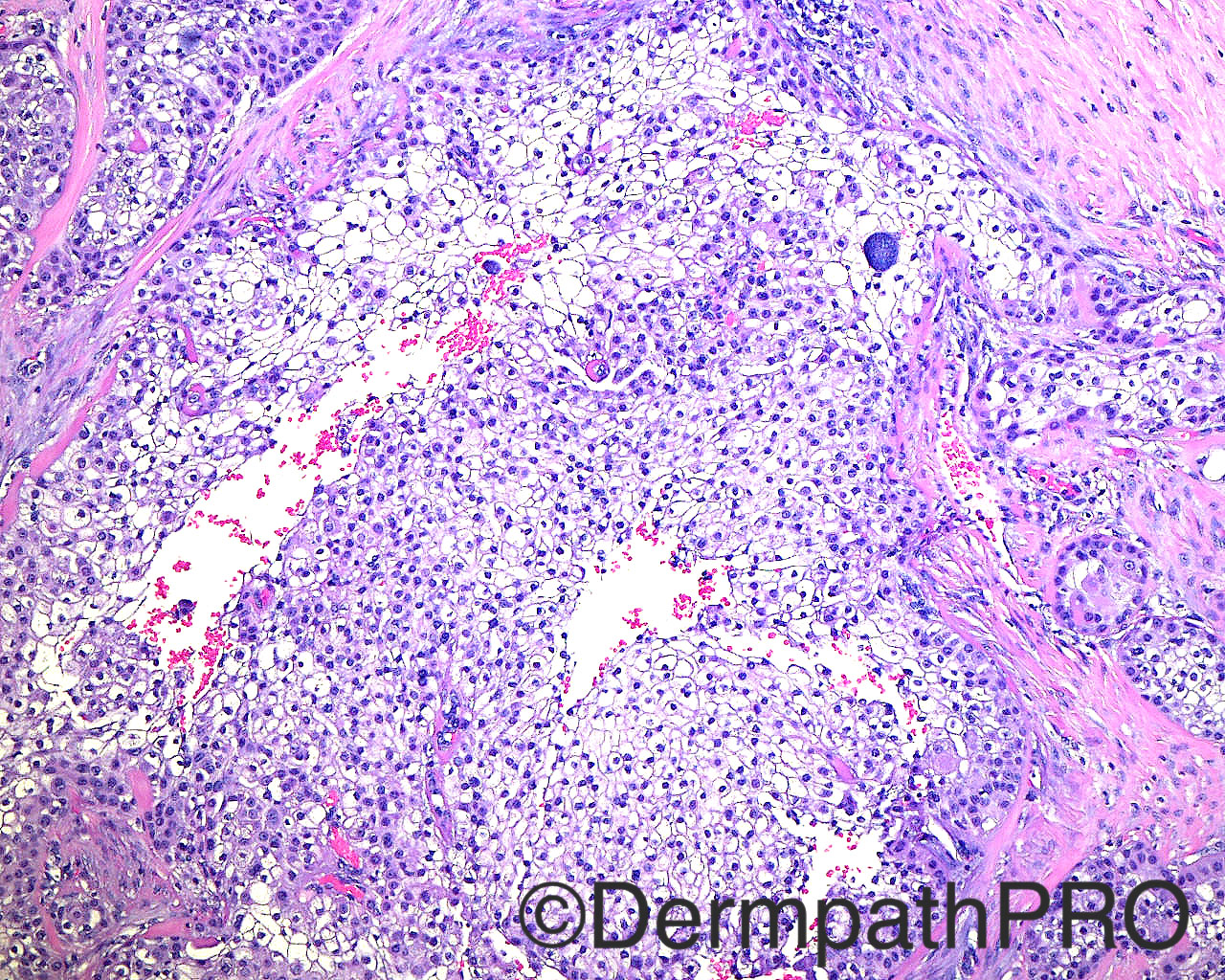
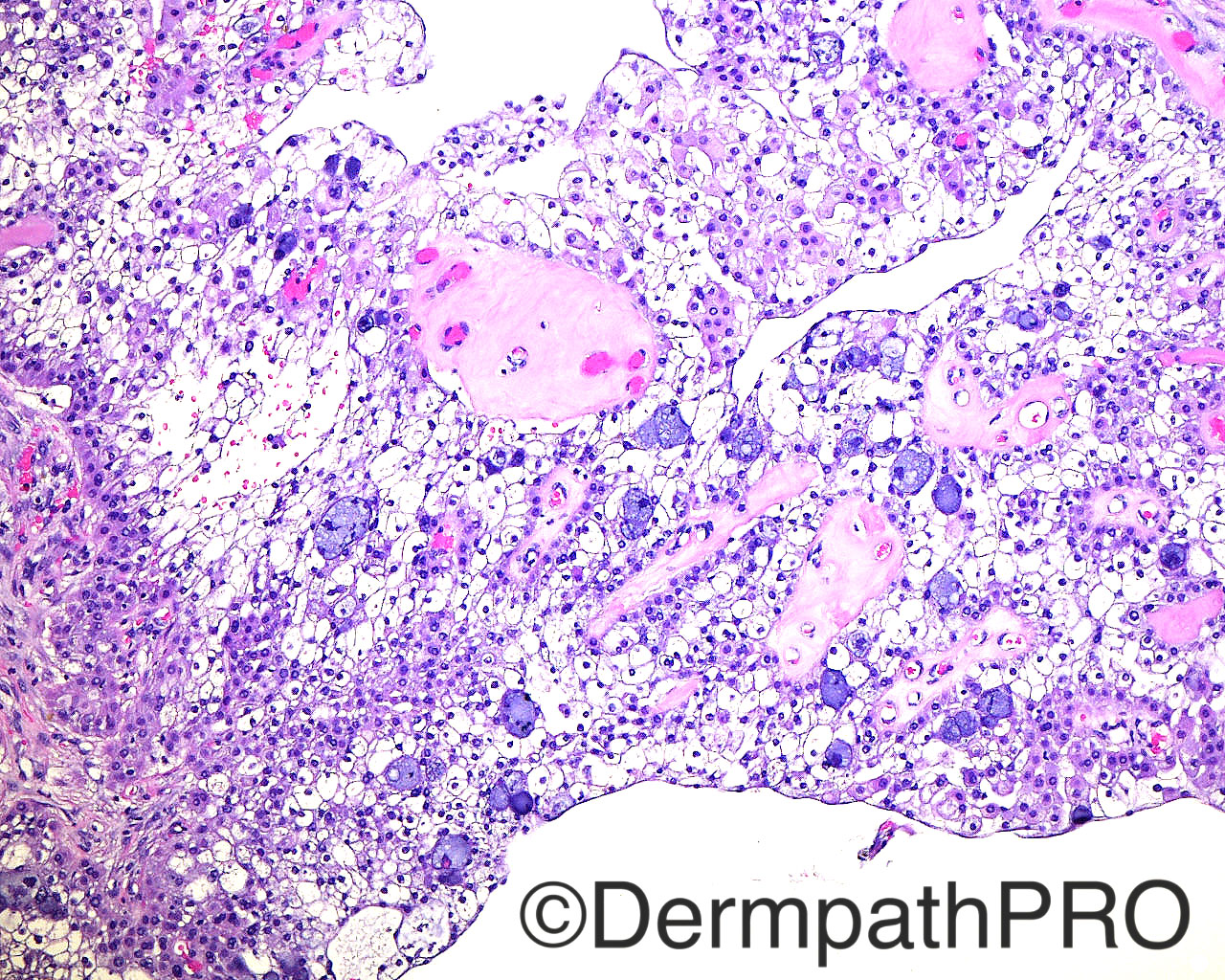
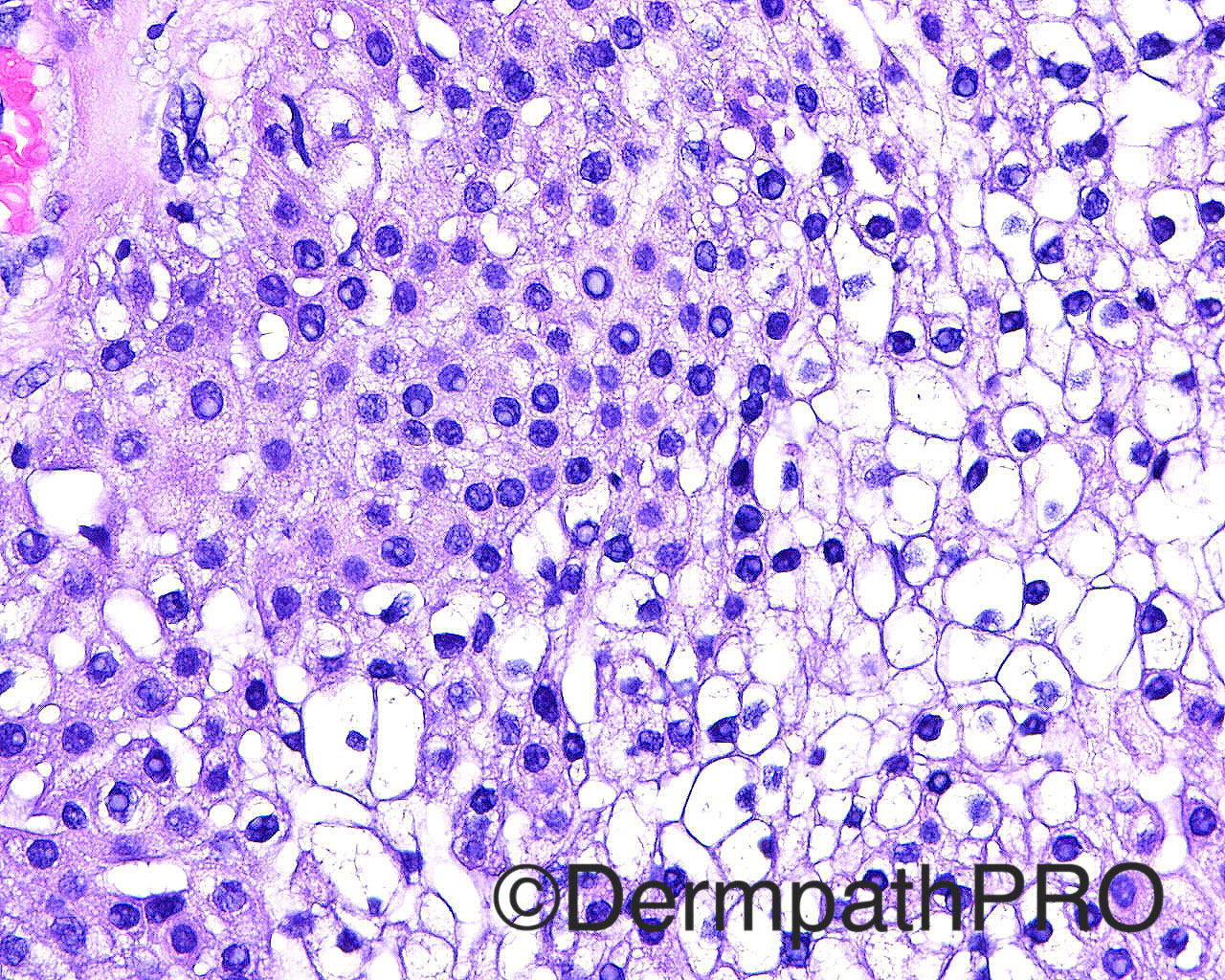
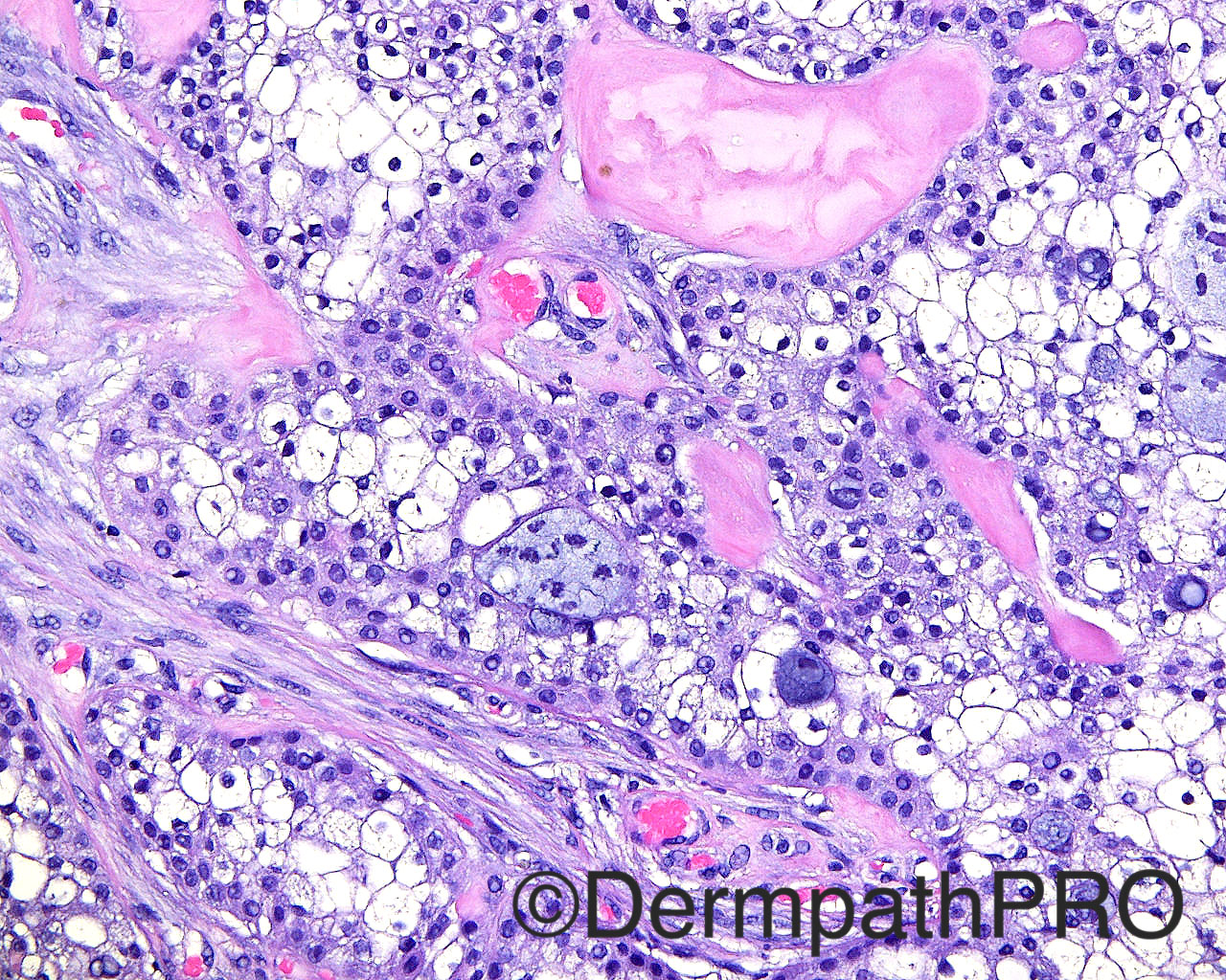
The patient is a 50 year old white man with an excision with margin exam if malignant of a fluid filled mass taken from the right posterior scalp.

Case posted by Dr Mark Hurt

  Report Record

User Feedback


vincenzo polizzi

Posted

Clear cell Hyadradenoma with goblet cell metaplasia 

Share this comment


Link to comment
share_externally

Raul Perret

Posted

I think there is loss of circumscription with areas of infiltrative pattern and deep extension. I dont see clear pleomorphism in these pictures neither mitosis but i would be very cautious with this lesion. I think it fits better with low grade hidradenocarcinoma or at least atypical like it is proposed by some authors: http://www.ncbi.nlm.nih.gov/pubmed/?term=Atypical+and+malignant+hidradenomas%3A+a+histological

Would perform ki67 and/or phh3. These tumors can be extremely unpredictable, I once saw a case that looked like a completely benign hidradenoma with only a small focus of loss of circumscription that thankfully was sent with a lymph node that the surgeon found suspicious during surgery. It turned out that the lymph node was metastatic so a dx of low grade hidradenocarcinoma was rendered.

Share this comment


Link to comment
share_externally

Dr. Mona Abdel-Halim

Posted

As Raul said, although not frankly pleomorphic and mitotic, one should deal with it in caution to avoid missing a hidradenocarcinoma

Share this comment


Link to comment
share_externally

vincenzo polizzi

Posted

Yes. The fig2 shows what looks like an infiltrative growth pattern but the other ones show a well circumscribed cystic lesion. Inside the cyst there is a perivascular extensive hyanilinization that could explain the infiltrative appearance. The Hydroadenocarcinoma is rare and usually starts ex Novo not by a malignant trasformation.

Share this comment


Link to comment
share_externally

vincenzo polizzi

Posted

But i agree with the Raul's sug in these cases.

Share this comment


Link to comment
share_externally


Join the conversation

You can post now and register later. If you have an account, sign in now to post with your account.

Guest
Add a comment...

×   Pasted as rich text.   Paste as plain text instead

  Only 75 emoji are allowed.

×   Your link has been automatically embedded.   Display as a link instead

×   Your previous content has been restored.   Clear editor

×   You cannot paste images directly. Upload or insert images from URL.

Loading...

×
×
  • Create New...