Jump to content
In this section we have spot diagnoses posted on a daily basis since June 2010, now over 4000! You can review the archived cases and read the suggested diagnoses by users and the final comment by the contributors.
Case are uploaded each week day by 10 am UK time with the correct diagnosis will generally be posted at 8 pm UK time. Why not view the most recent spot diagnosis and proffer a diagnosis?

Case Number : Case 1497- 21 March Posted By: Guest

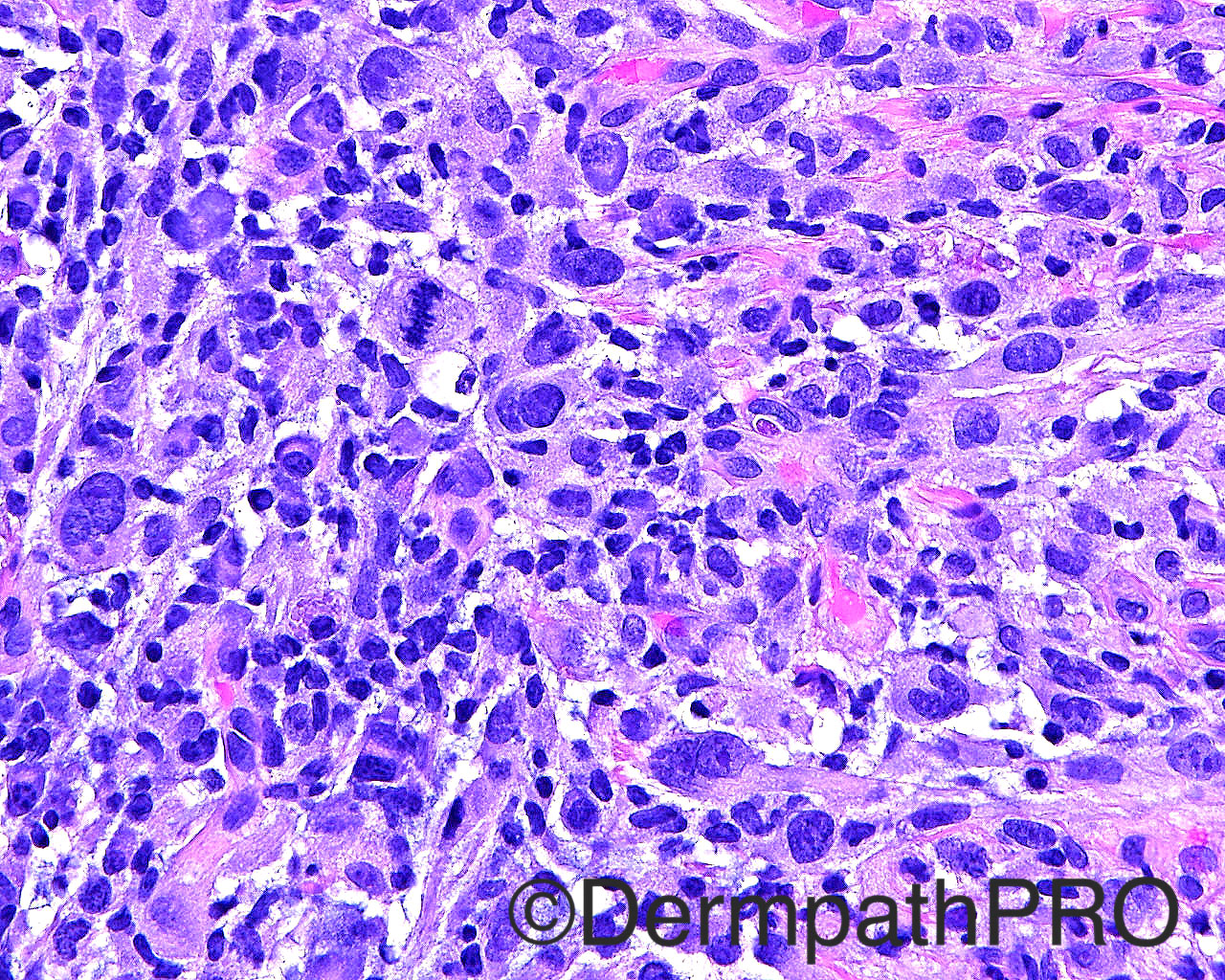
Please read the clinical history and view the images by clicking on them before you proffer your diagnosis.
Submitted Date :
   (0 reviews)

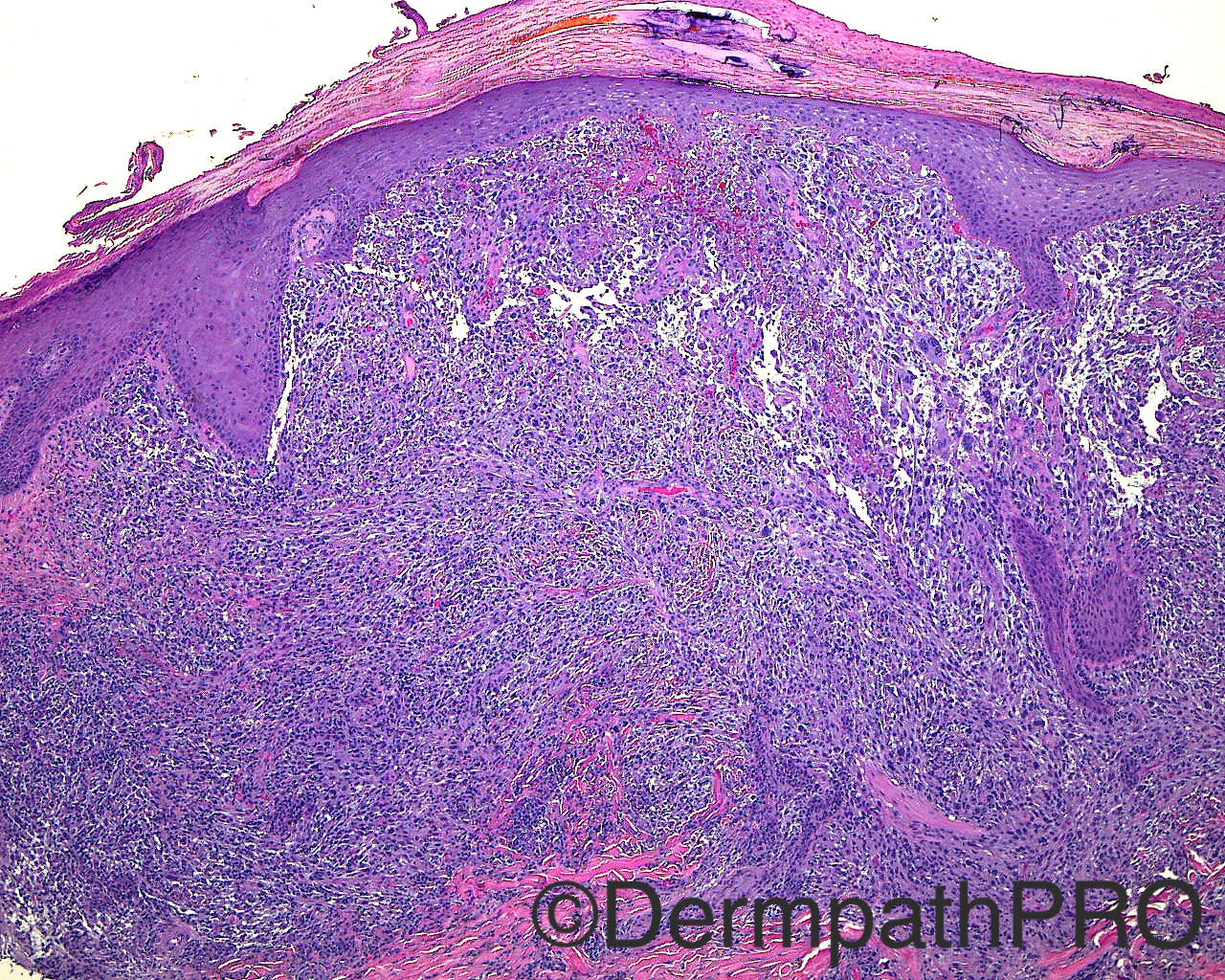
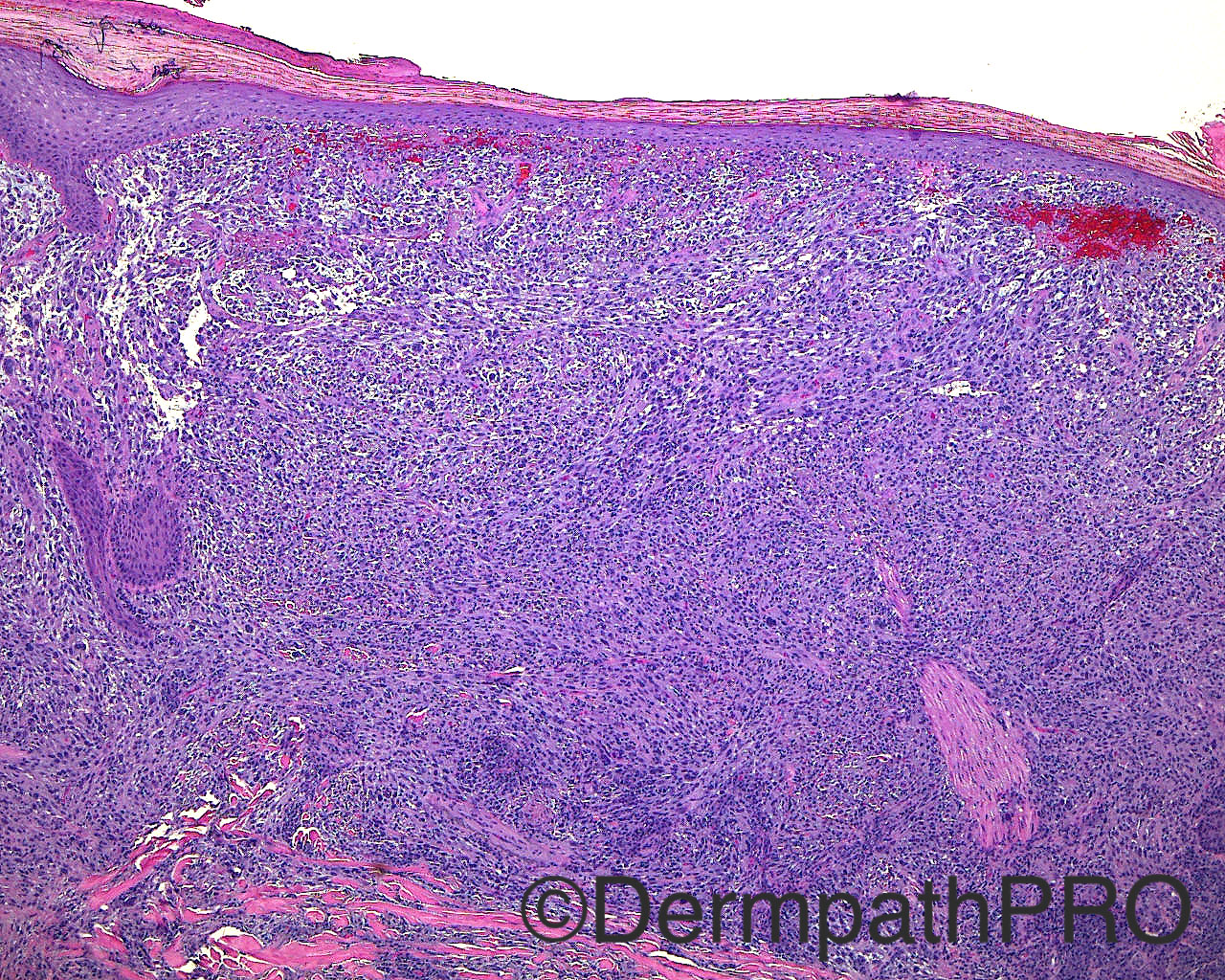
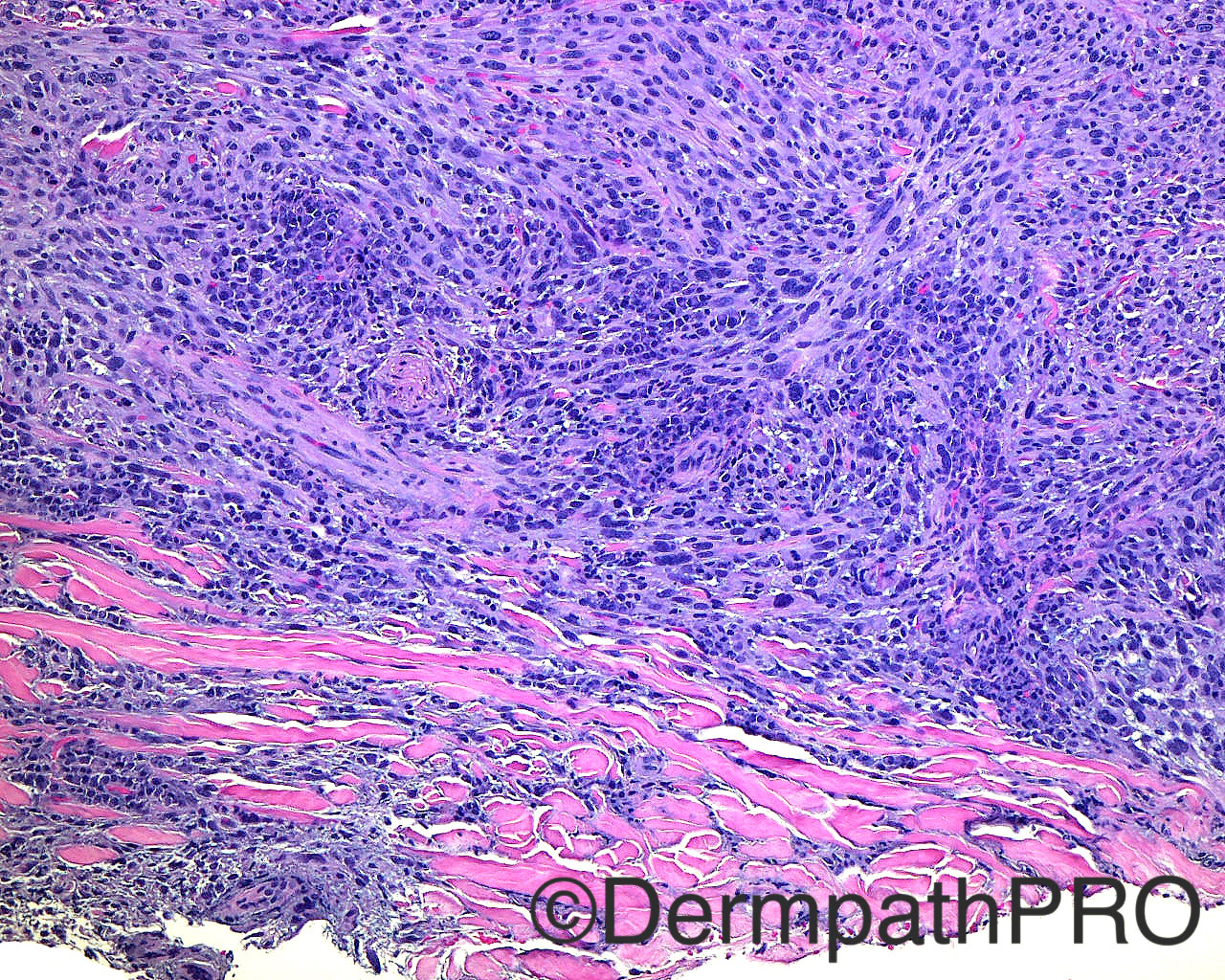
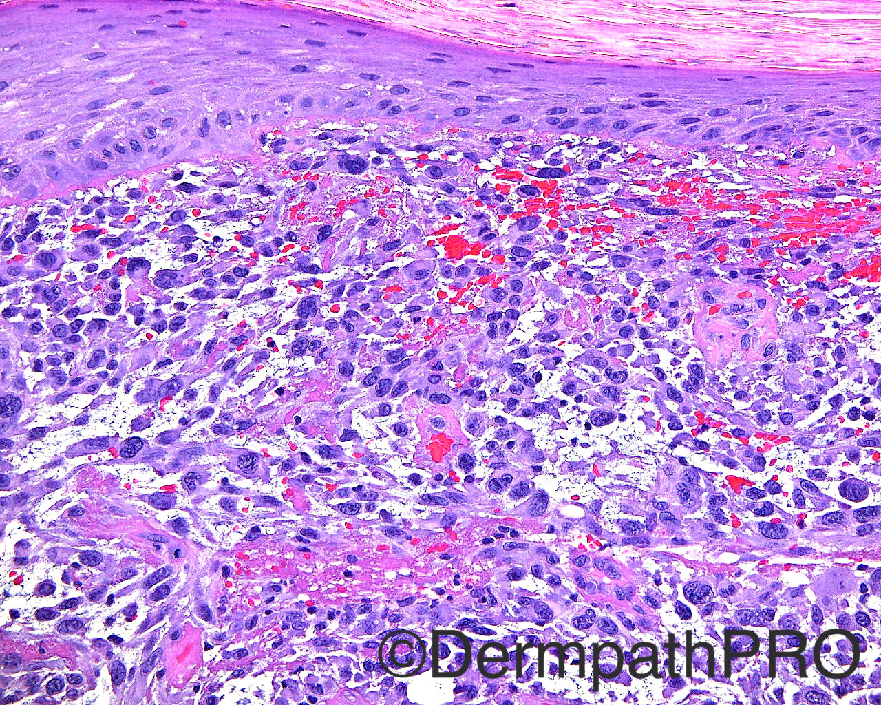
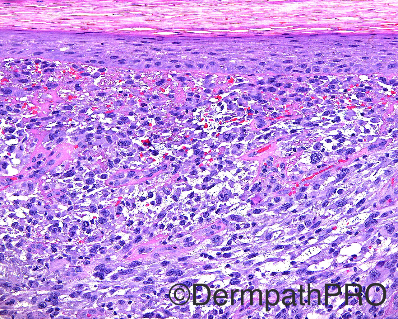
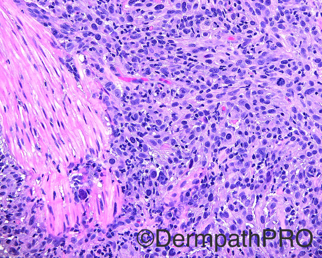
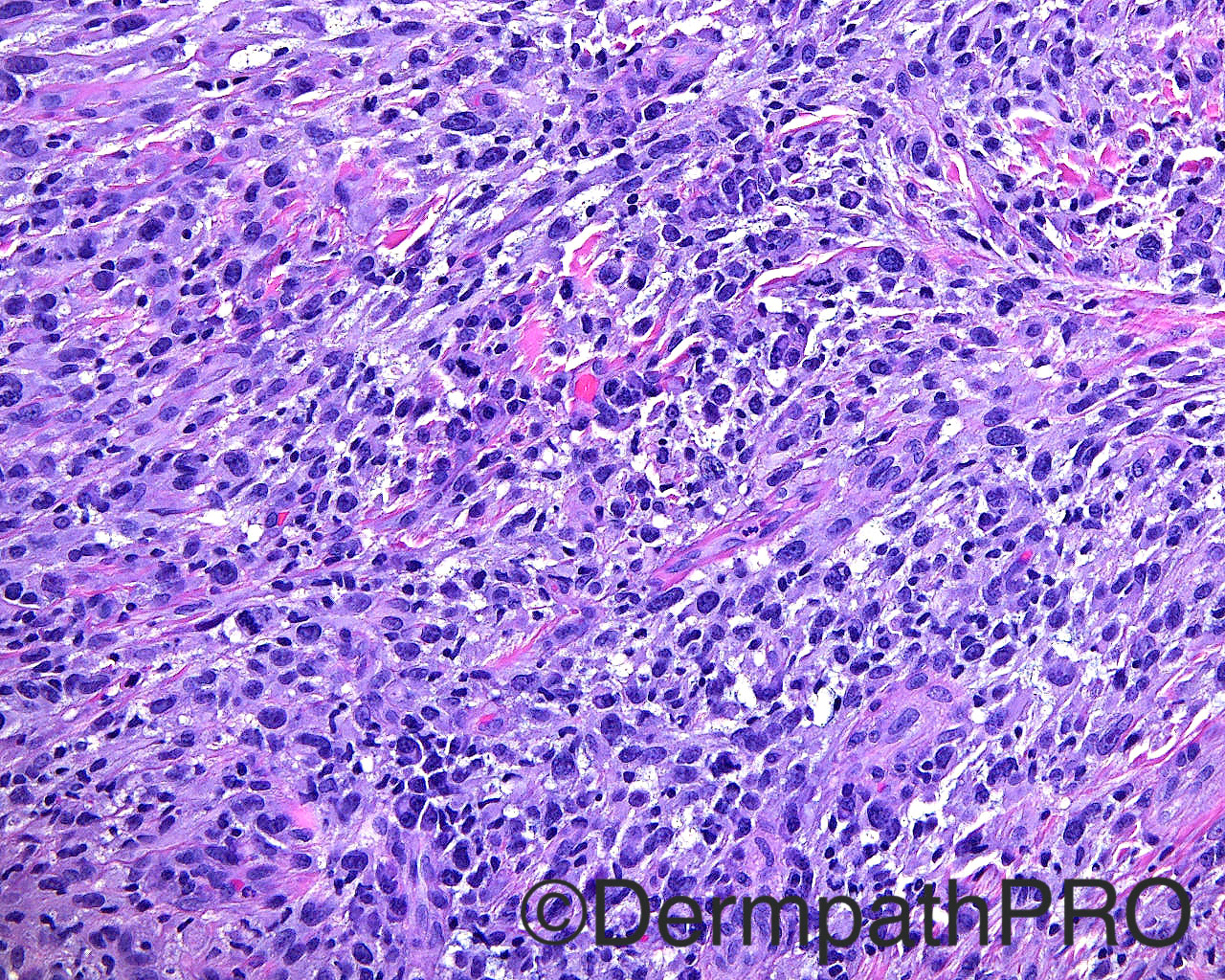
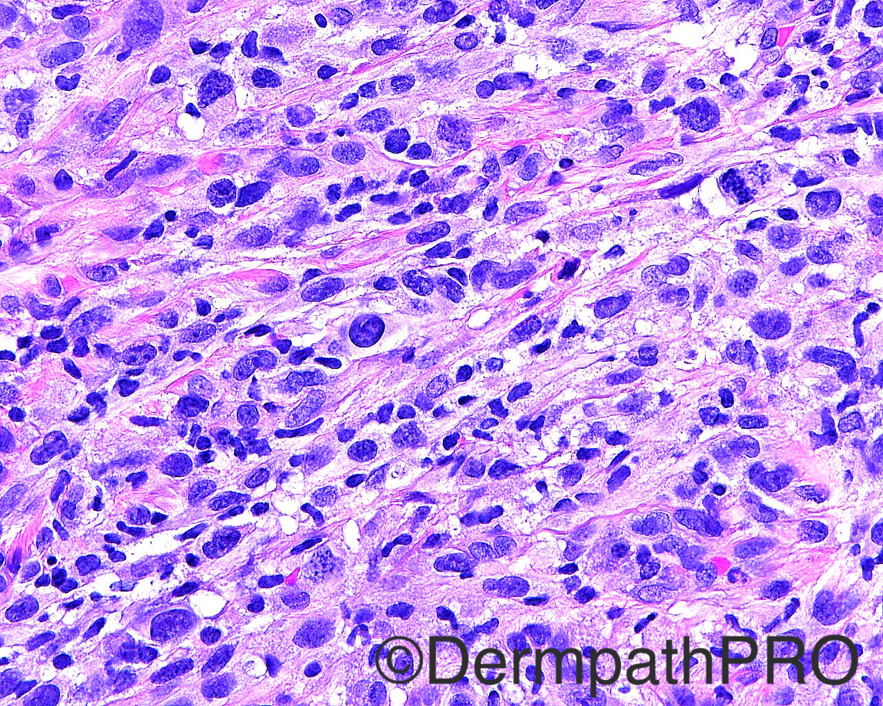
The patient is an 80 year old white man with a shave biopsy of a nonhealing bleeding lesion that gets crusted, present for three months, taken from the mid upper forehead.

Case posted by Dr Mark Hurt


  • Like 1
  Report Record

User Feedback


Raul Perret

Posted

Due to clinical information and morphology I would consider atypical fibroxanthoma, it bothers me that there is no grenz zone though. Of course a wide panel of immunos should be performed first (CK, P63 or P40, S-100, melanocytic markers, desmin, CD31, CD10). Picture 5 shows some basal membrane thickening I was wondering the cause...

Share this comment


Link to comment
share_externally

vincenzo polizzi

Posted

Pleomorphic tumor, sarcomatoid pattern, with perinuclear cytoplasmic clearing, Golgi-like, reminiscent of ALCL, sarcomatous type. Obviously it needs IHC.

Share this comment


Link to comment
share_externally

vincenzo polizzi

Posted

I may be wrong but i see several cells with abundant cytoplasm, horseshoe (“hallmark”) nucleus or wreath-like or multiple nuclei, perinuclear eosinophilic region...so i favor Anaplastic Large Cell Lymphoma.

Share this comment


Link to comment
share_externally

Dr. Mona Abdel-Halim

Posted

I thought based on morphology of ALCL, but complete IHC work up should be done

Share this comment


Link to comment
share_externally

Nitin Khirwadkar

Posted

Poorly differentiated tumour, requiring a good panel of IHC. My differentials, pleomorphic dermal sarcoma/AFX, angiosarcoma, pseudovascular SCC, melanoma (unlikely), LMS.

Share this comment


Link to comment
share_externally


Join the conversation

You can post now and register later. If you have an account, sign in now to post with your account.

Guest
Add a comment...

×   Pasted as rich text.   Paste as plain text instead

  Only 75 emoji are allowed.

×   Your link has been automatically embedded.   Display as a link instead

×   Your previous content has been restored.   Clear editor

×   You cannot paste images directly. Upload or insert images from URL.

Loading...

×
×
  • Create New...