Jump to content
In this section we have spot diagnoses posted on a daily basis since June 2010, now over 4000! You can review the archived cases and read the suggested diagnoses by users and the final comment by the contributors.
Case are uploaded each week day by 10 am UK time with the correct diagnosis will generally be posted at 8 pm UK time. Why not view the most recent spot diagnosis and proffer a diagnosis?

Case Number : Case 1498- 22 March Posted By: Guest

Please read the clinical history and view the images by clicking on them before you proffer your diagnosis.
Submitted Date :
   (0 reviews)

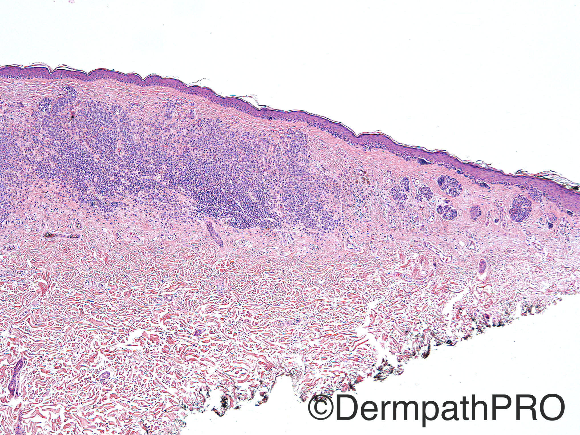
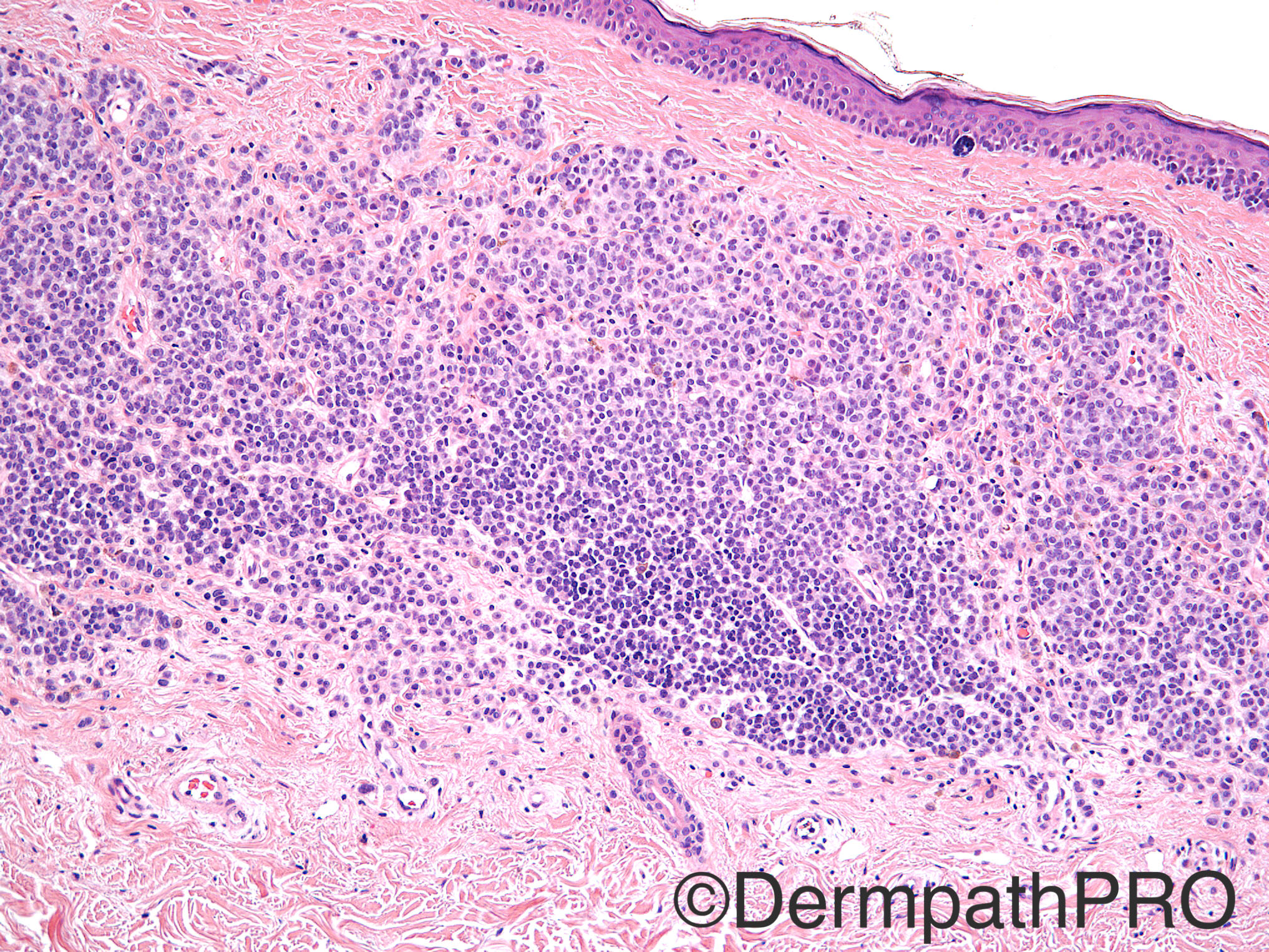
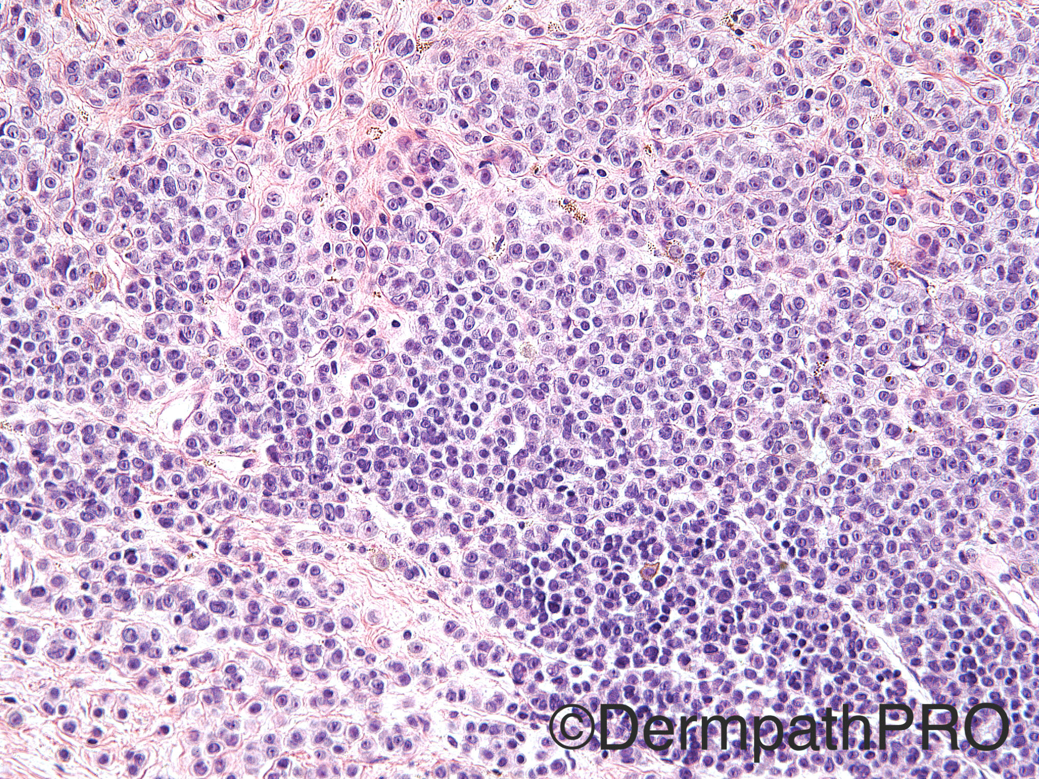
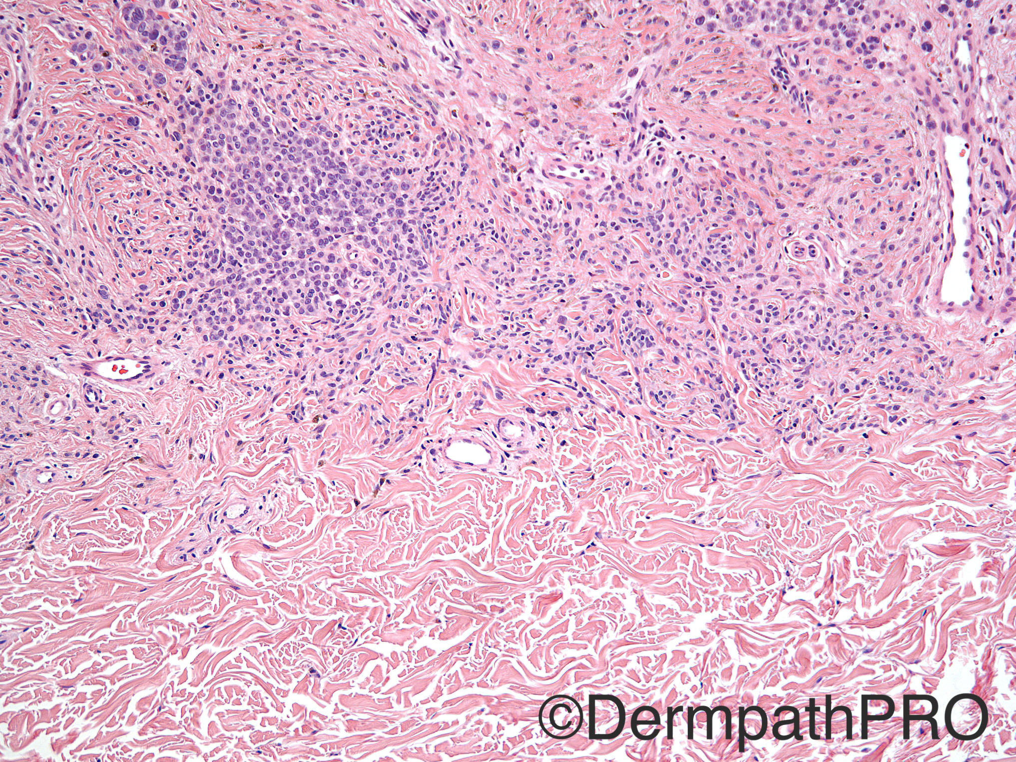
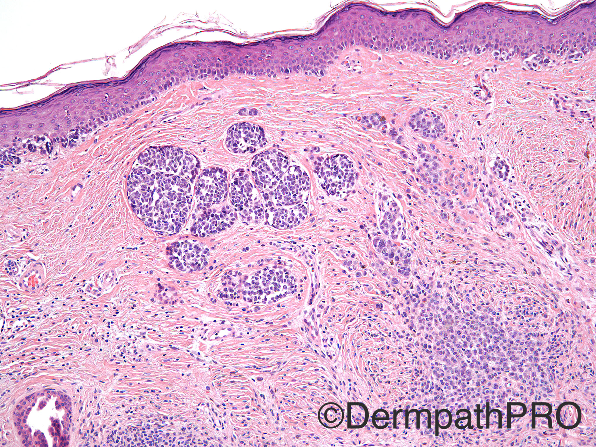
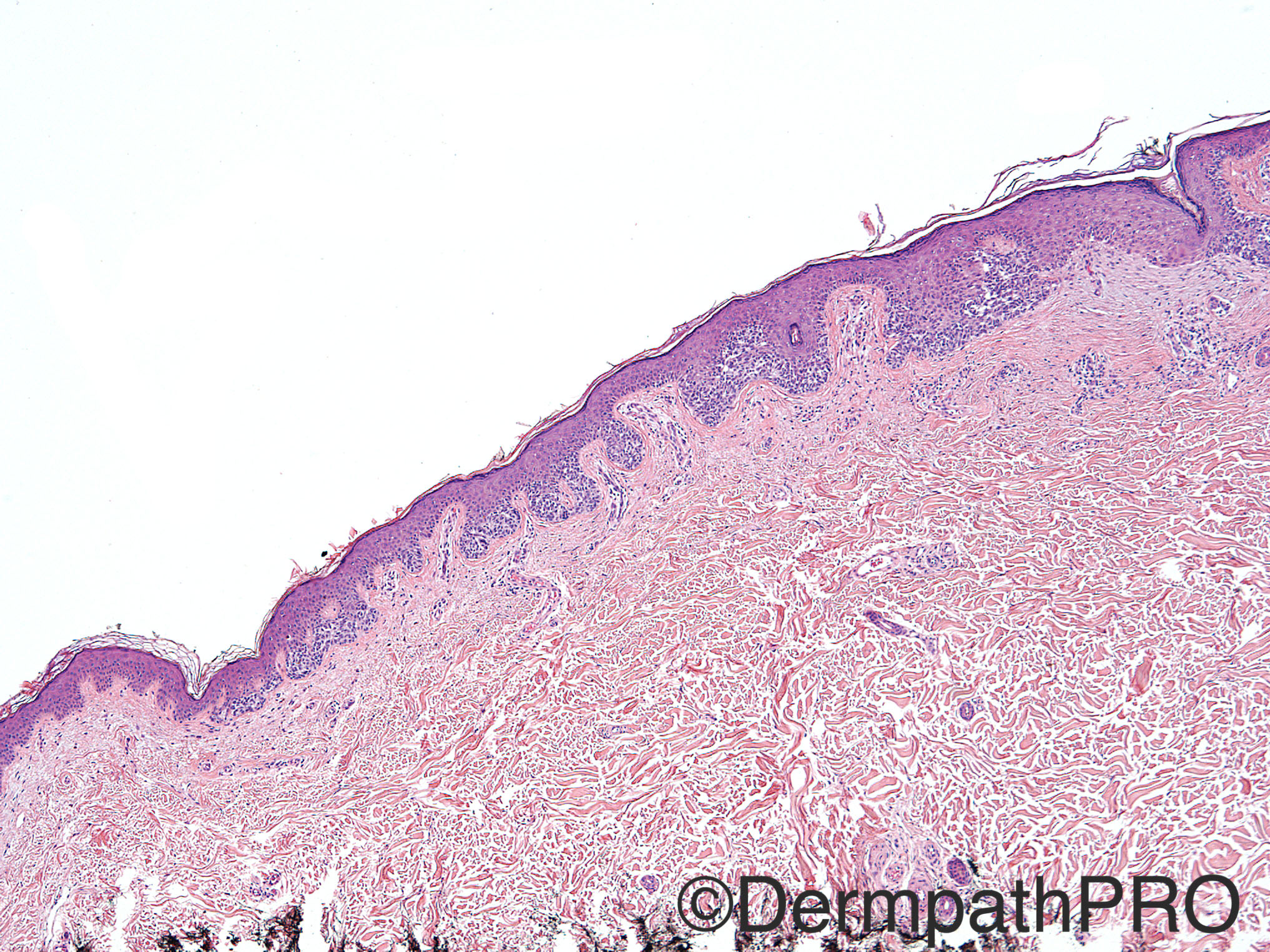
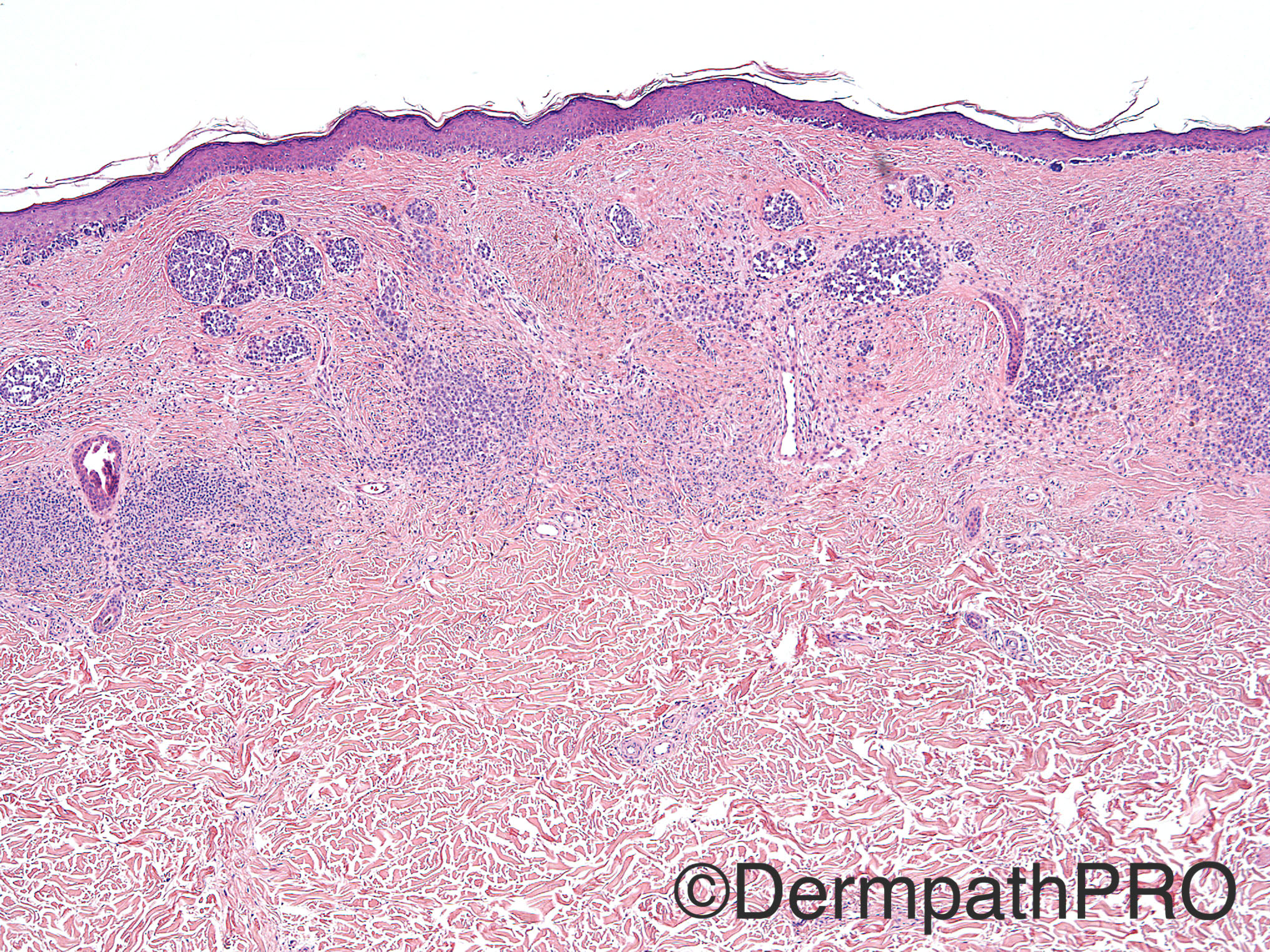
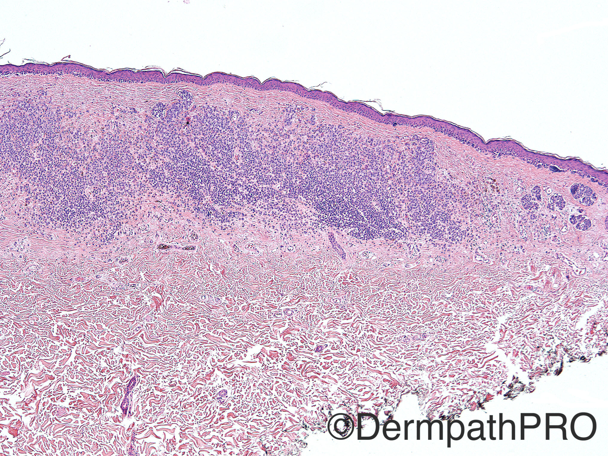
63 year old male with 1.5 x 1 cm atypical nevus on right abdomen.

Case posted by Dr Uma Sundram


  • Like 2
  Report Record

User Feedback


Raul Perret

Posted

Melanoma with concomitant banal nevus. Maybe developed on the last one but who knows...

Share this comment


Link to comment
share_externally

Dr. Mona Abdel-Halim

Posted

Melanoma on top of a nevus.

Share this comment


Link to comment
share_externally

vincenzo polizzi

Posted

There is a trizonal pattern, with a lentiginous junctional proliferation of atypical melanocytes at the top, a residual nevus at the bottom and a dermal scar in between. That would be reminiscent of a recurrent nevus...

Share this comment


Link to comment
share_externally

Nitin Khirwadkar

Posted

Looks like a melanoma (small cell)  that has developed within a precursor naevus. The junctional component is quite worrying. There is some dermal regression, aggregates of atypical melanocytes are noted, along with a banal component. A p16 may help in highlighting the precursor naevus.

Share this comment


Link to comment
share_externally

Uma Sundram

Posted

Nevoid melanoma. Very difficult case. The panel with the super small cells is the focus of residual nevus but even the areas that look like maturing nevus cells within the dermis consist of nevoid or small cell melanoma. Was sent out in consultation to UCSF who agreed, with array CGH analysis supporting a melanoma profile.

Share this comment


Link to comment
share_externally


Join the conversation

You can post now and register later. If you have an account, sign in now to post with your account.

Guest
Add a comment...

×   Pasted as rich text.   Paste as plain text instead

  Only 75 emoji are allowed.

×   Your link has been automatically embedded.   Display as a link instead

×   Your previous content has been restored.   Clear editor

×   You cannot paste images directly. Upload or insert images from URL.

Loading...

×
×
  • Create New...