Jump to content
In this section we have spot diagnoses posted on a daily basis since June 2010, now over 4000! You can review the archived cases and read the suggested diagnoses by users and the final comment by the contributors.
Case are uploaded each week day by 10 am UK time with the correct diagnosis will generally be posted at 8 pm UK time. Why not view the most recent spot diagnosis and proffer a diagnosis?

Case Number : Case 1501- 25 March Posted By: Guest

Please read the clinical history and view the images by clicking on them before you proffer your diagnosis.
Submitted Date :
   (0 reviews)

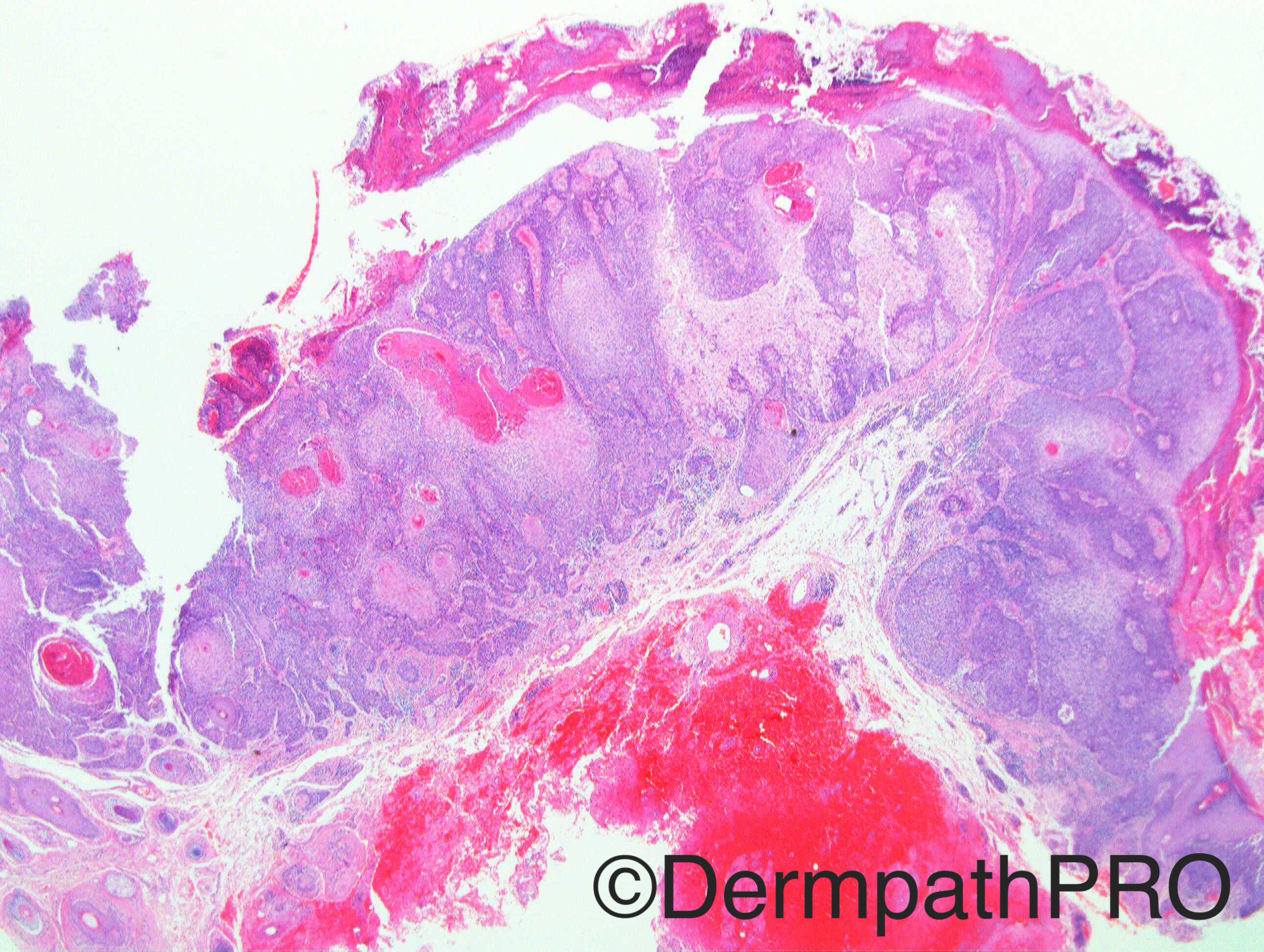
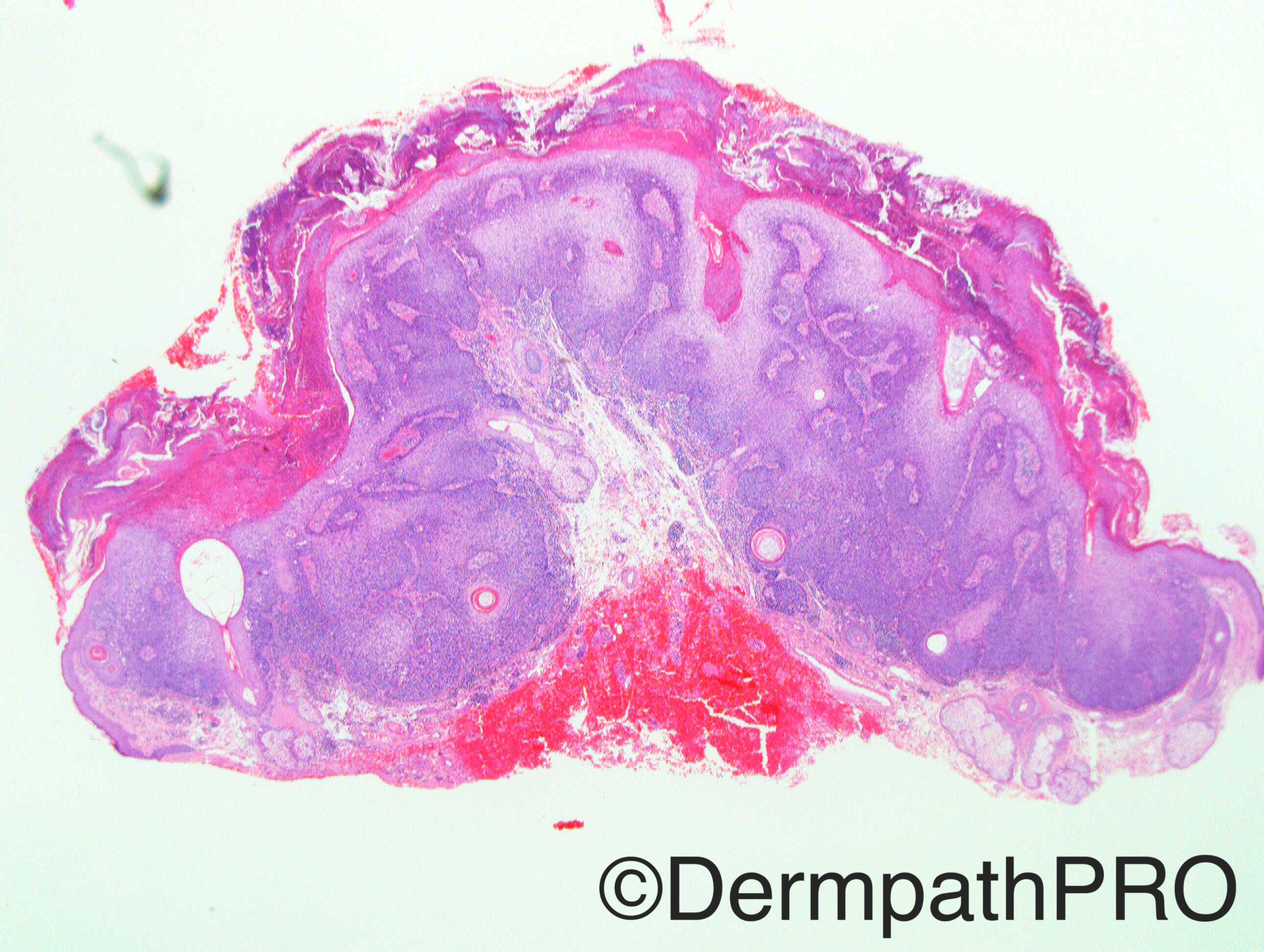
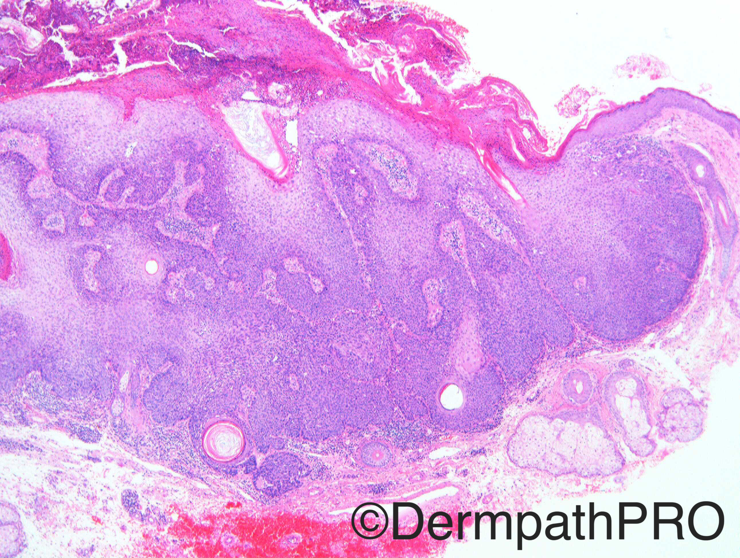
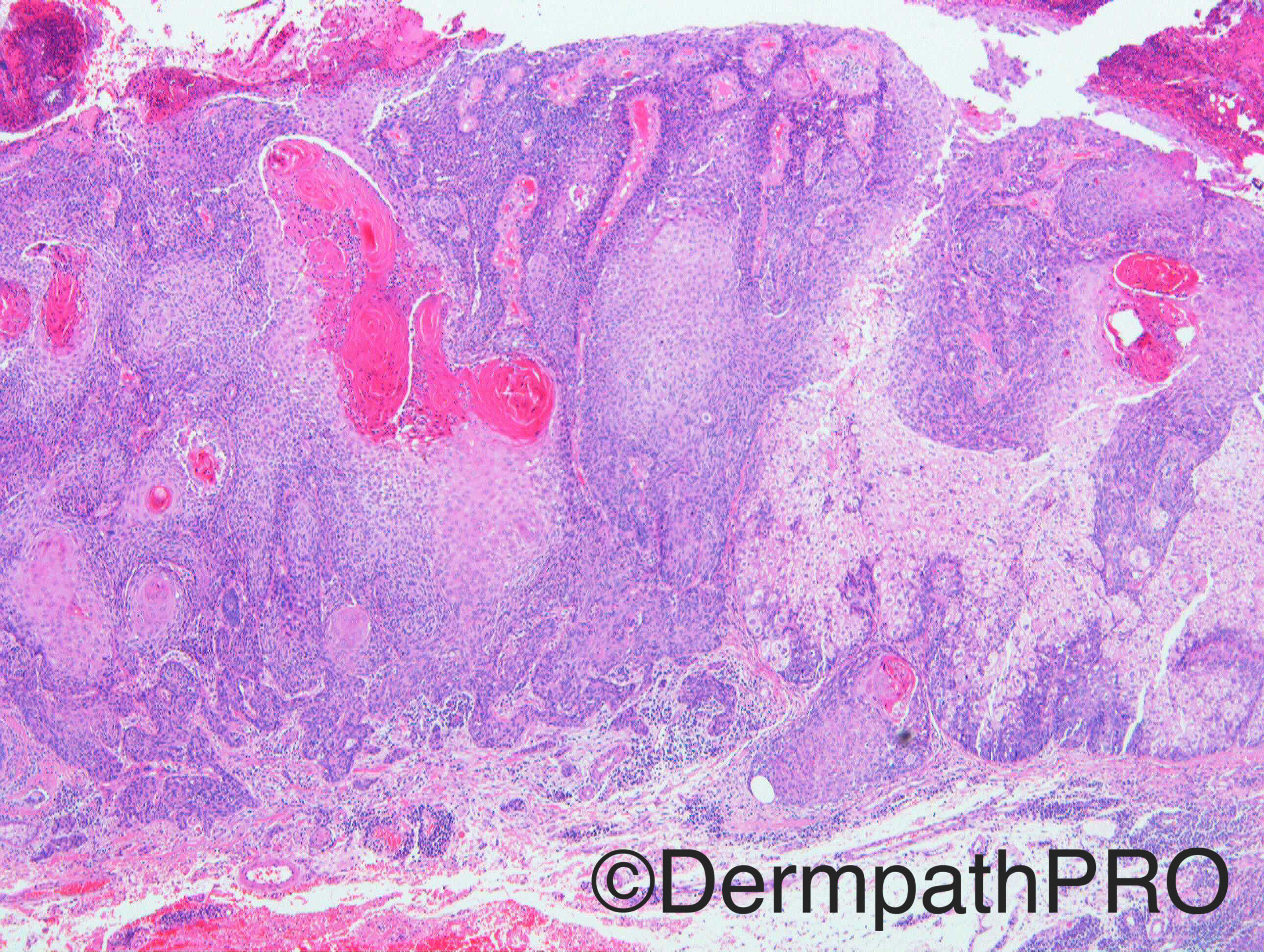
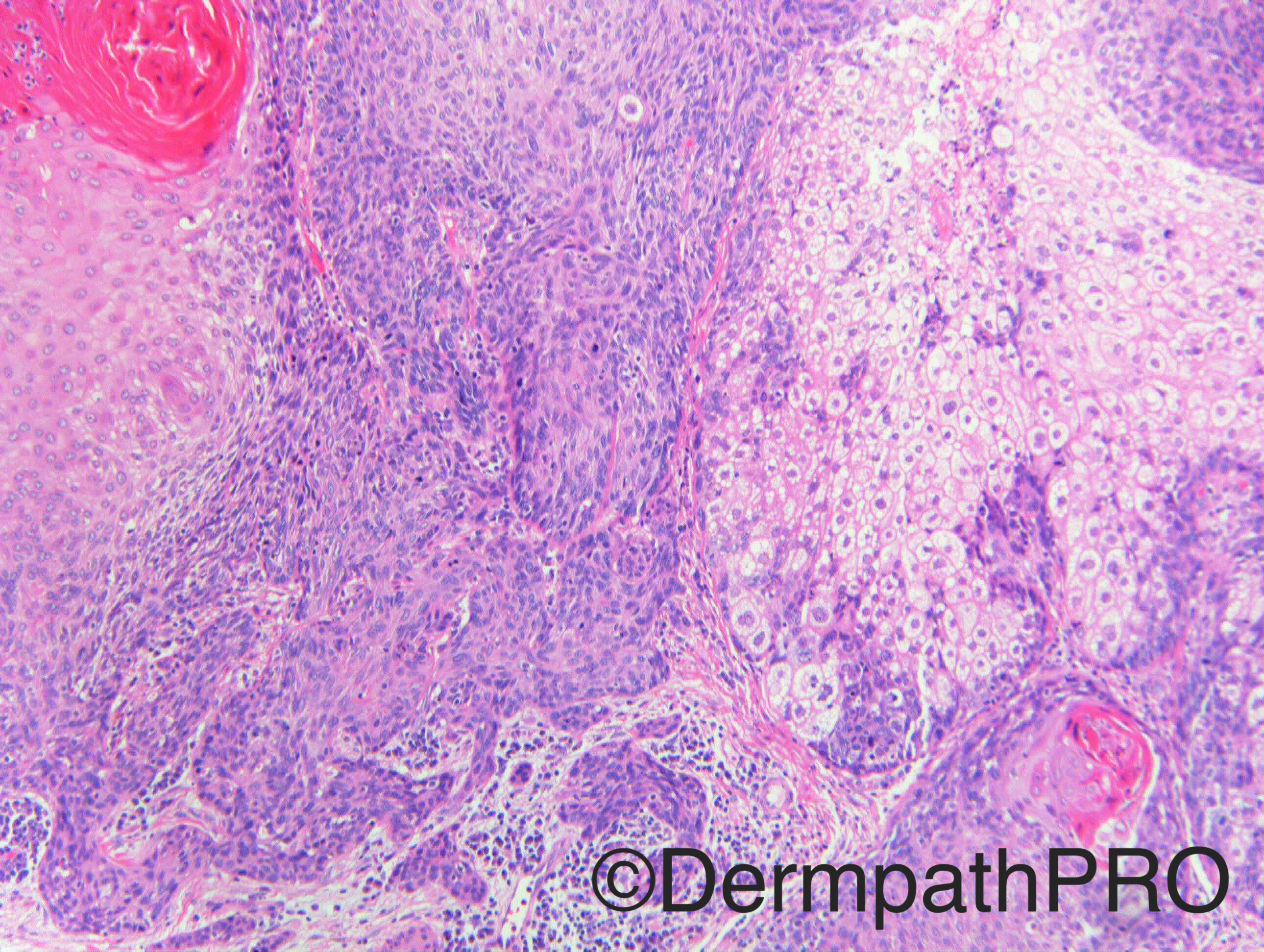
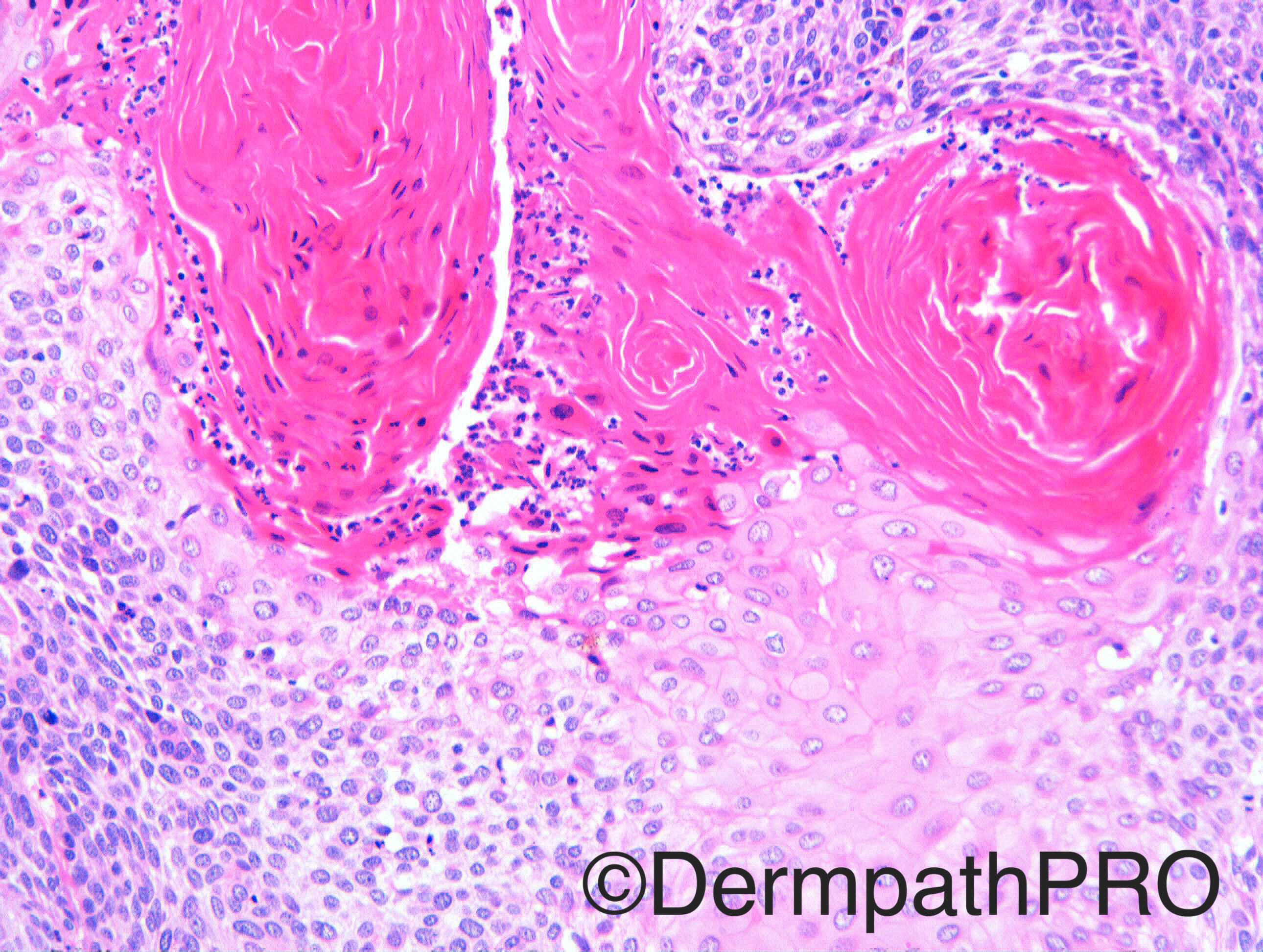
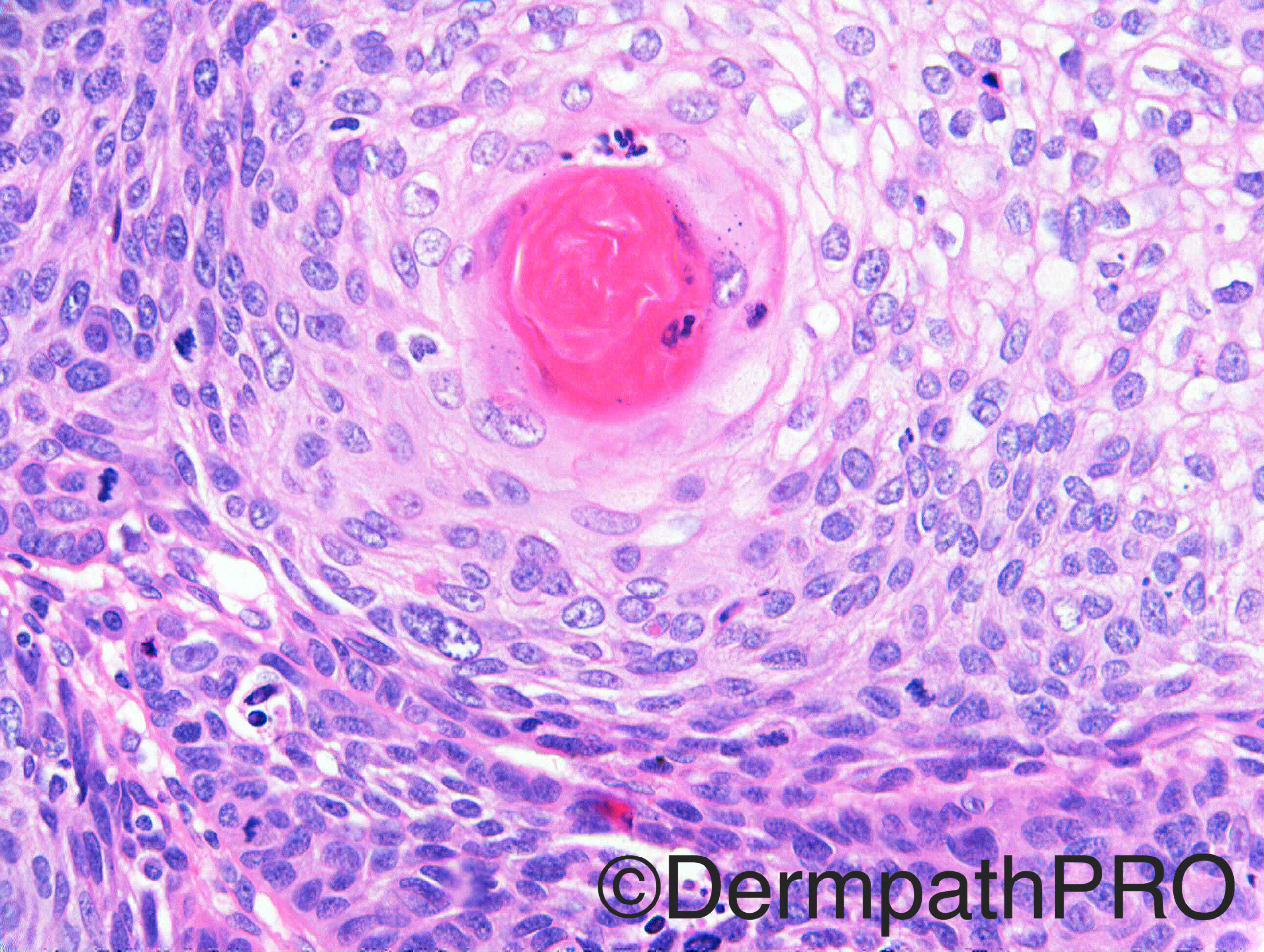
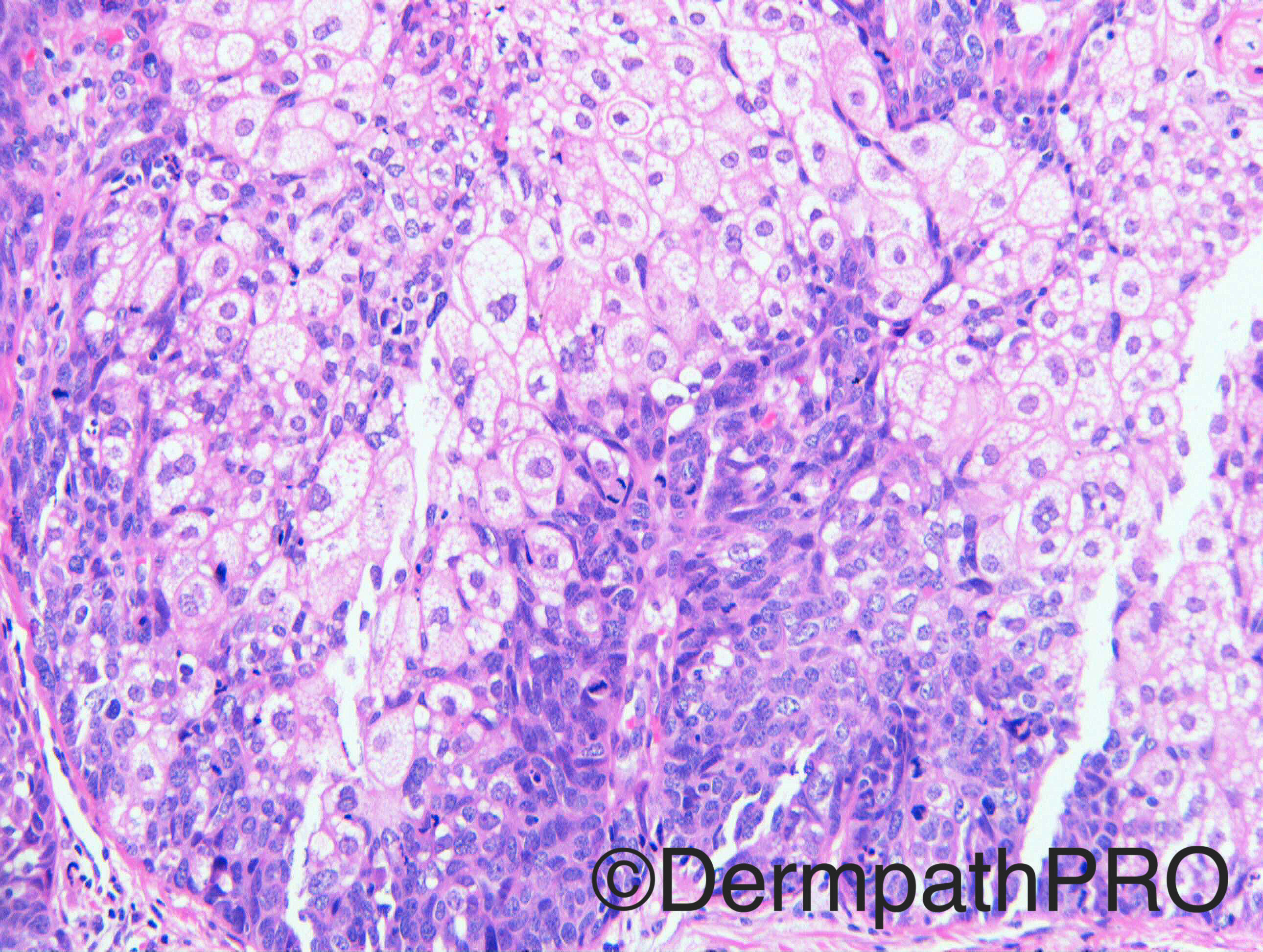
M80 Upper eyelid ?SCC, ?Viral wart. Case c/o Dr Veena Shinde

Case posted by Dr Richard Carr

  Report Record

User Feedback


Dr. Mona Abdel-Halim

Posted

Seb carcinoma with areas of squamous differentiation

Share this comment


Link to comment
share_externally

Raul Perret

Posted

Agree with Mona sebaceous carcinoma with squamous metaplasia. A differential could be a collision tumor but the áreas of squamous differentiation dont seem atypical in this case

Share this comment


Link to comment
share_externally

Guest Romualdo

Posted

I am also thinking of the possibility suggested by Raul: a collision tumor, sebaceous and squamous cell carcinoma.

Share this comment


Link to comment
share_externally

vincenzo polizzi

Posted

Trichilemmal carcinoma with sebaceous differentiation

Share this comment


Link to comment
share_externally

Guest Arash Daryakarr

Posted

I think this is sebaceous ca with areas of squamous metaplasia.

Share this comment


Link to comment
share_externally

Nitin Khirwadkar

Posted

Sebaceous carcinoma with squamous metaplasia. Initial images look like a sebaceoma. Has largely pushing borders, focally infiltrative.

Share this comment


Link to comment
share_externally

Sasi Attili

Posted

Agree, Sebaceous Carcinoma (infiltrative focally) with squamous differentiation. Maybe a bit too old but consider Muir Torre Syndrome? Have seen it in rare cases, present very late

Share this comment


Link to comment
share_externally

Dr. Richard Carr

Posted

Thanks for all the excellent responses.  I am on a little holiday hence delay in my post.  I called this sebaceous carcinoma with tricholemmal (follicular) carcinoma-like areas.  The lobulated, folliculocentric pattern is seen in the low powers and I suspect the lesion is overwhelmingly in situ so should have a very good prognosis.  I guess this is another example of a hybrid sebaceous-follicular-apocrine duct unit neoplasm. Of course the sebaceous duct drains in to the hair follicle so this combination is not entirely surprising or perhaps it is to be expected.  I don't recall having seen a case like this previously where the two differentiations were so well formed but also intermixed. It is funny when we see a tumour with squamous differentiation and another differentiation e.g. sebaceous or glandular the latter defines the classification.  Perhaps it is more precise to acknowledge the hybrids e.g. Carcinoma with sebaceous and follicular tricholemmal differentiation. The age and site would be most unusual for Muir Torre but no harm in mentioning that it is unlikely to be associated with Muir Torre in the report to remind the clinical colleague to think of the possibility.

Share this comment


Link to comment
share_externally


Join the conversation

You can post now and register later. If you have an account, sign in now to post with your account.

Guest
Add a comment...

×   Pasted as rich text.   Paste as plain text instead

  Only 75 emoji are allowed.

×   Your link has been automatically embedded.   Display as a link instead

×   Your previous content has been restored.   Clear editor

×   You cannot paste images directly. Upload or insert images from URL.

Loading...

×
×
  • Create New...