Jump to content
In this section we have spot diagnoses posted on a daily basis since June 2010, now over 4000! You can review the archived cases and read the suggested diagnoses by users and the final comment by the contributors.
Case are uploaded each week day by 10 am UK time with the correct diagnosis will generally be posted at 8 pm UK time. Why not view the most recent spot diagnosis and proffer a diagnosis?

Case Number : Case 1527 - 02 May Posted By: Guest

Please read the clinical history and view the images by clicking on them before you proffer your diagnosis.
Submitted Date :
   (0 reviews)

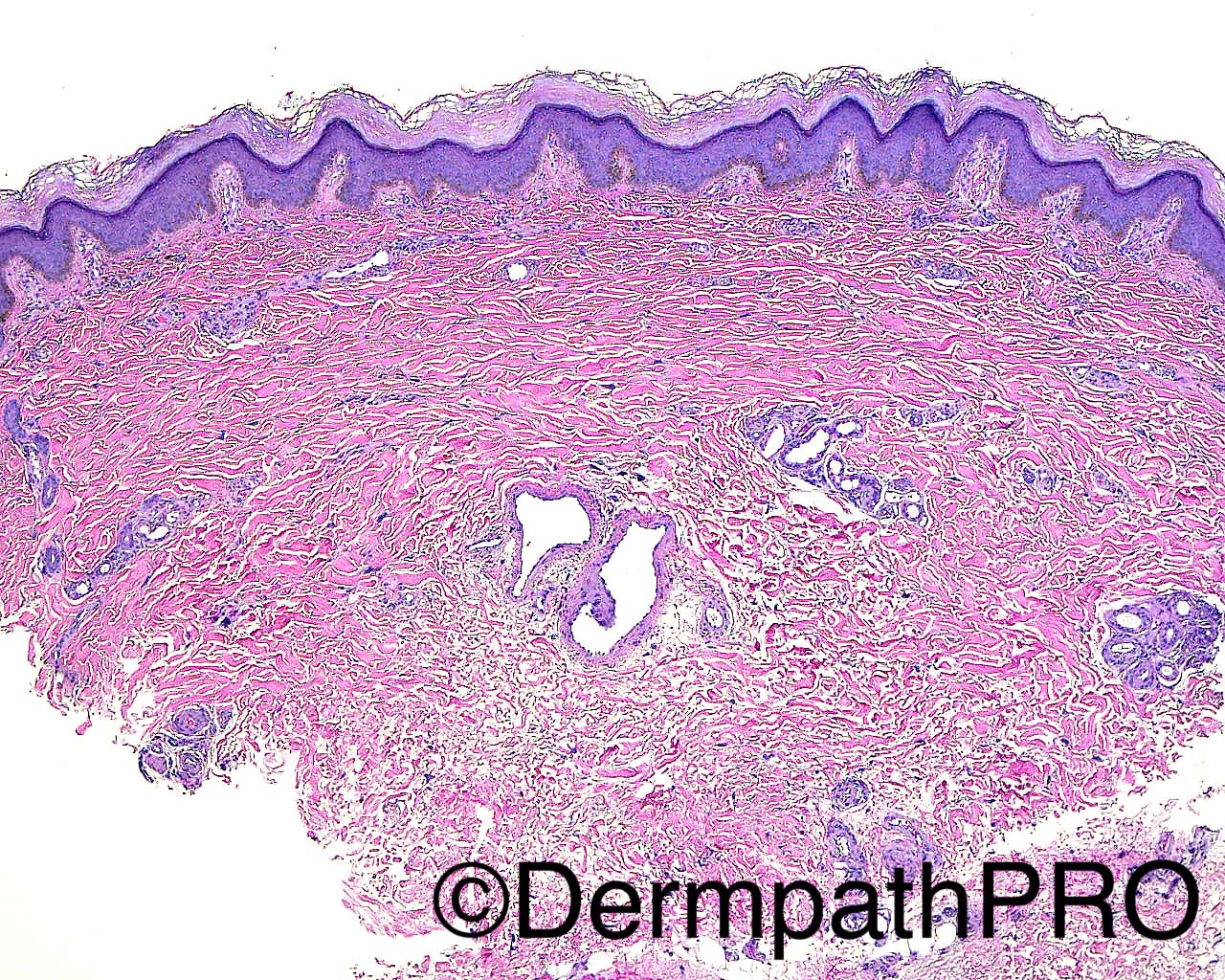
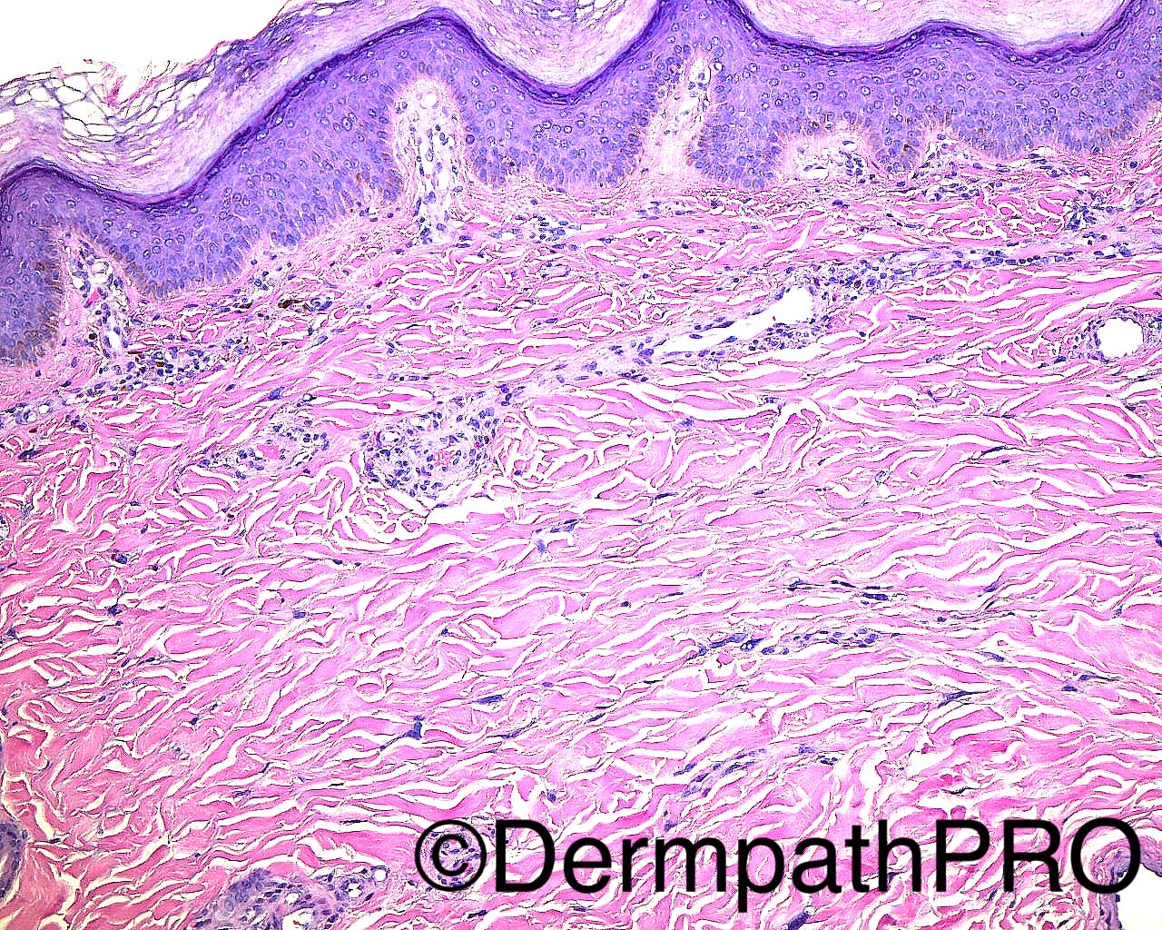
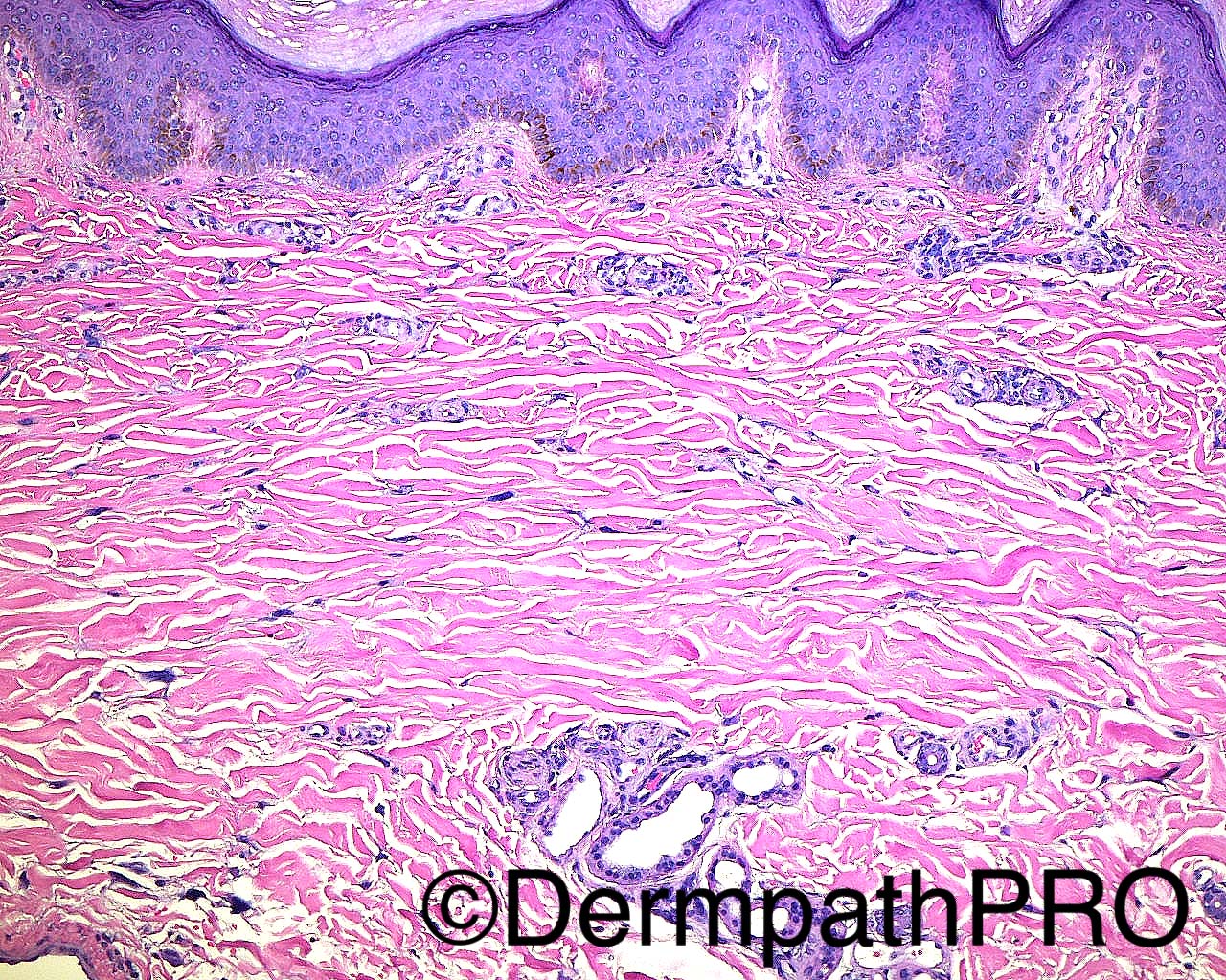
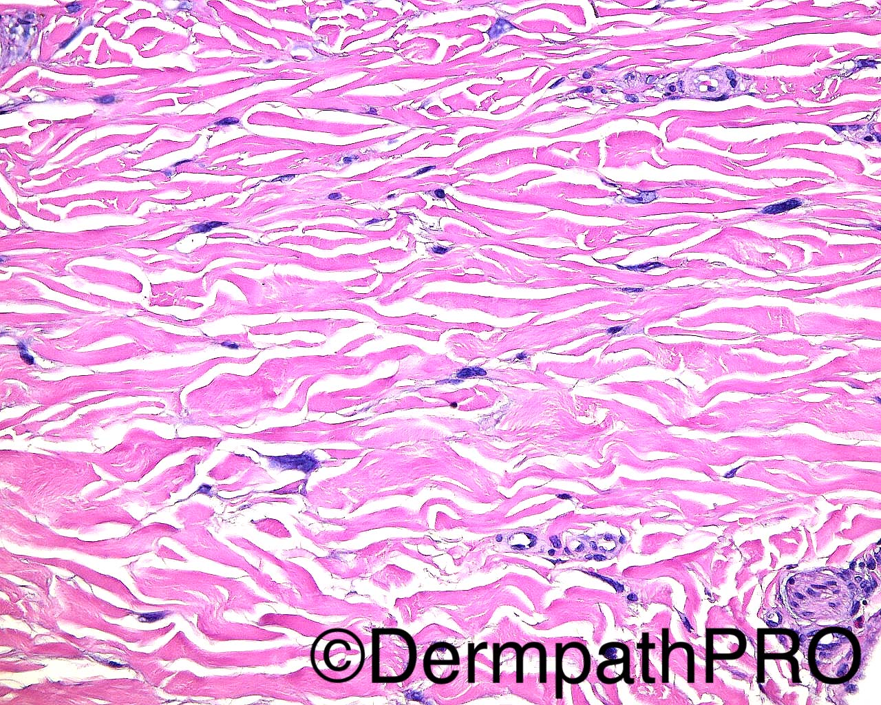
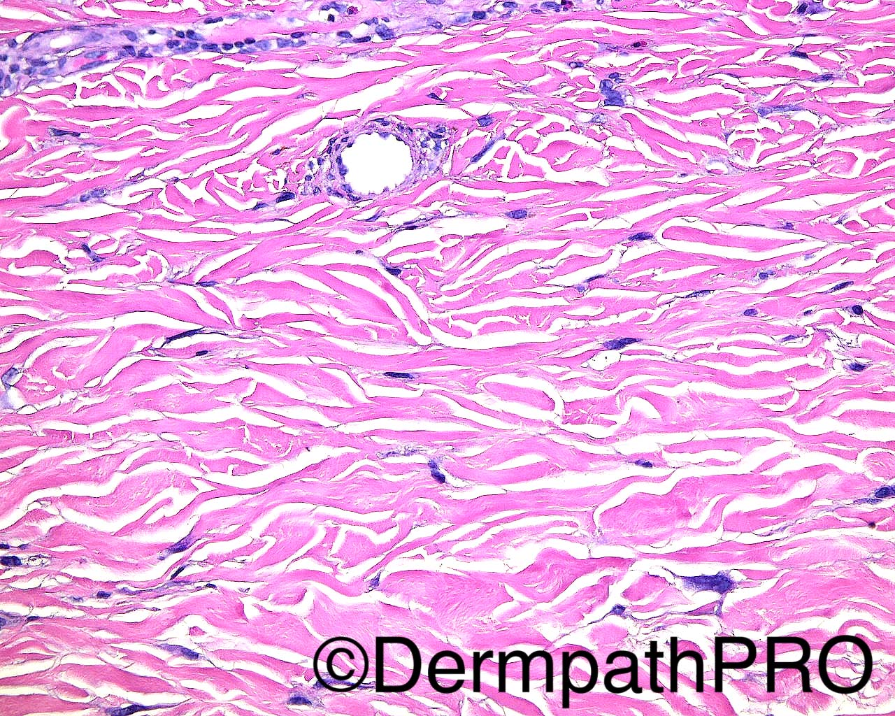
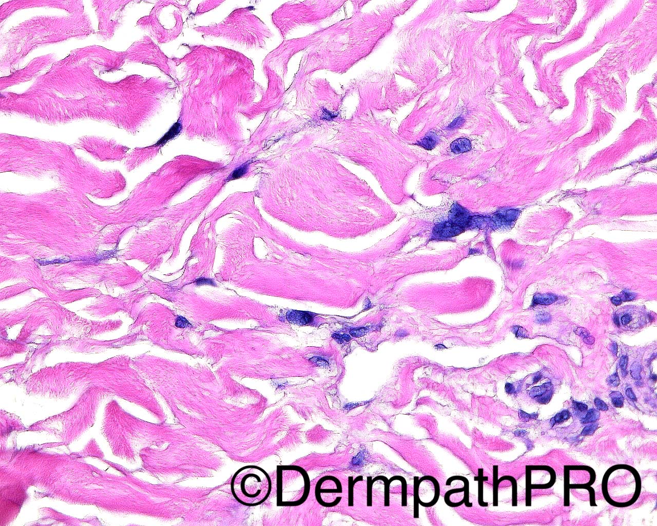
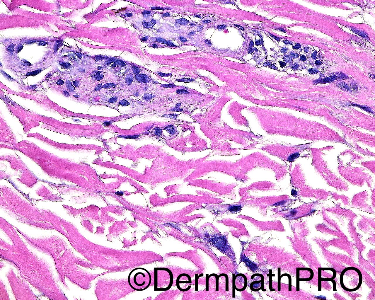
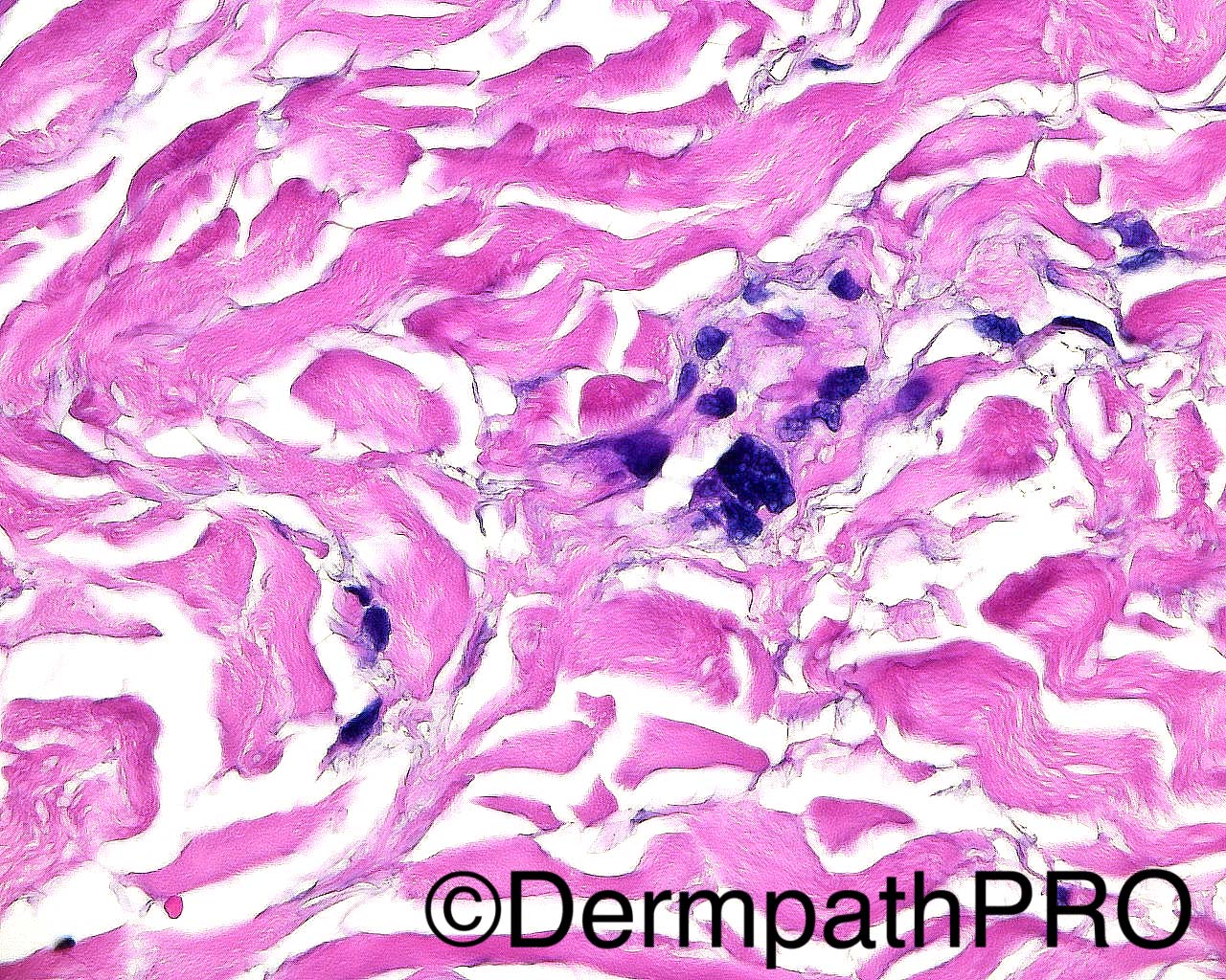
The patient is a 49 year old woman with a history of Raynaud's. A punch biopsy of brown, velvet-like plaques is taken from the right dorsal index MCP joint. Clinical Diagnosis: acanthosis nigricans vs post-inflammatory pigmentary changes vs lichenoid dermatitis.

Dr Mark Hurt.


  • Thanks 1
  Report Record

User Feedback


Raul Perret

Posted

Multiple differential diagnosis crossed my mind including pleomorphic fibroma, sclerotic fibroma, rare presentation of kaposi, atrophic dermatofibroma. But the clinical history suggests multiple lesions in an acral location, I dont see findings to support the proposed clinical diagnosis. I would perform some immunos to unveil the nature of those cells. The clínical setting of multiple lesions, unilateral acral, female with an histology showing dilated vascular vessels with some pericytes, fusiform hypercromatic cells some looking multinucleated as well as some fibrosis makes me consider in first place multinucleate cell angiohistiocytoma.

Share this comment


Link to comment
share_externally

Dr. Mona Abdel-Halim

Posted

This is very difficult..
The striking feature to me is the thick sclerotic collagen background together with the plump spindle cells, putting this with Raynaud's disease, I am thinking of a collagen disease. Thought of Fibroblastic rheumatism but the clinical is missing the arthritis and the contractures.. Also thought of Gottron's but there is no interface although there are few melanophages, but Gottron's do not show these plump fibroblasts which seems to be highlighted for a purpose. I did not perceive them as giant cells actually. Multinucleate cell angiohistiocytoma is different clinically also. It is difficult :-(

Share this comment


Link to comment
share_externally

Interstitial granuloma annulare vs. scleromyxoedema/lichen myxoedematosus. 

Share this comment


Link to comment
share_externally

Nitin Khirwadkar

Posted

Difficult case! Sclerosing dermopathy, favour scleromyxoedema. Occasional vessels slightly thick. Not sure what the scattered eosinophils mean. Could be totally off track!

Share this comment


Link to comment
share_externally

Arti Bakshi

Posted

Agree with dermal sclerosis being the main pathology. There appears to be increased pigmentation of the basal layer of epidermis And some telagiectasia. Coupled with history of Raynaud's, this could be a limited form of systemic sclerosis.( although 'velvety plaques' a bit odd). Scleromyxoedema is a good d/d.

Share this comment


Link to comment
share_externally

Raul Perret

Posted

The only case that I ever saw on clinical practice was last year during residency training and the cells were exactly like here with hypercromasia and a degenerative type of atypia and just some dilated vessels, I guess the pictures we see on the books are sometimes the exception rather than the rule

Share this comment


Link to comment
share_externally

Arti Bakshi

Posted

Well done, Raul!...u were right all along!

Share this comment


Link to comment
share_externally

vincenzo polizzi

Posted

Well done, Raul! 

Share this comment


Link to comment
share_externally

Dr. Mona Abdel-Halim

Posted

👍🏻👍🏻 Raul :-)

Share this comment


Link to comment
share_externally

Dr. Richard Carr

Posted

Last Friday's was Lichen nitidus by the way with a typical clinical history. Apologies for delay - it was a national holiday in UK yesterday.

Share this comment


Link to comment
share_externally


Join the conversation

You can post now and register later. If you have an account, sign in now to post with your account.

Guest
Add a comment...

×   Pasted as rich text.   Paste as plain text instead

  Only 75 emoji are allowed.

×   Your link has been automatically embedded.   Display as a link instead

×   Your previous content has been restored.   Clear editor

×   You cannot paste images directly. Upload or insert images from URL.

Loading...

×
×
  • Create New...