Jump to content
In this section we have spot diagnoses posted on a daily basis since June 2010, now over 4000! You can review the archived cases and read the suggested diagnoses by users and the final comment by the contributors.
Case are uploaded each week day by 10 am UK time with the correct diagnosis will generally be posted at 8 pm UK time. Why not view the most recent spot diagnosis and proffer a diagnosis?

Case Number : Case 1650 - 21 October Posted By: Guest

Please read the clinical history and view the images by clicking on them before you proffer your diagnosis.
Submitted Date :
   (0 reviews)

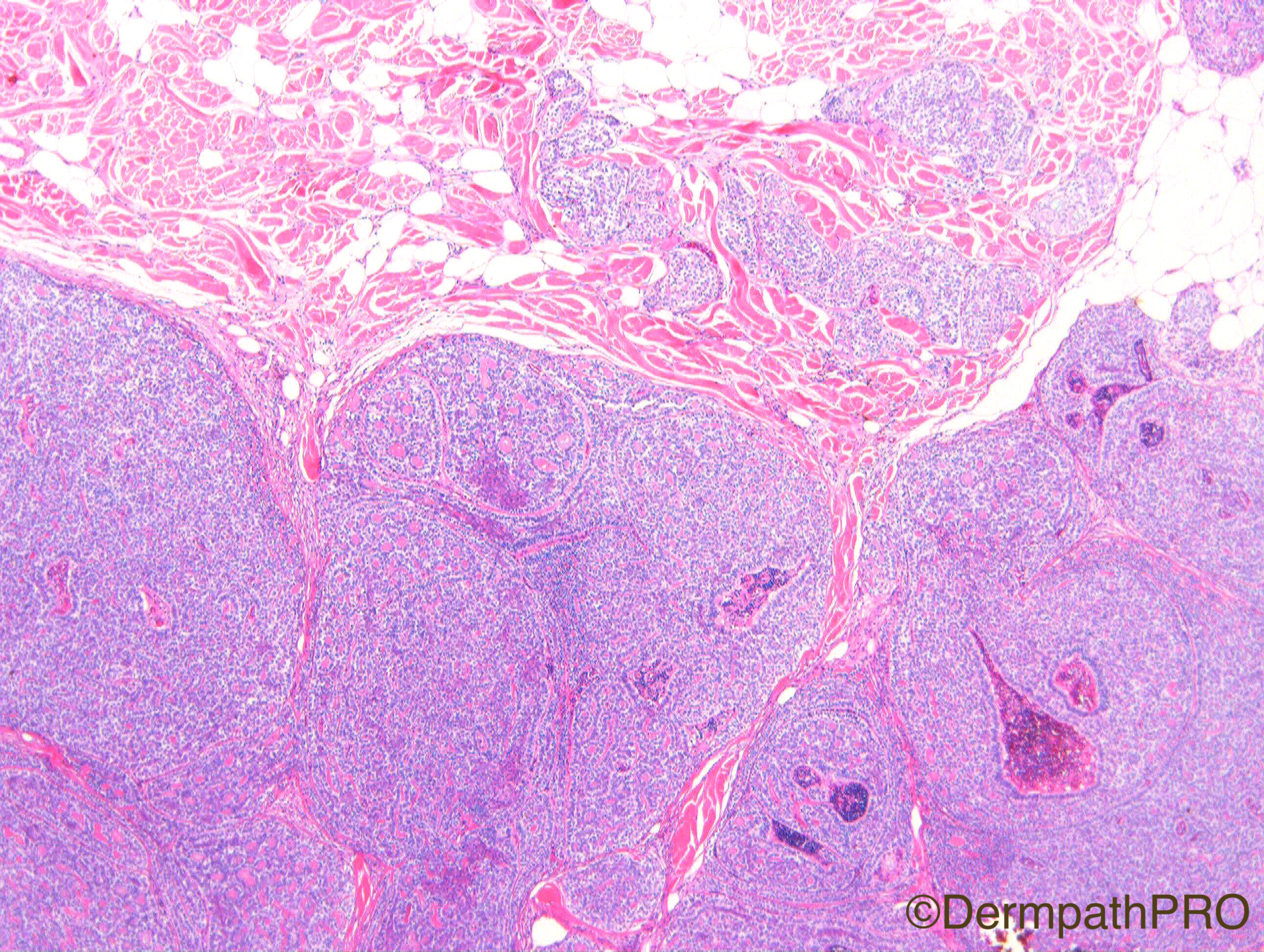
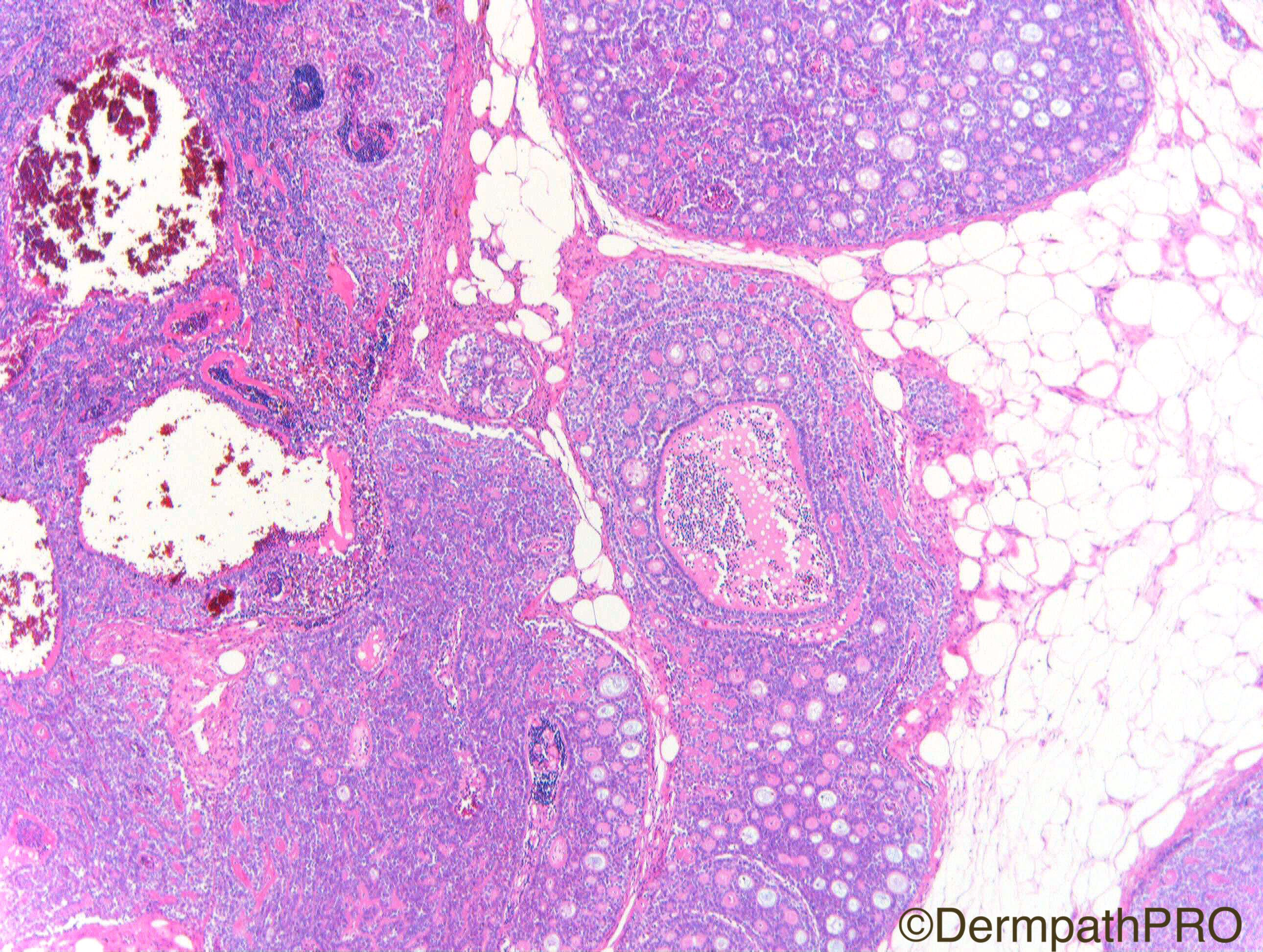
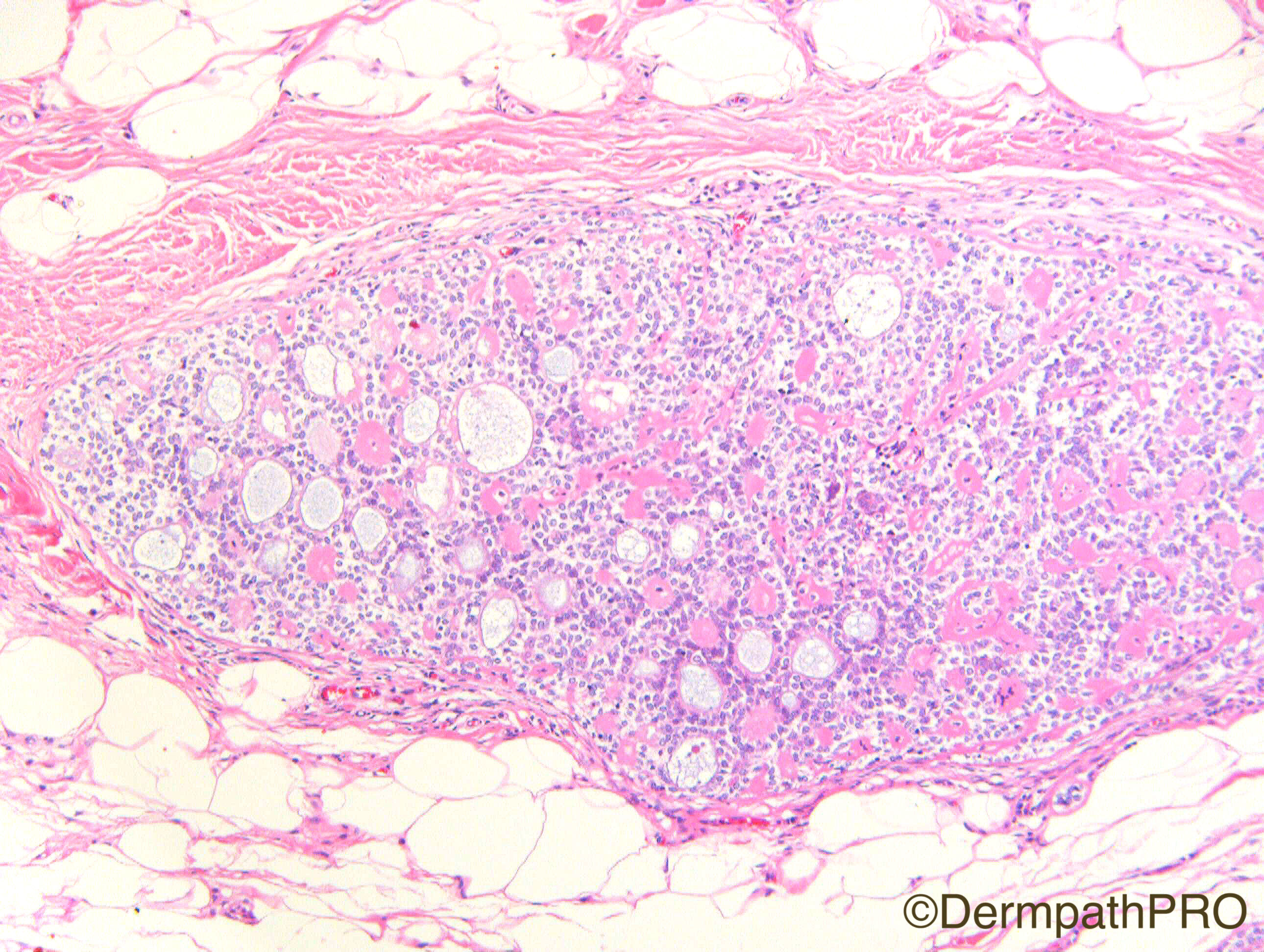
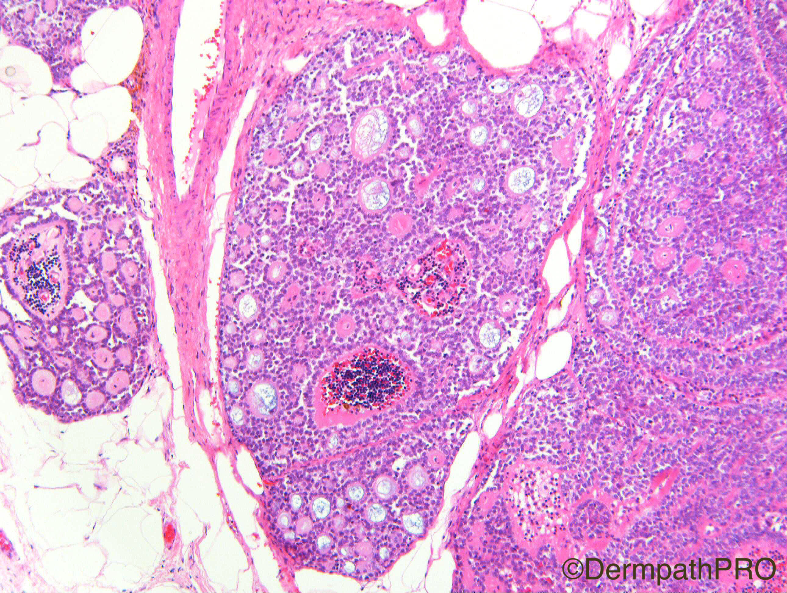
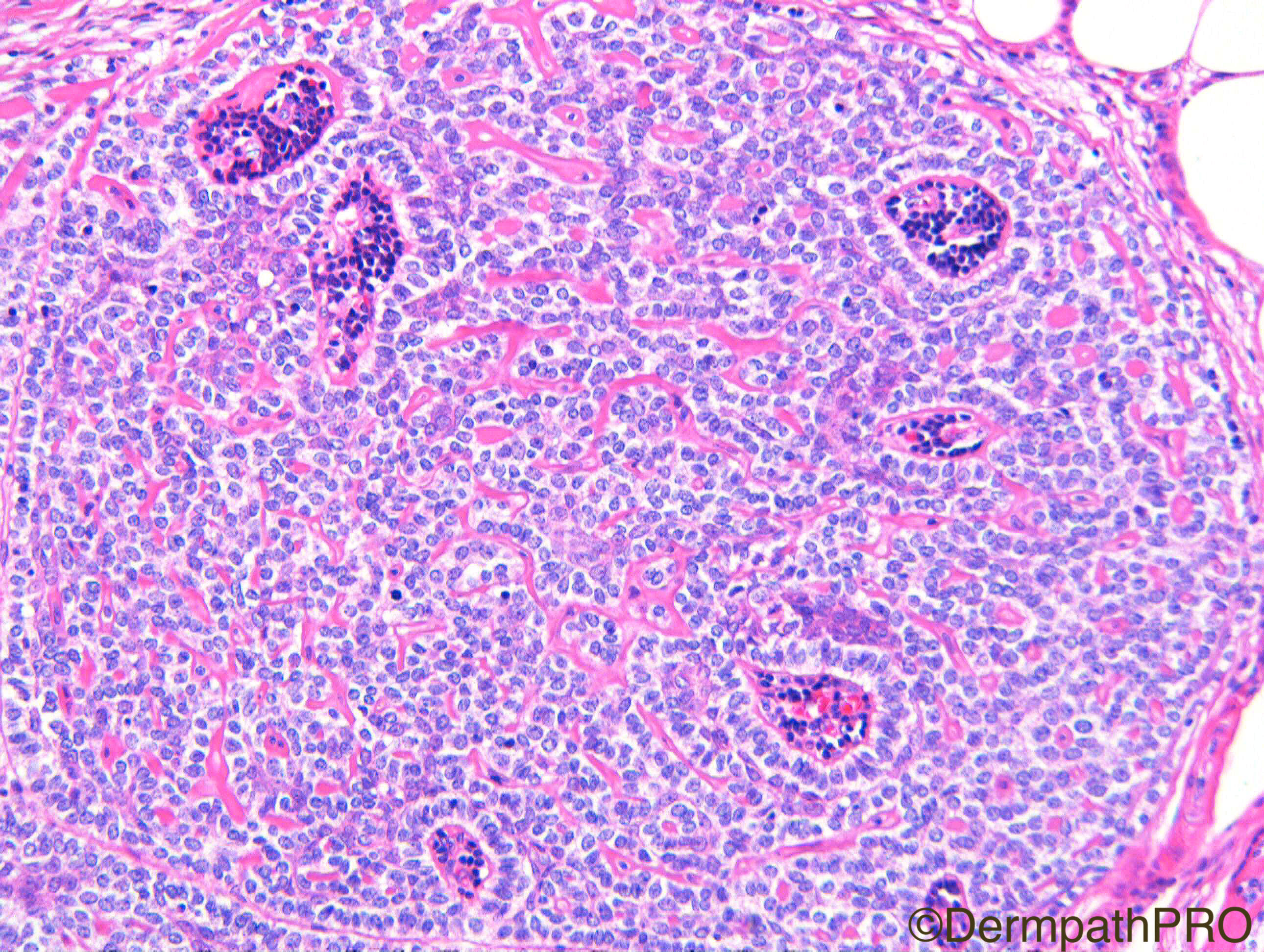
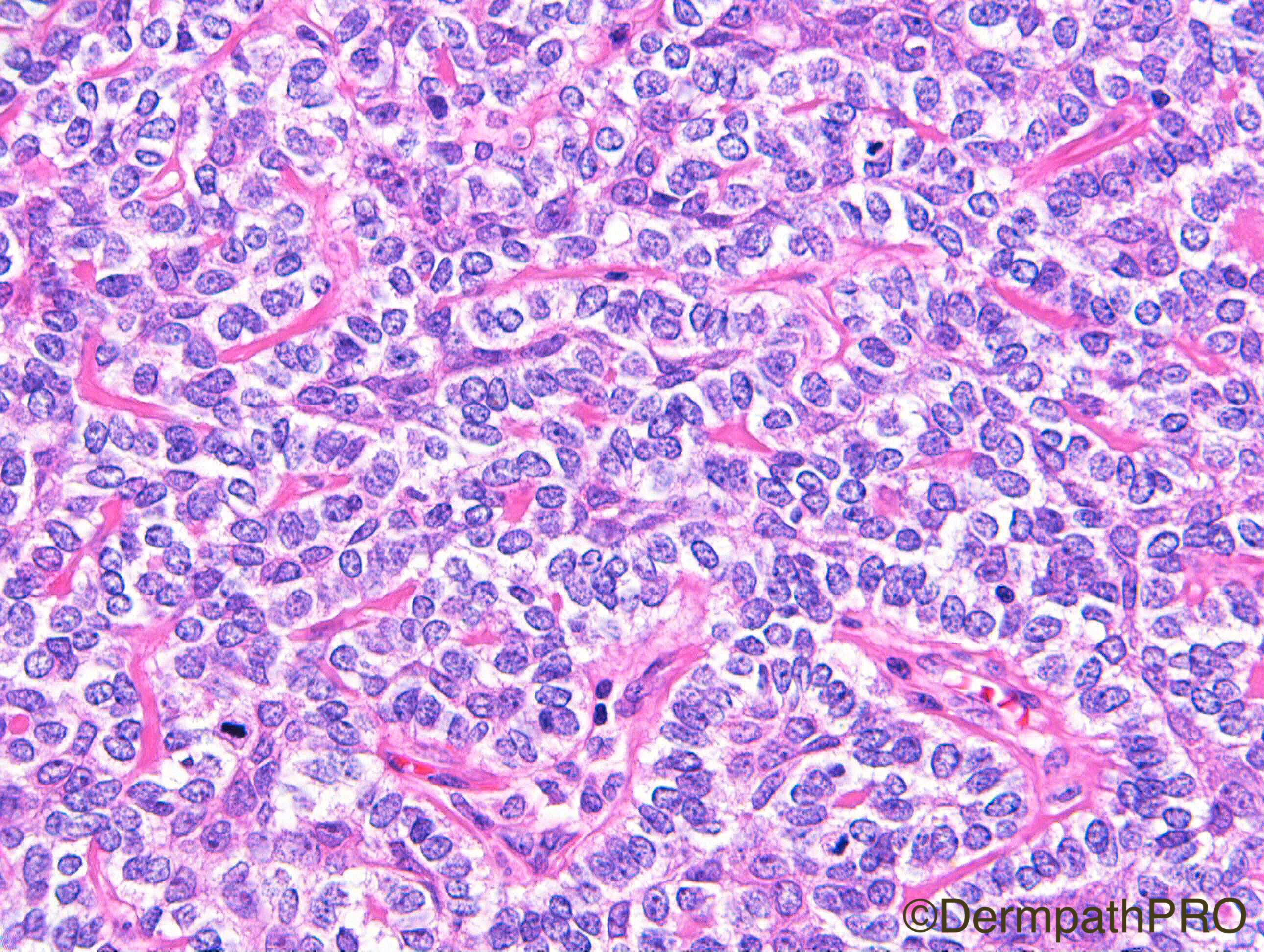
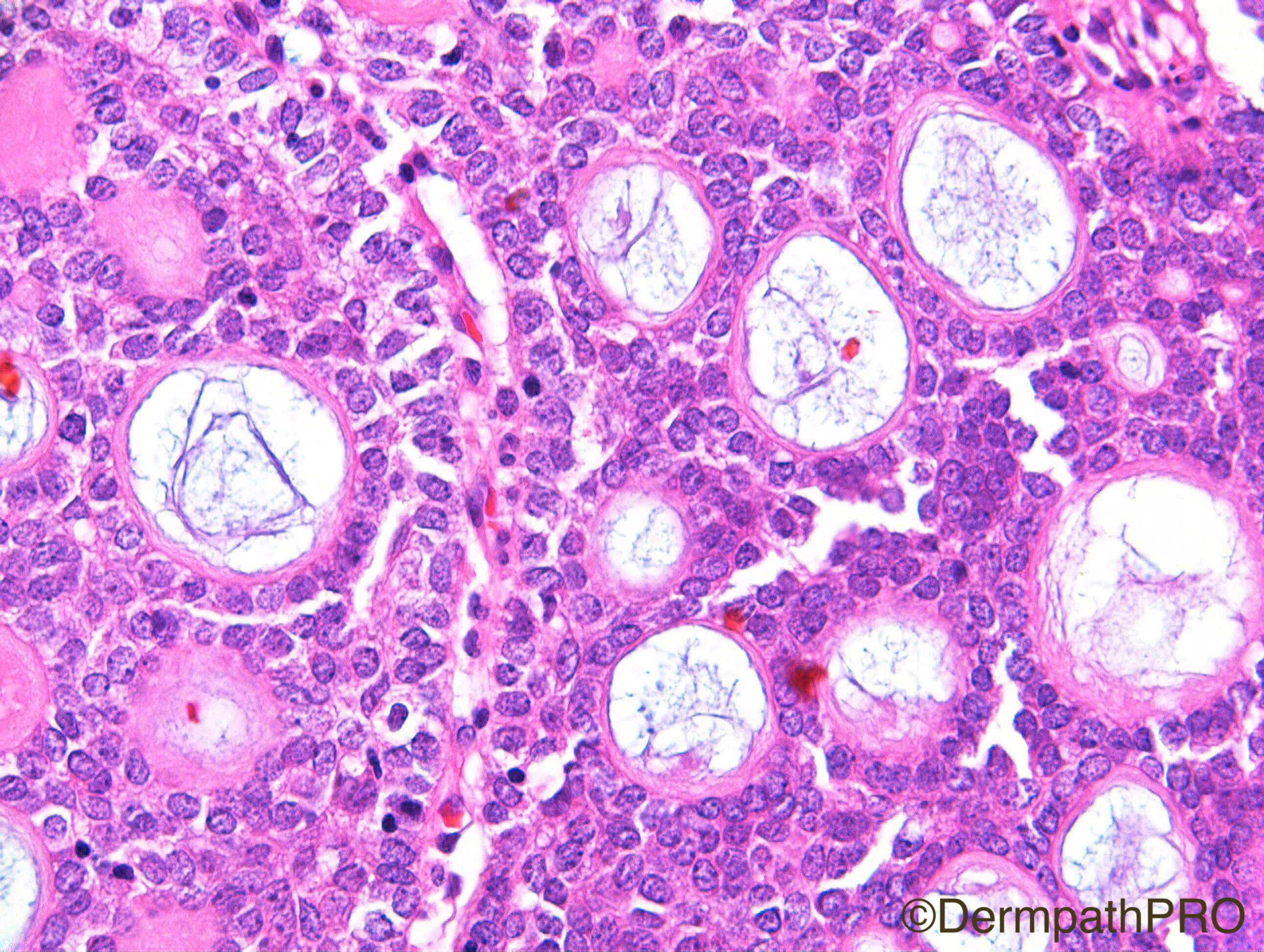
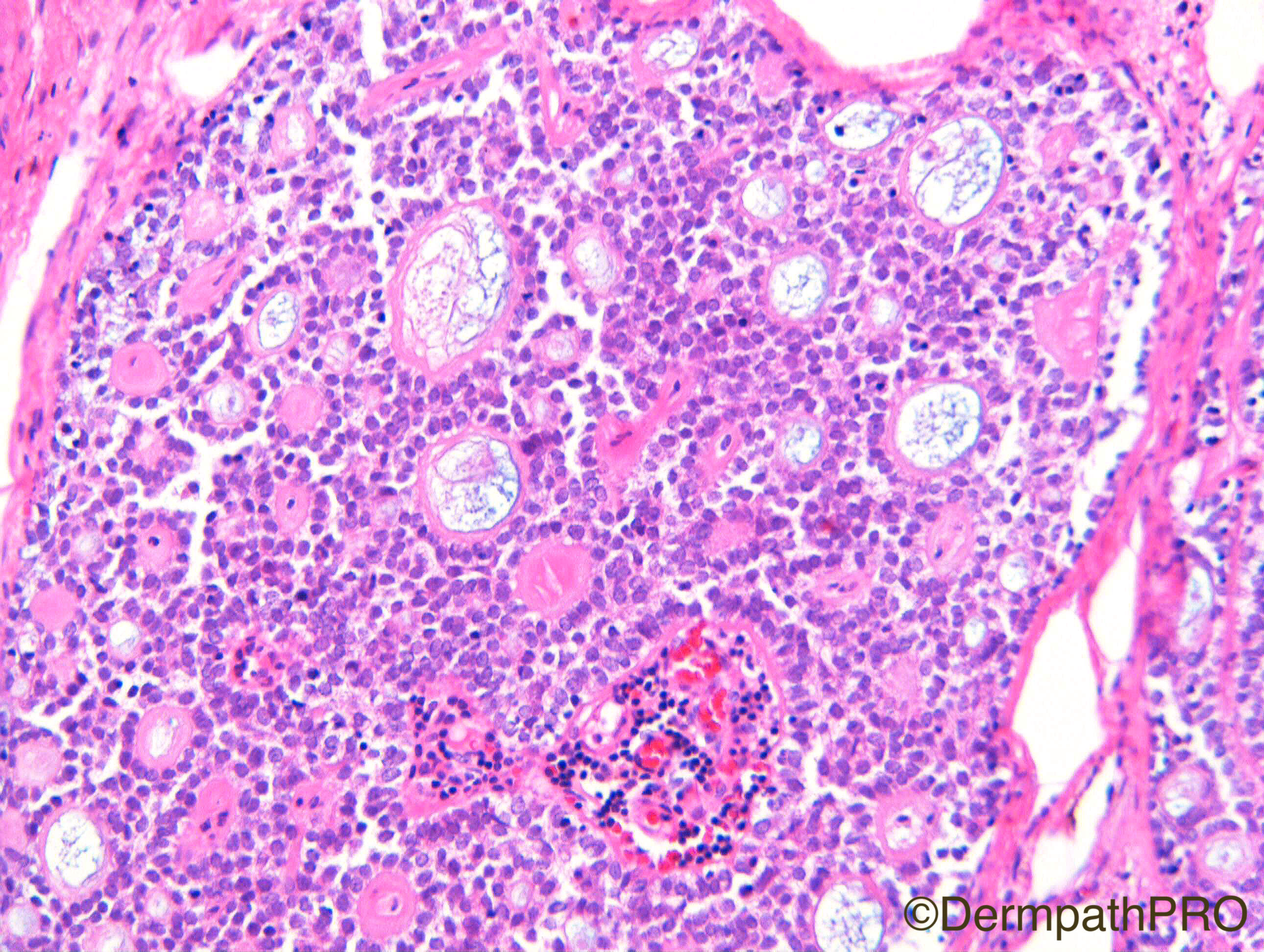
Clinical Details: F50. Back. ?Epidermoid cyst.

Case Posted by Dr Richard A Carr

  Report Record

User Feedback


Admin_Dermpath

Posted

Another beautiful case from Dr Richard A Carr to round off the week.


Cheers, Geoff

Share this comment


Link to comment
share_externally

Dr. Mona Abdel-Halim

Posted

Spiradenocylindroma maybe better than just spiradenoma !

Share this comment


Link to comment
share_externally

Raul Perret

Posted

Richard, you showed this case to Dr. Kazakov when we were in Pilsen right?


Share this comment


Link to comment
share_externally

Spiradenoma (maybe spiradenocylindroma) with too much tubular differentiation

Share this comment


Link to comment
share_externally

Eman El-Nabarawy

Posted

Spiradenoma with excessive hyaline deposits and duct all differentiation.

Share this comment


Link to comment
share_externally

vincenzo polizzi

Posted

I see only myoepithelial-like cells with cylindromatous arrangement...but sincerely I don't know what it is! Waiting for the diagnosis.

Share this comment


Link to comment
share_externally

vincenzo polizzi

Posted

...and I see thymoma- like lymphocytes rich perivascular spaces but... again I'm waiting for the diagnosis. 

Share this comment


Link to comment
share_externally

Dr. Richard Carr

Posted

Both Raul and Nitin are correct they have seen the case in Pilsen and Warwick respectfully - I suspect this is a reflection of how dermpathpro is making connections across the planet! Vincenzo - you are quite correct - there are no tubules visible in these images - at least I could not see them on H&E.

Share this comment


Link to comment
share_externally

Arti Bakshi

Posted

There is a prominent cribriform pattern with pseudo cysts containing mucinous material. Other areas more typically spiradenomatous.

Maybe a spiradenoma with 'adenoid cystic Ca' like areas. 

(Cant enlarge images...is that a problem others are facing too?)

Share this comment


Link to comment
share_externally

Dr. Richard Carr

Posted

Yes well done all, and especially Arti, this is a spiradenoma with adenoid cystic carcinoma-like areas.  It was extremely pauci-luminal (even on BerEP4 only very sparse weak central ductal cells but no clear cut lumina). Case also seen by Dmitry Kazakov in Pilsen who thought it was a benign lesion.

Share this comment


Link to comment
share_externally


Join the conversation

You can post now and register later. If you have an account, sign in now to post with your account.

Guest
Add a comment...

×   Pasted as rich text.   Paste as plain text instead

  Only 75 emoji are allowed.

×   Your link has been automatically embedded.   Display as a link instead

×   Your previous content has been restored.   Clear editor

×   You cannot paste images directly. Upload or insert images from URL.

Loading...

×
×
  • Create New...