Jump to content
In this section we have spot diagnoses posted on a daily basis since June 2010, now over 4000! You can review the archived cases and read the suggested diagnoses by users and the final comment by the contributors.
Case are uploaded each week day by 10 am UK time with the correct diagnosis will generally be posted at 8 pm UK time. Why not view the most recent spot diagnosis and proffer a diagnosis?

Case Number : Case 1620 - 9 September Posted By: Guest

Please read the clinical history and view the images by clicking on them before you proffer your diagnosis.
Submitted Date :
   (0 reviews)

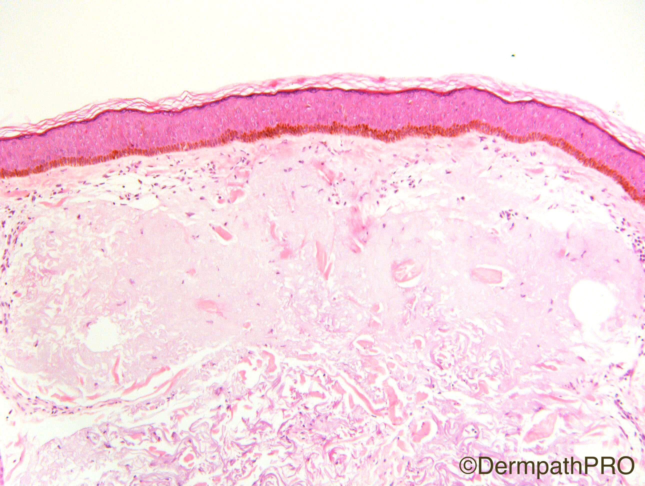
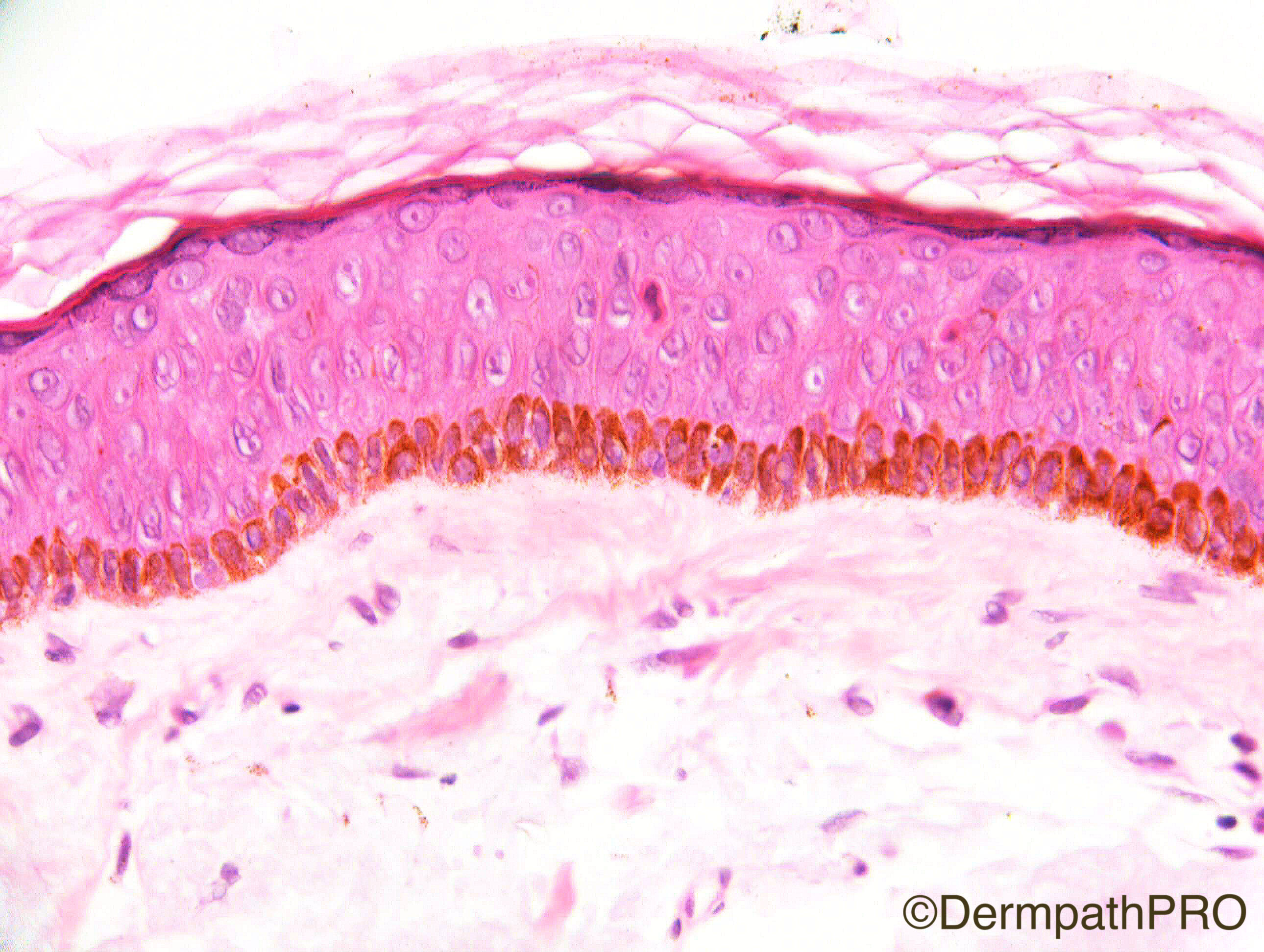
Male 85. Right frontal lesion ?lentigo maligna ?solar lentigo

Case Posted by Dr Richard Carr


  Report Record

User Feedback


vincenzo polizzi

Posted

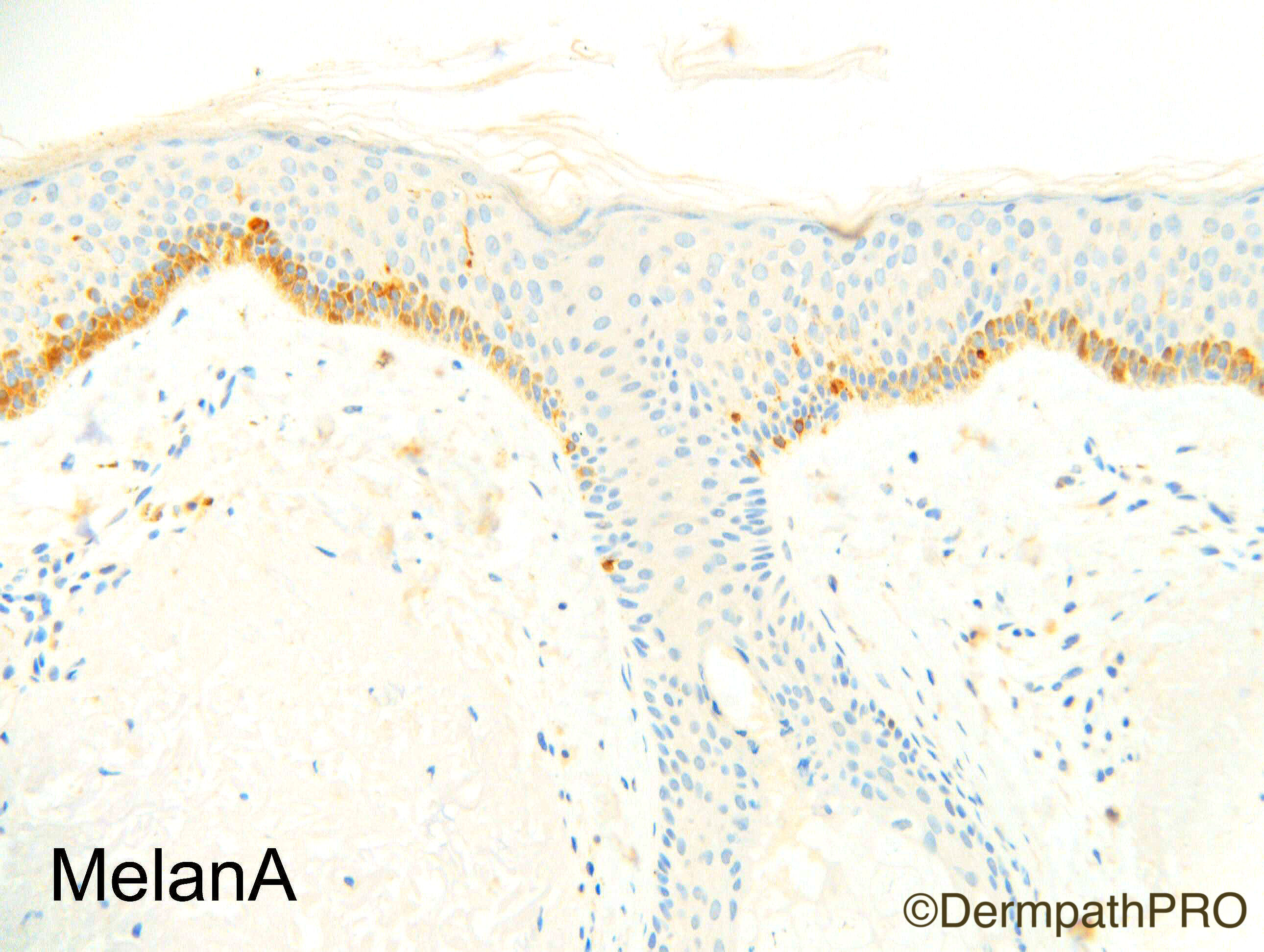
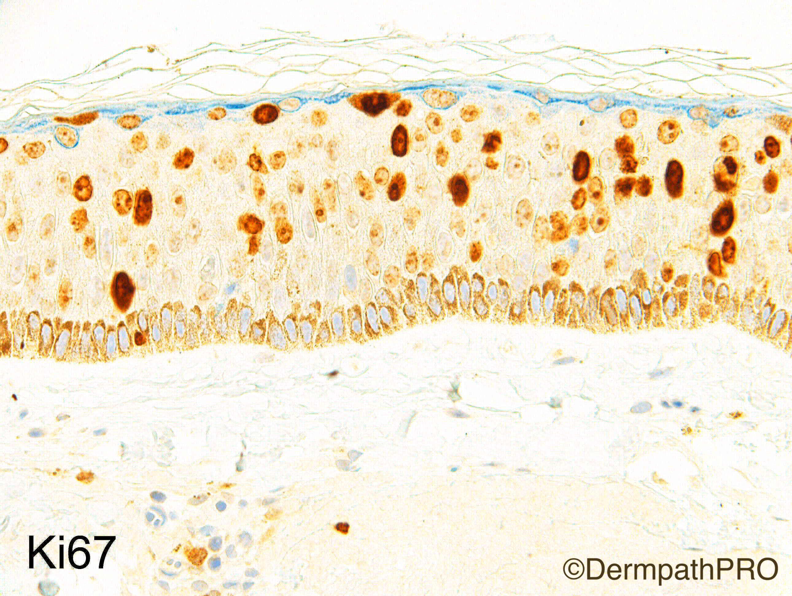
The full-thickness p53-positivity with a ki67 increased expression, also in upper epidermis, fit well with a mild keratinocytic atypia and diskeratosis morphologically observed. The acrothrychium and acrosyringyum are spared with an abrupt border, and the basal layer is hyperpigmented,without significative increased number of melanocytes. These findings seem consistent with "bowenoid actinic keratosis", pigmented variant.

Share this comment


Link to comment
share_externally

Dr. Richard Carr

Posted

Hmm!   Seems this case was a little spoilt.  I was hoping you would have had a chance to give opinions on the H&E's without the IHC.  You should note the lack of maturation of the cells (even at low power the epithelium looks a little to mauve due to high nuclear cytoplasmic ratio). A higher magnification one can appreciate the subtle lack of maturation, occasional prematurely keratinising cells and no doubt you will find mitoses at all levels if you search carefully. The sudden and very abruptly orthokeratinisation near the surface without parakeratosis evident might fool you into no considering actinic keratosis here.  So please be aware of this particularly subtle type of bowenoid dysplasia.  It is morphologically similar to cases that can be seen in genital sites and also very easily missed. 

My diagnosis: Pigmented atrophic pattern of subtle bowenoid actinic keratosis!

Share this comment


Link to comment
share_externally


Join the conversation

You can post now and register later. If you have an account, sign in now to post with your account.

Guest
Add a comment...

×   Pasted as rich text.   Paste as plain text instead

  Only 75 emoji are allowed.

×   Your link has been automatically embedded.   Display as a link instead

×   Your previous content has been restored.   Clear editor

×   You cannot paste images directly. Upload or insert images from URL.

Loading...

×
×
  • Create New...