Jump to content
In this section we have spot diagnoses posted on a daily basis since June 2010, now over 4000! You can review the archived cases and read the suggested diagnoses by users and the final comment by the contributors.
Case are uploaded each week day by 10 am UK time with the correct diagnosis will generally be posted at 8 pm UK time. Why not view the most recent spot diagnosis and proffer a diagnosis?

Case Number : Case 1882 - 15 Aug - Dr Uma Sundram Posted By: Guest

Please read the clinical history and view the images by clicking on them before you proffer your diagnosis.
Submitted Date :
   (0 reviews)

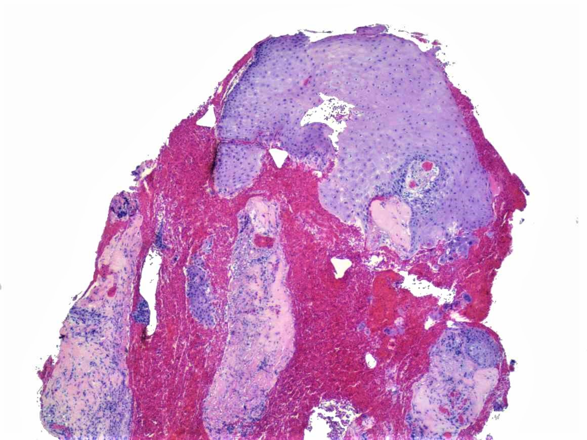
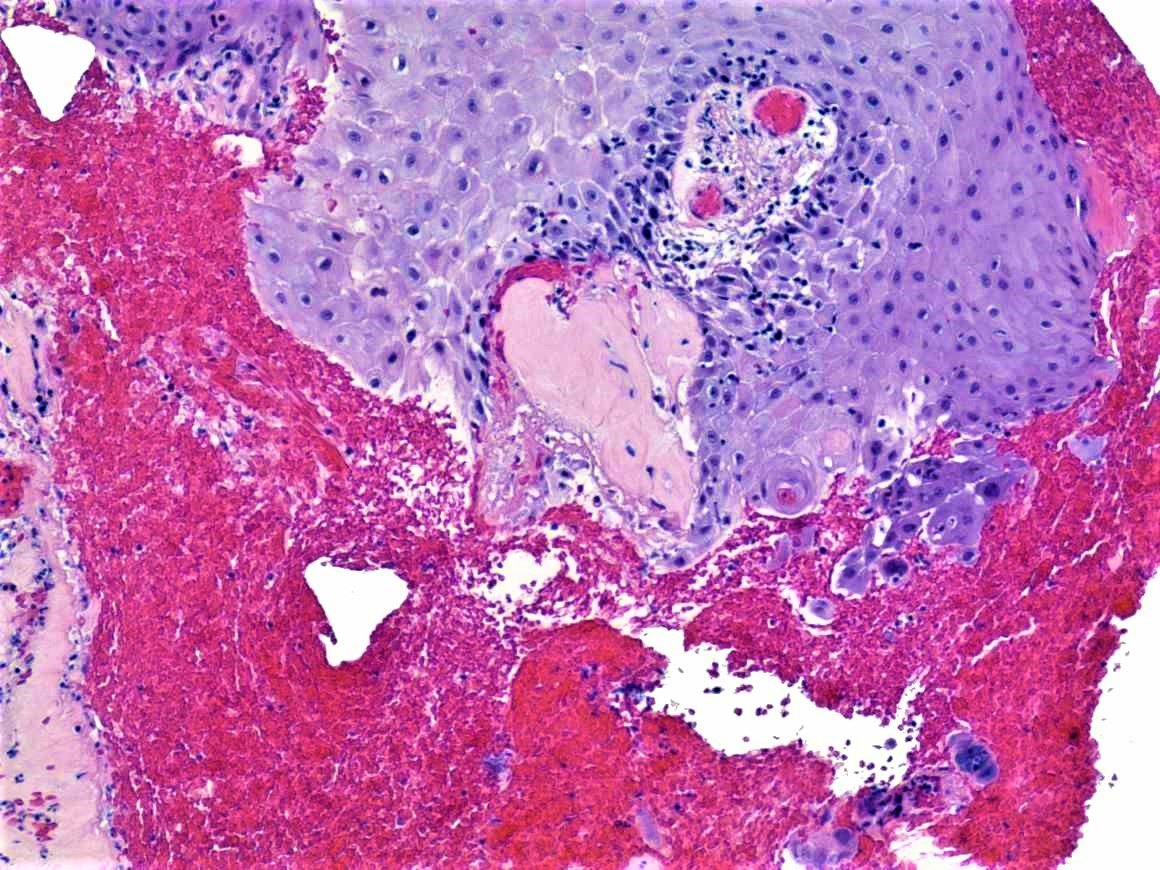
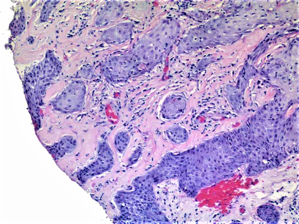
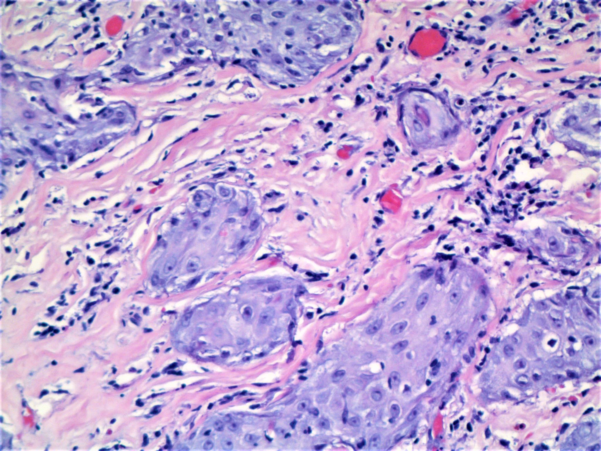
63 year old woman with lesion on vulva. No other history is provided.


  Report Record

User Feedback


Anil Patki

Posted

Islands of cells some of which are dark and some showing pale cytoplasm, along with the site of lesion (vulva) makes me think of syringoma showing clear cell change in some areas

Share this comment


Link to comment
share_externally

Raul Perret

Posted

For me there is an acanthotic epidermis with areas of dysmaturation and premature keratinization overlaying a dermis with broad condensated collagen.  This is consistent with differentiated VIN and lichen sclerosus. The main problem of the biopsy is the bad orientation that can simulate invasive areas. But considering the highly pleomorphic cells and irregular nests with jagged edges from my point of view there are real areas suspicious of invasion.

Share this comment


Link to comment
share_externally

Cem Leblebici

Posted

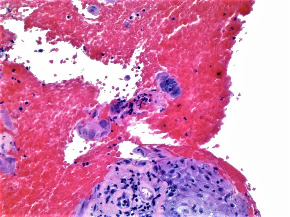
In Figure 4, epithelial cells showed enlarged nuclei with ground glass appearance. I suspect  pseudoepitheliomatous hyperplasia associated with herpes infection mimicking invasive carcinoma.

Share this comment


Link to comment
share_externally

Robledo F. Rocha

Posted

Herpesvirus infection in the setting of lichen sclerosus.

Share this comment


Link to comment
share_externally

Uma Sundram

Posted

Herpes mimicking invasive SCC.  The biopsy is misoriented which may make the dermis seem more sclerotic.

Share this comment


Link to comment
share_externally

Anil Patki

Posted

A symptomatic herpes infection should show ballooning degeneration of epidermis, multinucleated keratinocytes and inflammatory infiltrate.

Share this comment


Link to comment
share_externally

Anil Patki

Posted

Can it be Bowenoid papulosis? A clinician hardly ever biopsies a lesion of herpes on the genitals. The clinical features are so distinctive and we have serology and viral culture for confirmation.

Share this comment


Link to comment
share_externally


Join the conversation

You can post now and register later. If you have an account, sign in now to post with your account.

Guest
Add a comment...

×   Pasted as rich text.   Paste as plain text instead

  Only 75 emoji are allowed.

×   Your link has been automatically embedded.   Display as a link instead

×   Your previous content has been restored.   Clear editor

×   You cannot paste images directly. Upload or insert images from URL.

Loading...

×
×
  • Create New...