Jump to content
In this section we have spot diagnoses posted on a daily basis since June 2010, now over 4000! You can review the archived cases and read the suggested diagnoses by users and the final comment by the contributors.
Case are uploaded each week day by 10 am UK time with the correct diagnosis will generally be posted at 8 pm UK time. Why not view the most recent spot diagnosis and proffer a diagnosis?

Case Number : Case 1895 - 01 Sept - Dr Richard Carr Posted By: Guest

Please read the clinical history and view the images by clicking on them before you proffer your diagnosis.
Submitted Date :
   (0 reviews)

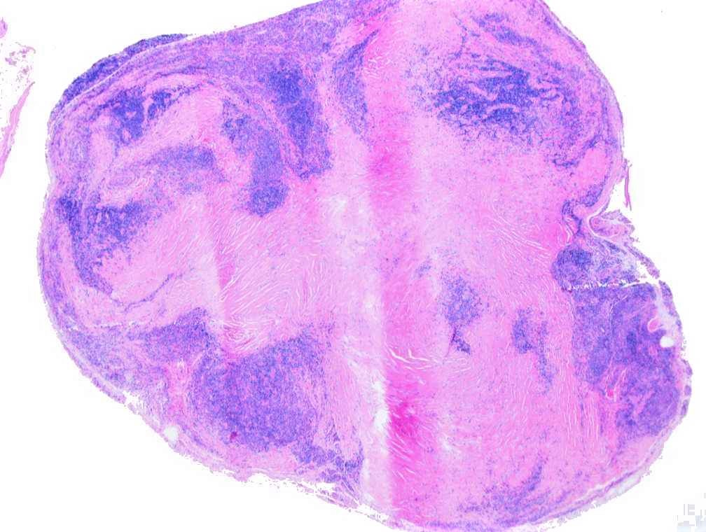
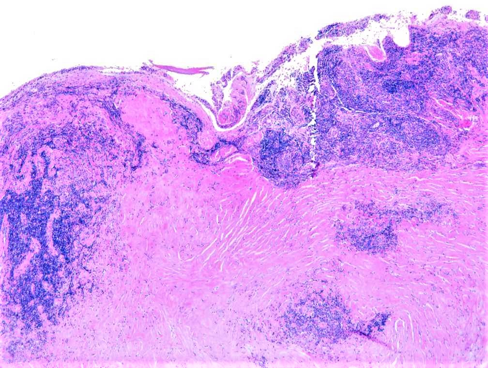
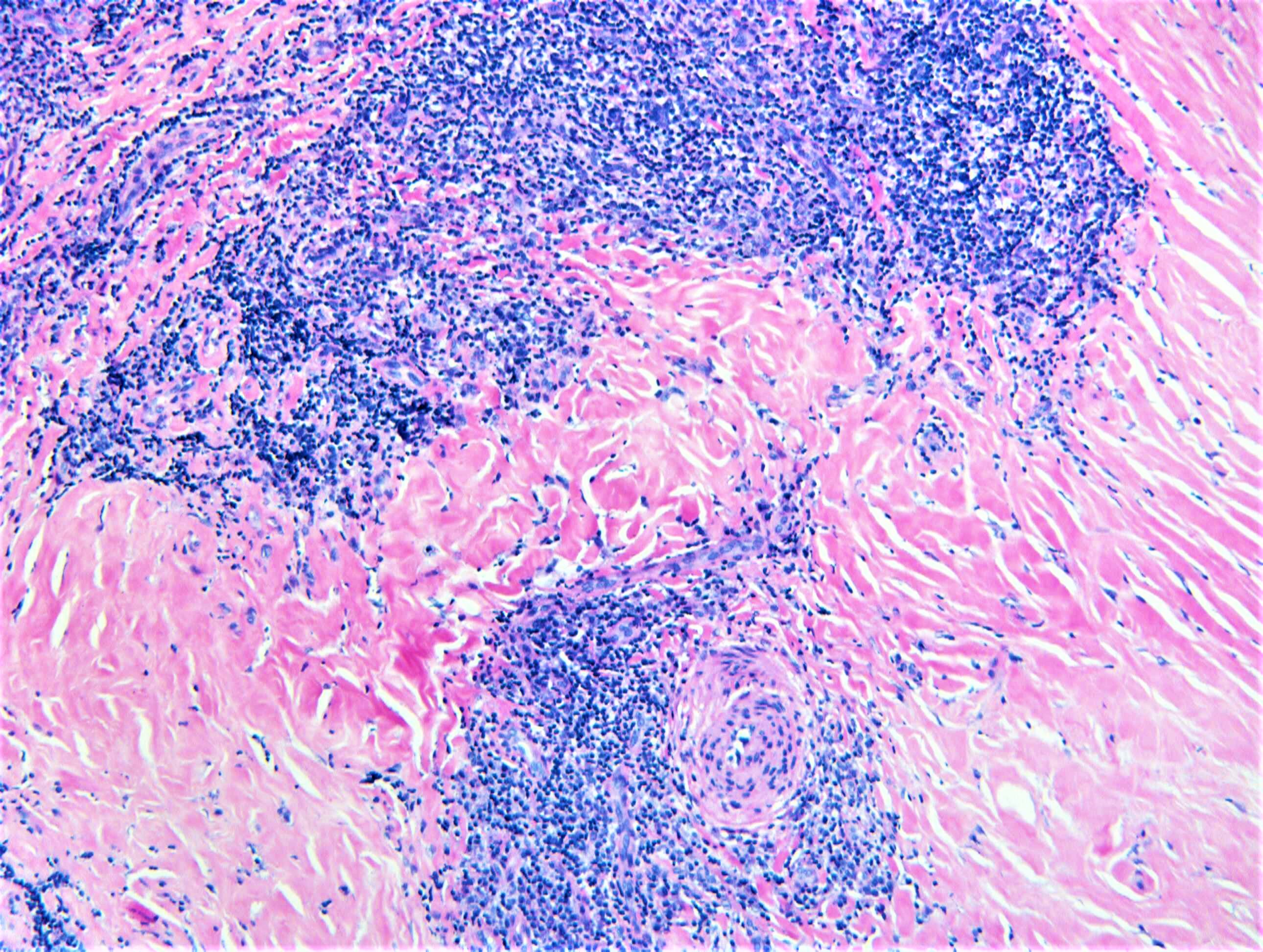
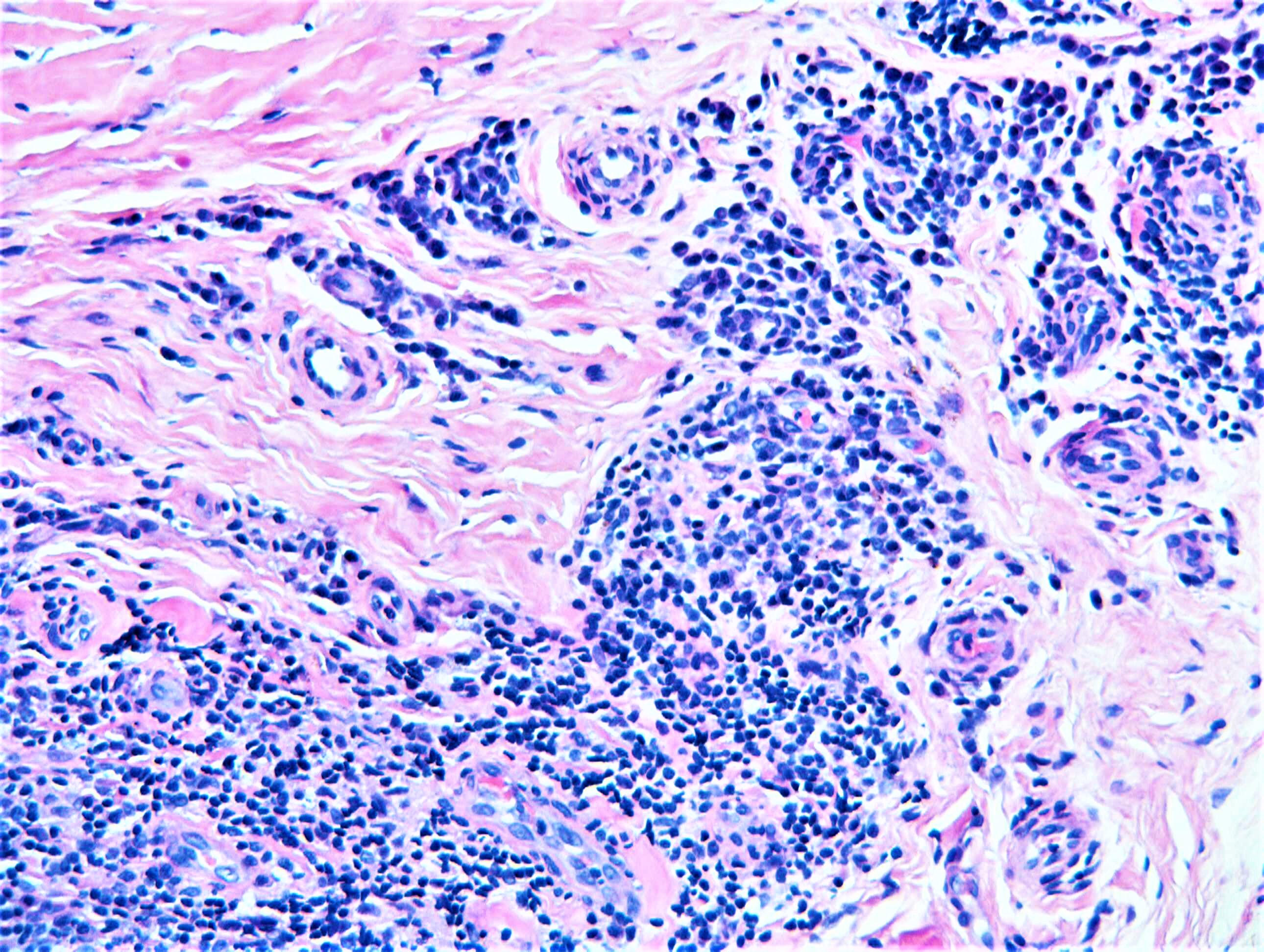
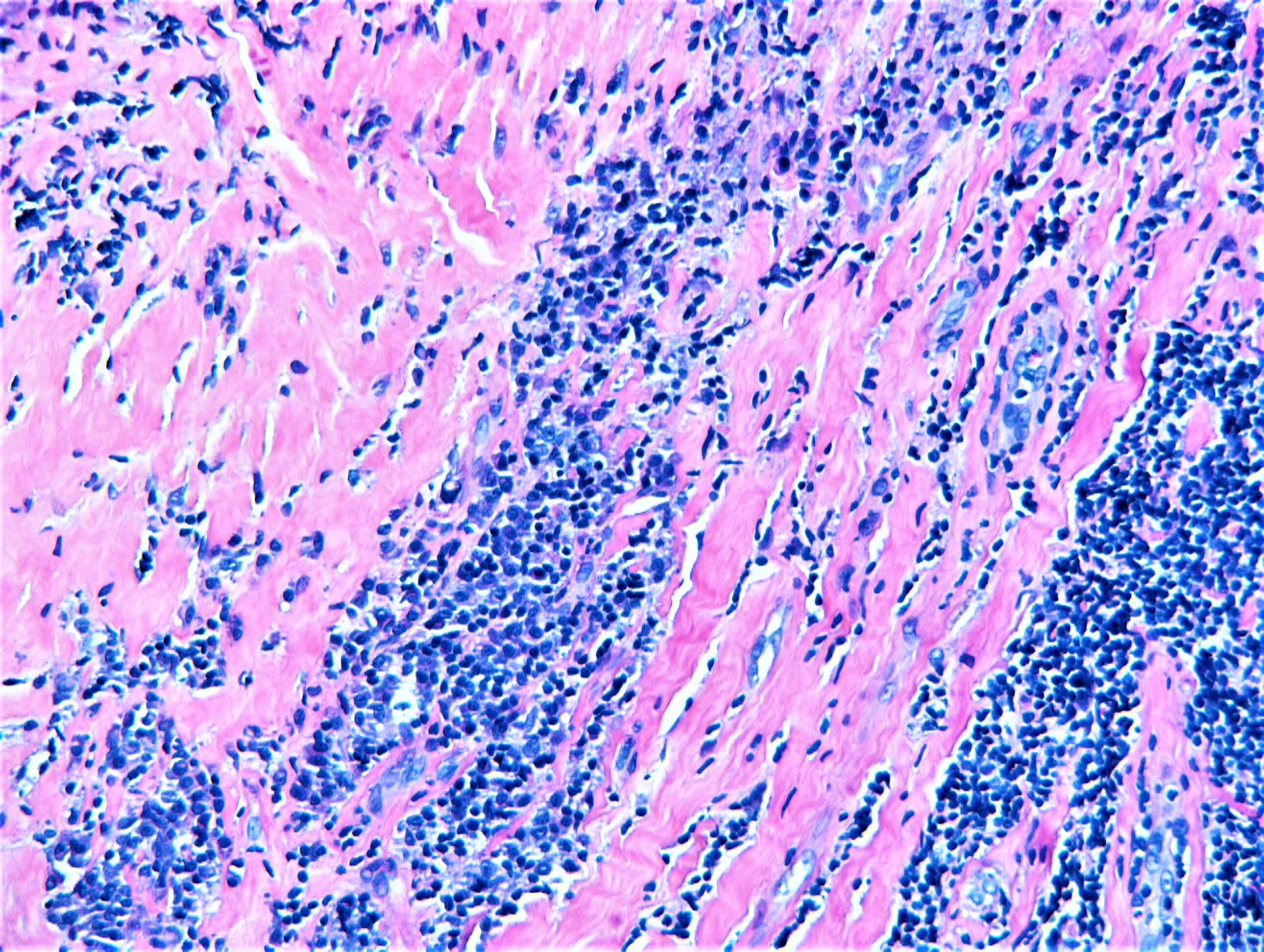
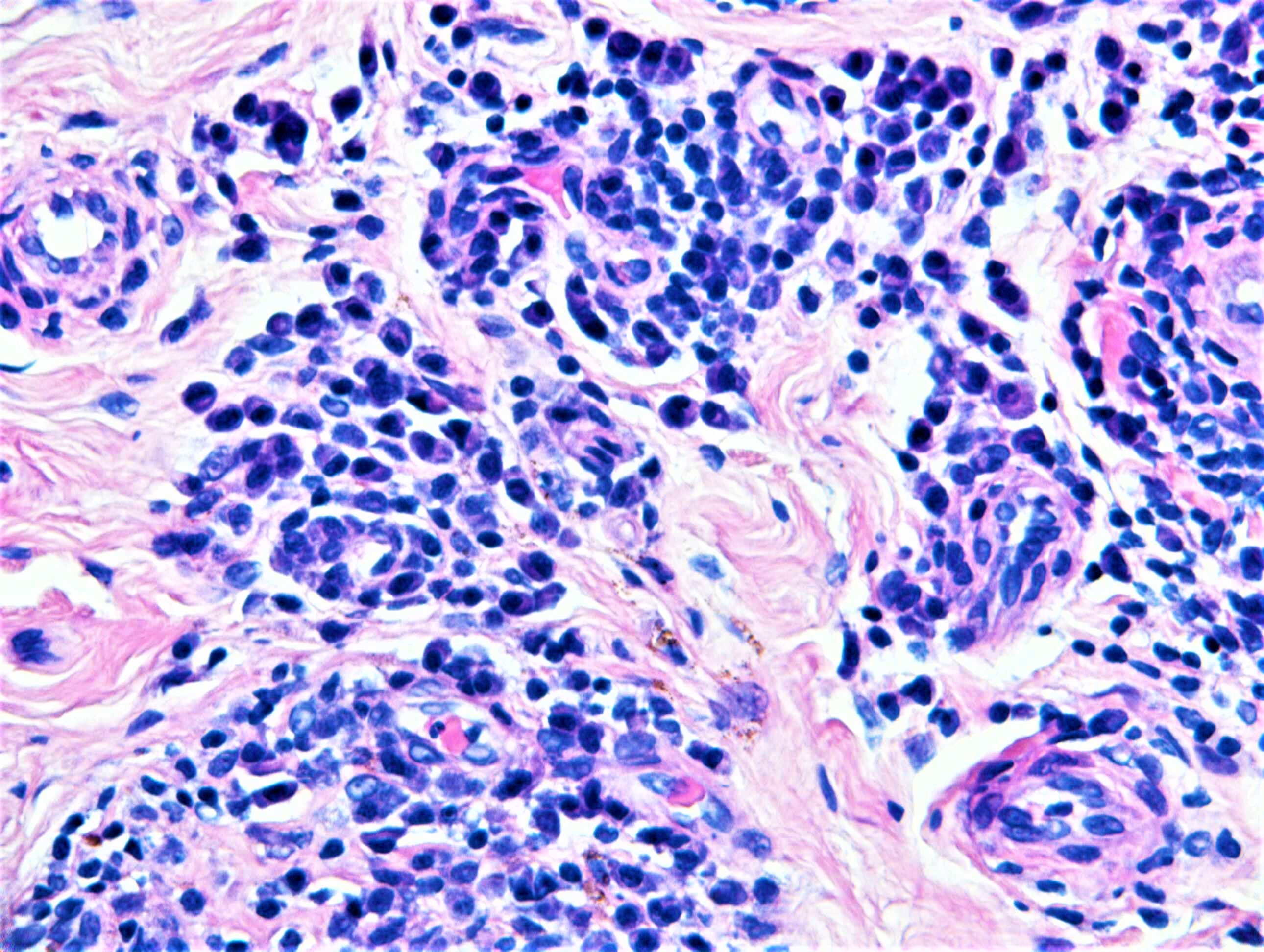
M50. Big toe. Ganglion.


  Report Record

User Feedback


urmilapandey

Posted

would favour a benign fibroblastic/myofibroblastic lesion. given the lympho/plasmacytic component, would consider inflammatory myofibroblastic tumour of soft tissue, odd myofibroma..

Share this comment


Link to comment
share_externally

vincenzo polizzi

Posted

Agree with Urmilapandey and Mona: Inflammatory Myofibroblastic Tumor, Plasma Cell Granuloma type.

Share this comment


Link to comment
share_externally

Raul Perret

Posted

agree with sclerosing type of inflammatory myofibroblastic tumor as spot diagnosis. Although I would perform some immunos before giving the dx, mainly in paucicellular lesions like this one

Share this comment


Link to comment
share_externally

Arti Bakshi

Posted

yes, inflammatory myofibroblastic tumour my first thought too.

I have put the final diagnosis for case 1894 (apologies for the delay!!)


Share this comment


Link to comment
share_externally

Dr. Richard Carr

Posted

Hmm!  How about infections? Lot's of plasma cells. 

Share this comment


Link to comment
share_externally

Arti Bakshi

Posted

wow!..did not think infection at all...could this be a juxta-articular fibrous nodule related to Borrelia??

Share this comment


Link to comment
share_externally

Dr. Richard Carr

Posted

This histology (a sclerotic juxta-articular located nodule with promoinent lymphoid containing abundant plasma cells) is very typical for a juxta-articular fibroid nodule of late Lyme disease, case s/b Professor Werner Kempf who agreed and kindly performed PCR which was negative. Prof. Kempf noted that late lesions of Lyme can be PCR negative but serology is usually positive (the reverse in early Lyme lesions). I'm still awaiting results of serology. Hope you all recognise the pattern of this rare manifestation/variant next time you see it.

Share this comment


Link to comment
share_externally

vincenzo polizzi

Posted

Thanks for this very interesting case, Dr Richard!

Share this comment


Link to comment
share_externally

Dr. Richard Carr

Posted

Thumbs up for Arti - no like button these days it appears.

Share this comment


Link to comment
share_externally

urmilapandey

Posted

22 hours ago, Dr. Richard Carr said:

This histology (a sclerotic juxta-articular located nodule with promoinent lymphoid containing abundant plasma cells) is very typical for a juxta-articular fibroid nodule of late Lyme disease, case s/b Professor Werner Kempf who agreed and kindly performed PCR which was negative. Prof. Kempf noted that late lesions of Lyme can be PCR negative but serology is usually positive (the reverse in early Lyme lesions). I'm still awaiting results of serology. Hope you all recognise the pattern of this rare manifestation/variant next time you see it.

wow! thanks indeed for sharing

Share this comment


Link to comment
share_externally


Join the conversation

You can post now and register later. If you have an account, sign in now to post with your account.

Guest
Add a comment...

×   Pasted as rich text.   Paste as plain text instead

  Only 75 emoji are allowed.

×   Your link has been automatically embedded.   Display as a link instead

×   Your previous content has been restored.   Clear editor

×   You cannot paste images directly. Upload or insert images from URL.

Loading...

×
×
  • Create New...