Jump to content
In this section we have spot diagnoses posted on a daily basis since June 2010, now over 4000! You can review the archived cases and read the suggested diagnoses by users and the final comment by the contributors.
Case are uploaded each week day by 10 am UK time with the correct diagnosis will generally be posted at 8 pm UK time. Why not view the most recent spot diagnosis and proffer a diagnosis?

Case Number : Case 2422 - 11 October 2019 Posted By: Dr. Richard Carr

Please read the clinical history and view the images by clicking on them before you proffer your diagnosis.
Submitted Date :
   (0 reviews)

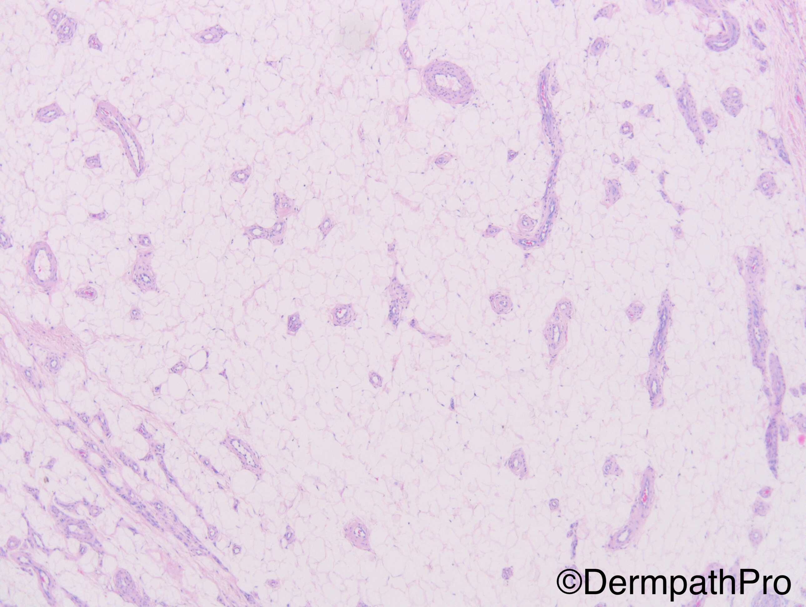
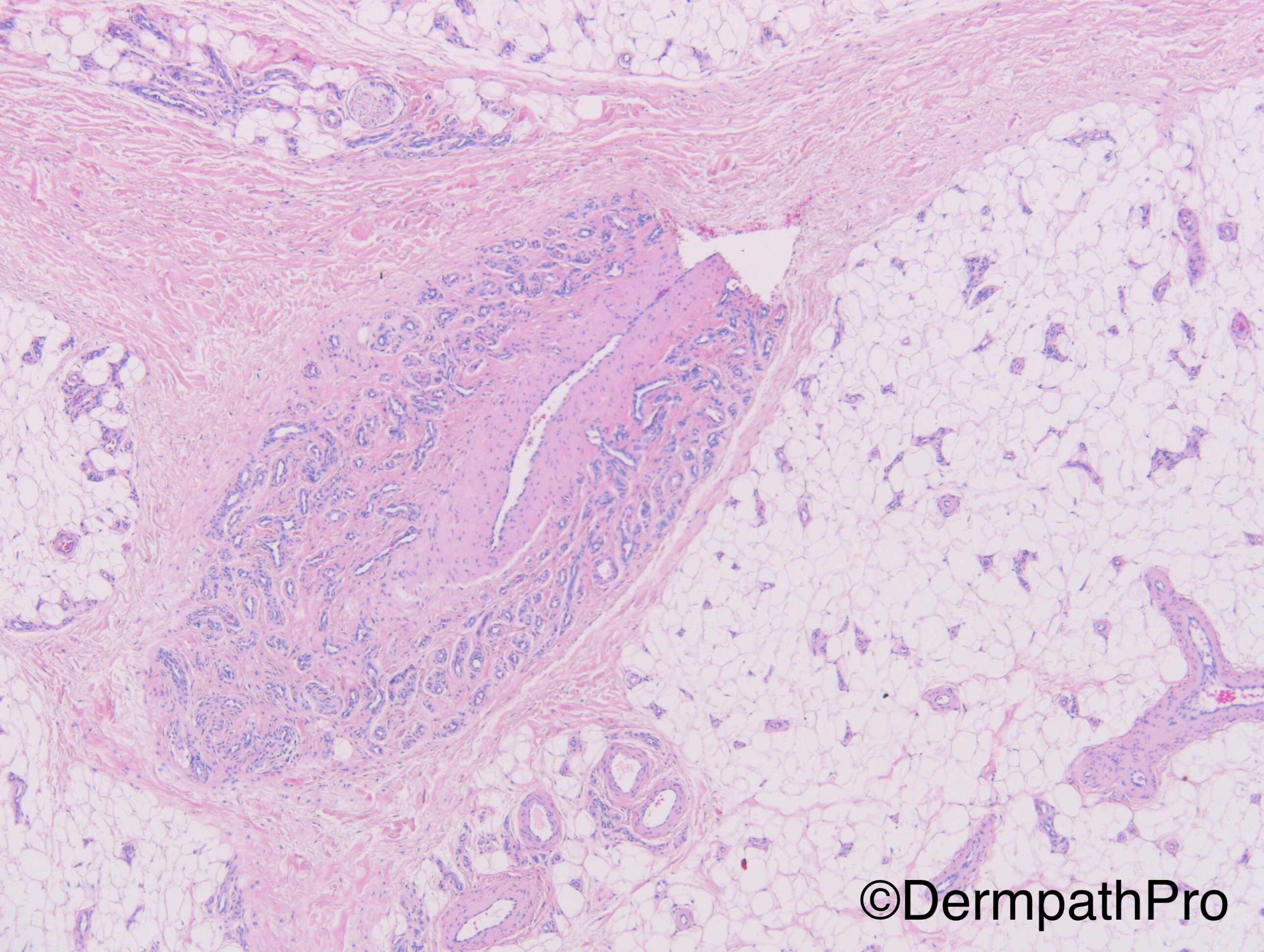
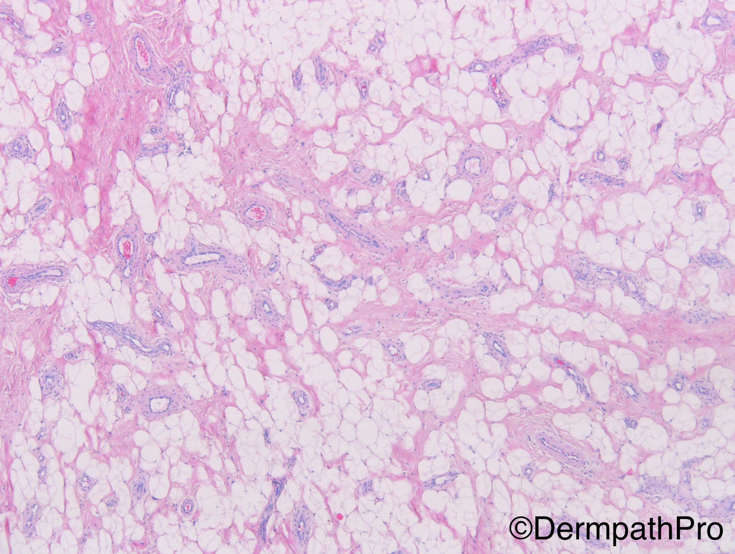
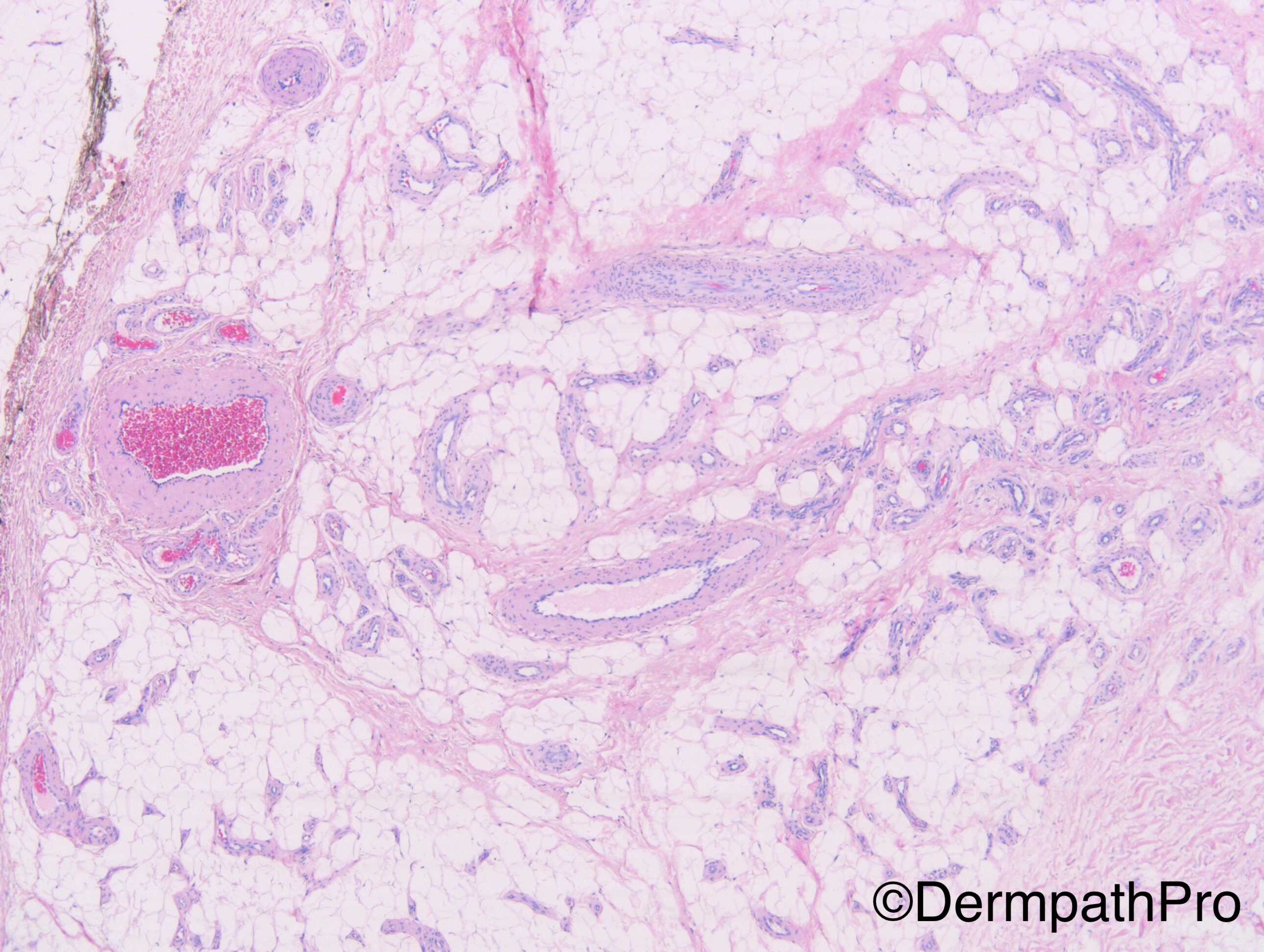
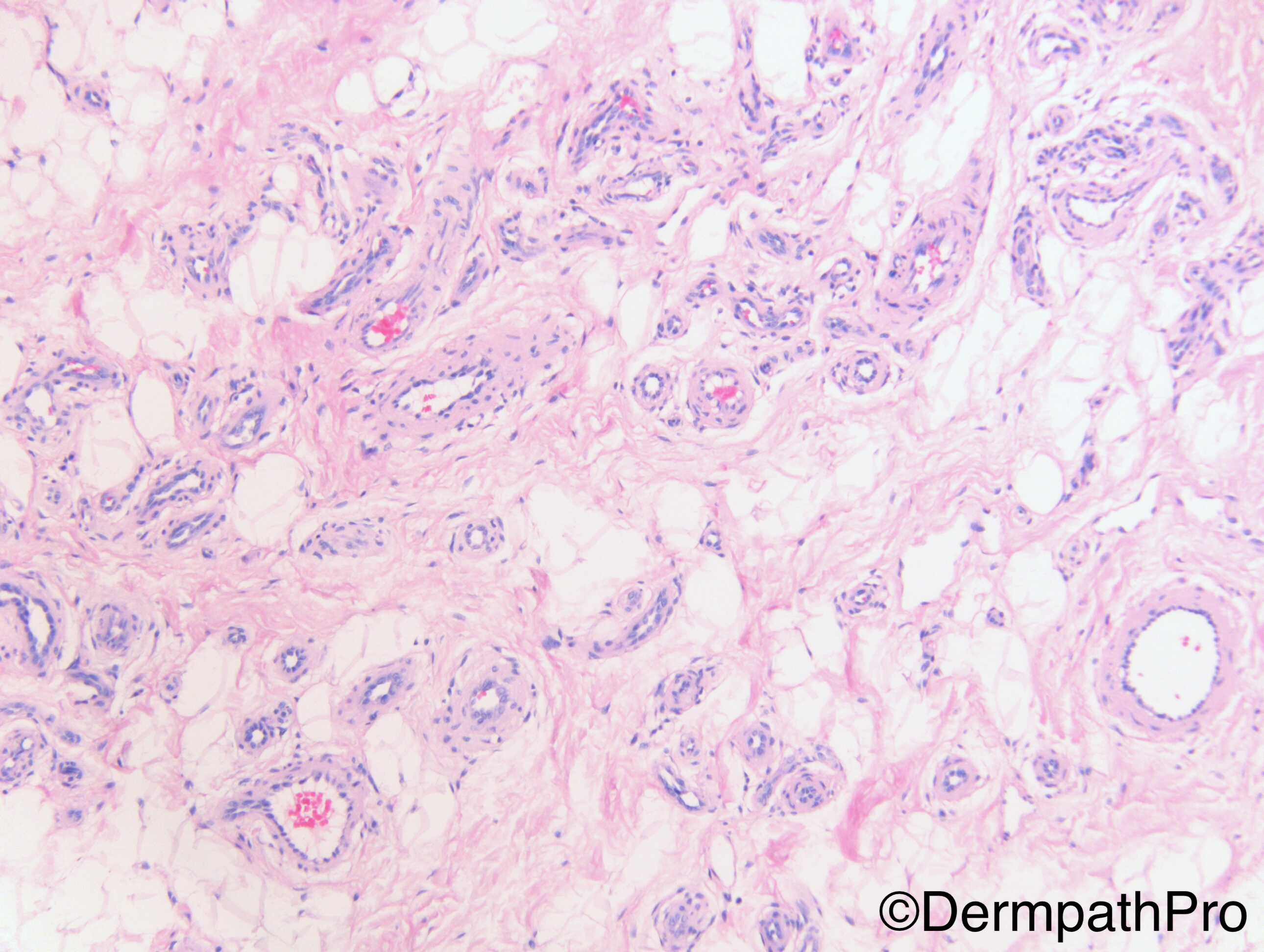
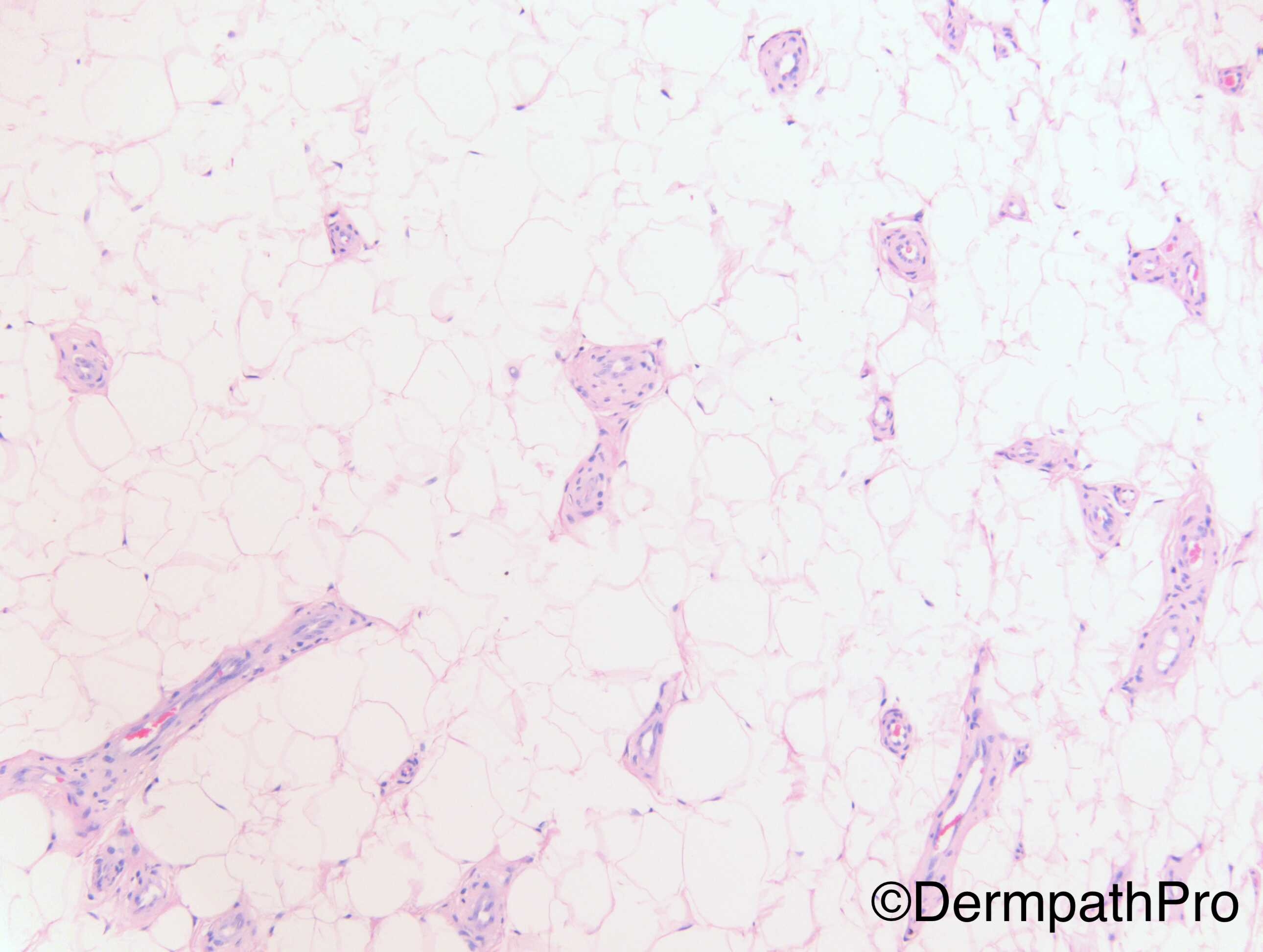
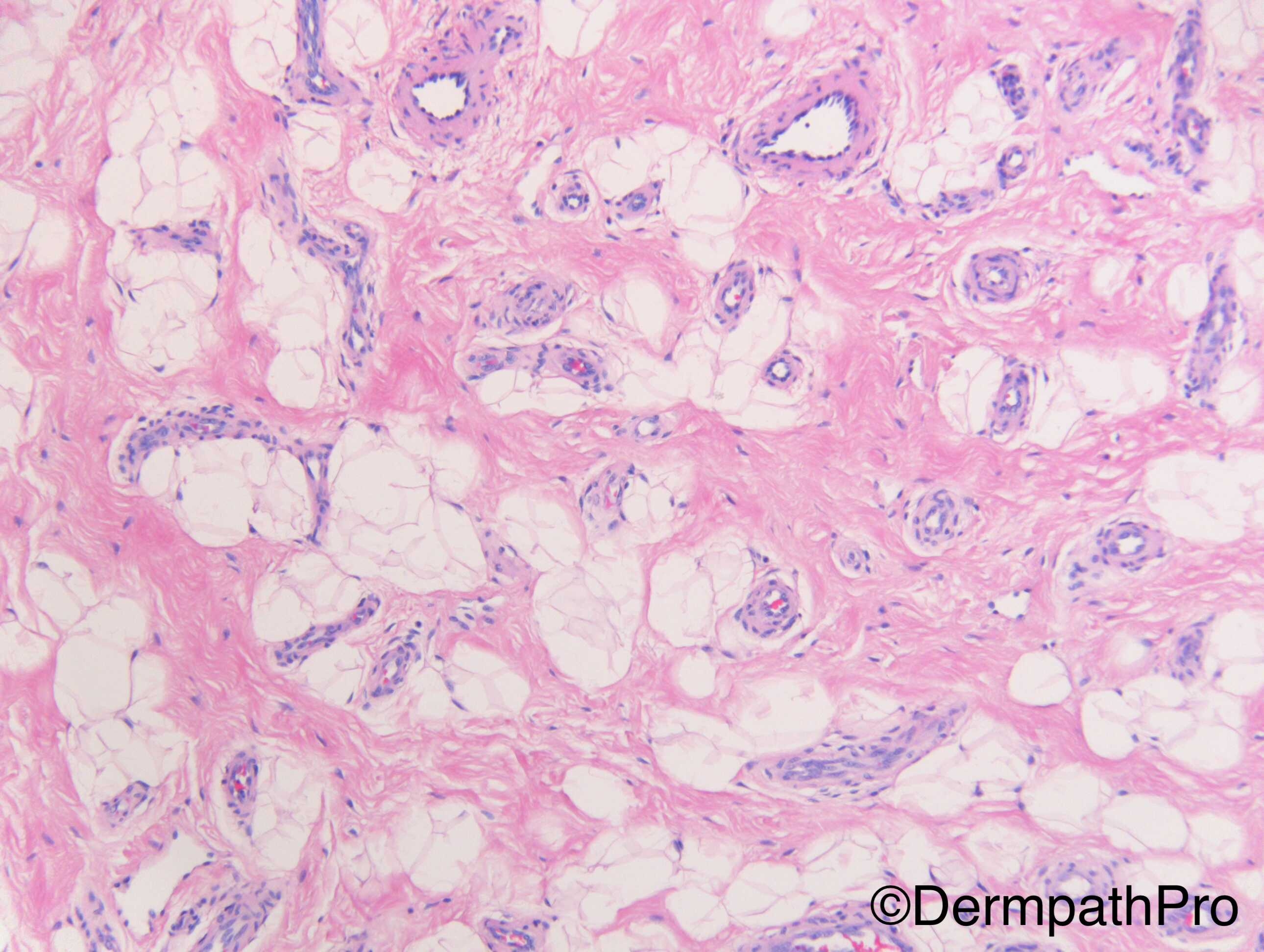
Recurrent lipoma forearm


  Report Record

User Feedback


In my experience, in angiolipoma, the vessels are usually dilated and have thin walls like sinuses. Some authors even require intravascular fibrin thrombi as a component of angiolipoma. The vessels in this lesion are thick-walled of various calibers and I do not appreciate fibrin thrombi in any of the vessels. 

It is a lesion with lipomatous and vascular tissue. The question is whether the lipomatous tissue is part of the lesion or is only a bystander. I suspect this is more likely a vascular lesion involving adipose tissue. Because of the presence of vascular structures of various calibers and types, it would consider the possibility of a vascular malformation or soft tissue angiomatosis. Clinical information such as size of the lion and age of the patient might be helpful. Also, what was the original diagnosis?

Share this comment


Link to comment
share_externally

Dr. Richard Carr

Posted

Dear Anh. M39.

Chris Fletcher kindly took a look at the images and also favoured a vascular malformation and commented on the possibility of further local recurrence. Your points are all well made. I was struggling to label it and was happy to accept the lesion for what you have indicated (I too dismissed a conventional angiolipoma). I remember seeing intramuscular haemangiomas (or malformations) many moons ago with lots of adipose tissue.

Share this comment


Link to comment
share_externally


Join the conversation

You can post now and register later. If you have an account, sign in now to post with your account.

Guest
Add a comment...

×   Pasted as rich text.   Paste as plain text instead

  Only 75 emoji are allowed.

×   Your link has been automatically embedded.   Display as a link instead

×   Your previous content has been restored.   Clear editor

×   You cannot paste images directly. Upload or insert images from URL.

Loading...

×
×
  • Create New...