Jump to content
In this section we have spot diagnoses posted on a daily basis since June 2010, now over 4000! You can review the archived cases and read the suggested diagnoses by users and the final comment by the contributors.
Case are uploaded each week day by 10 am UK time with the correct diagnosis will generally be posted at 8 pm UK time. Why not view the most recent spot diagnosis and proffer a diagnosis?

Case Number : Case 3072 - 15 April 2022 Posted By: Dr. Richard Carr

Please read the clinical history and view the images by clicking on them before you proffer your diagnosis.
Submitted Date :
   (0 reviews)

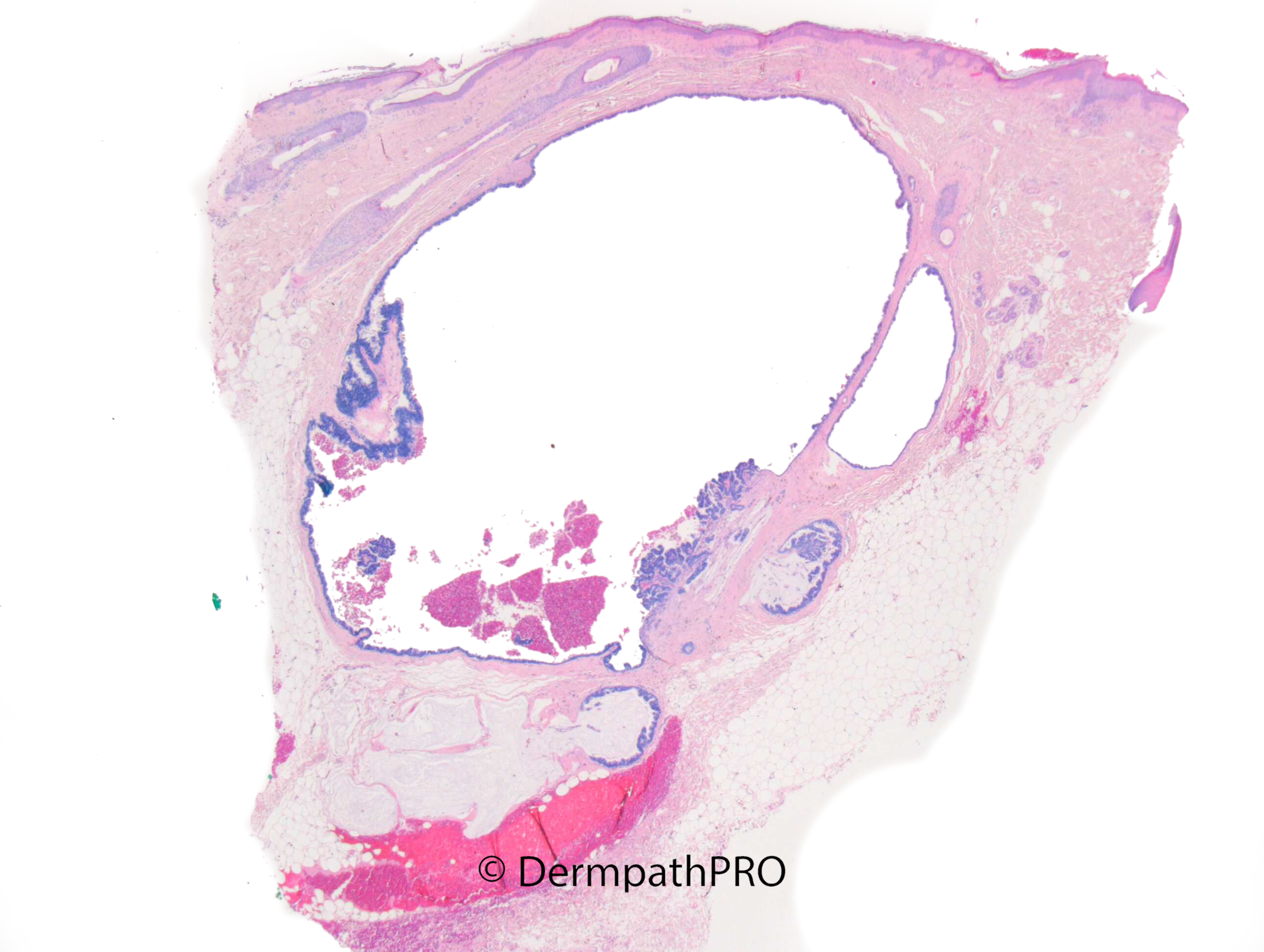
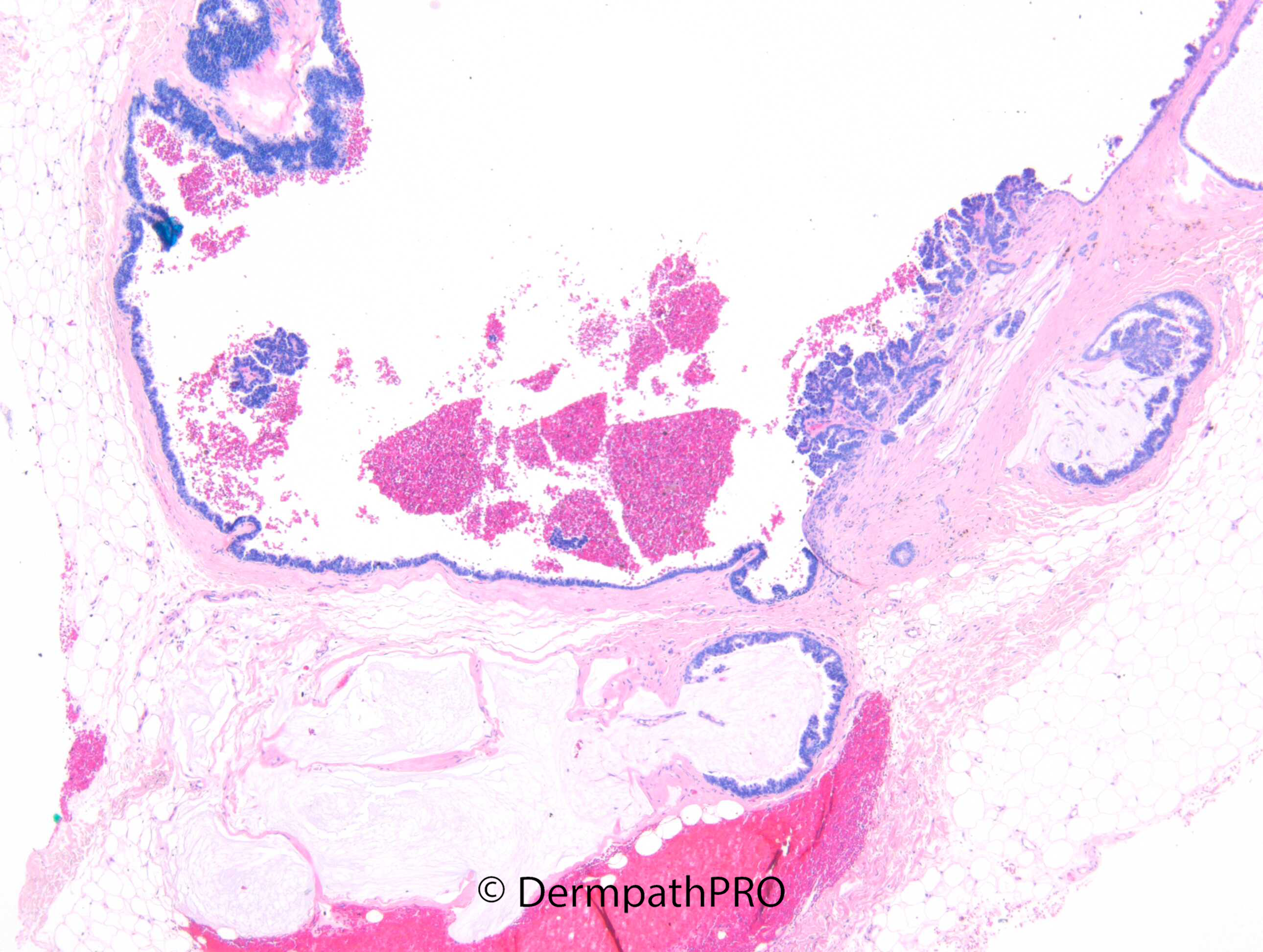
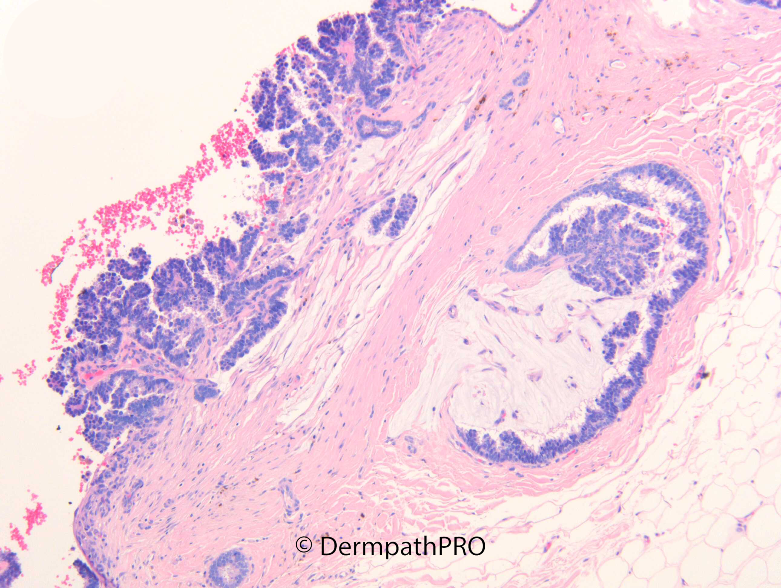
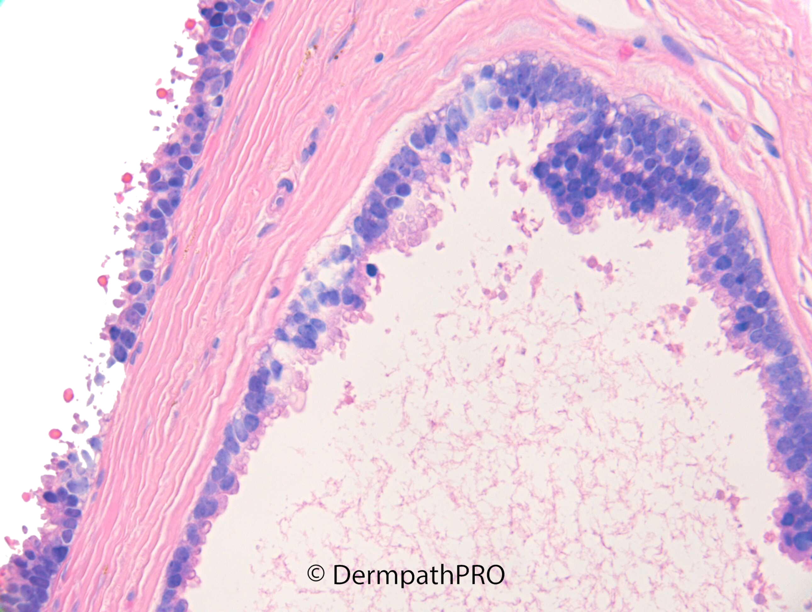
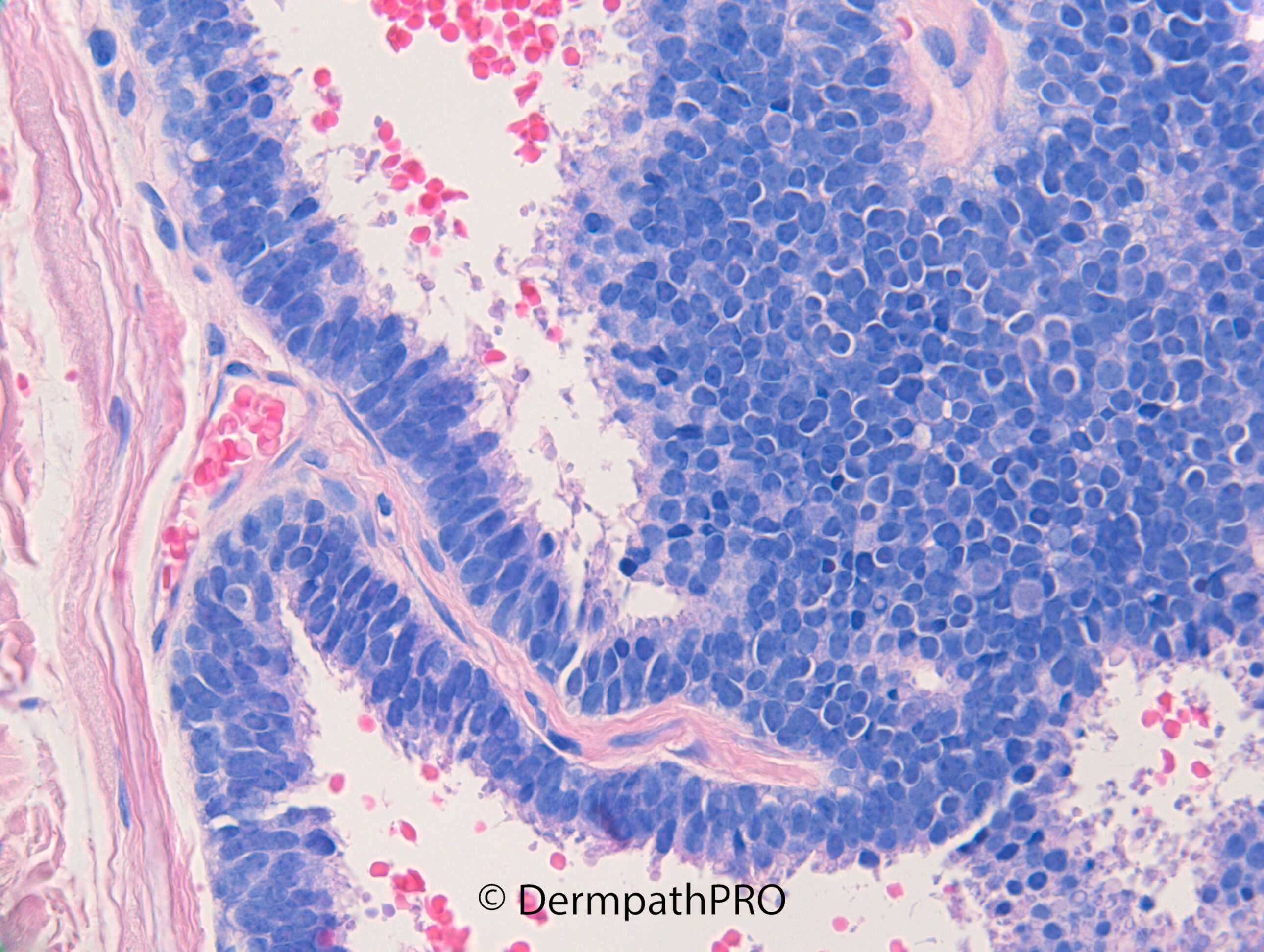
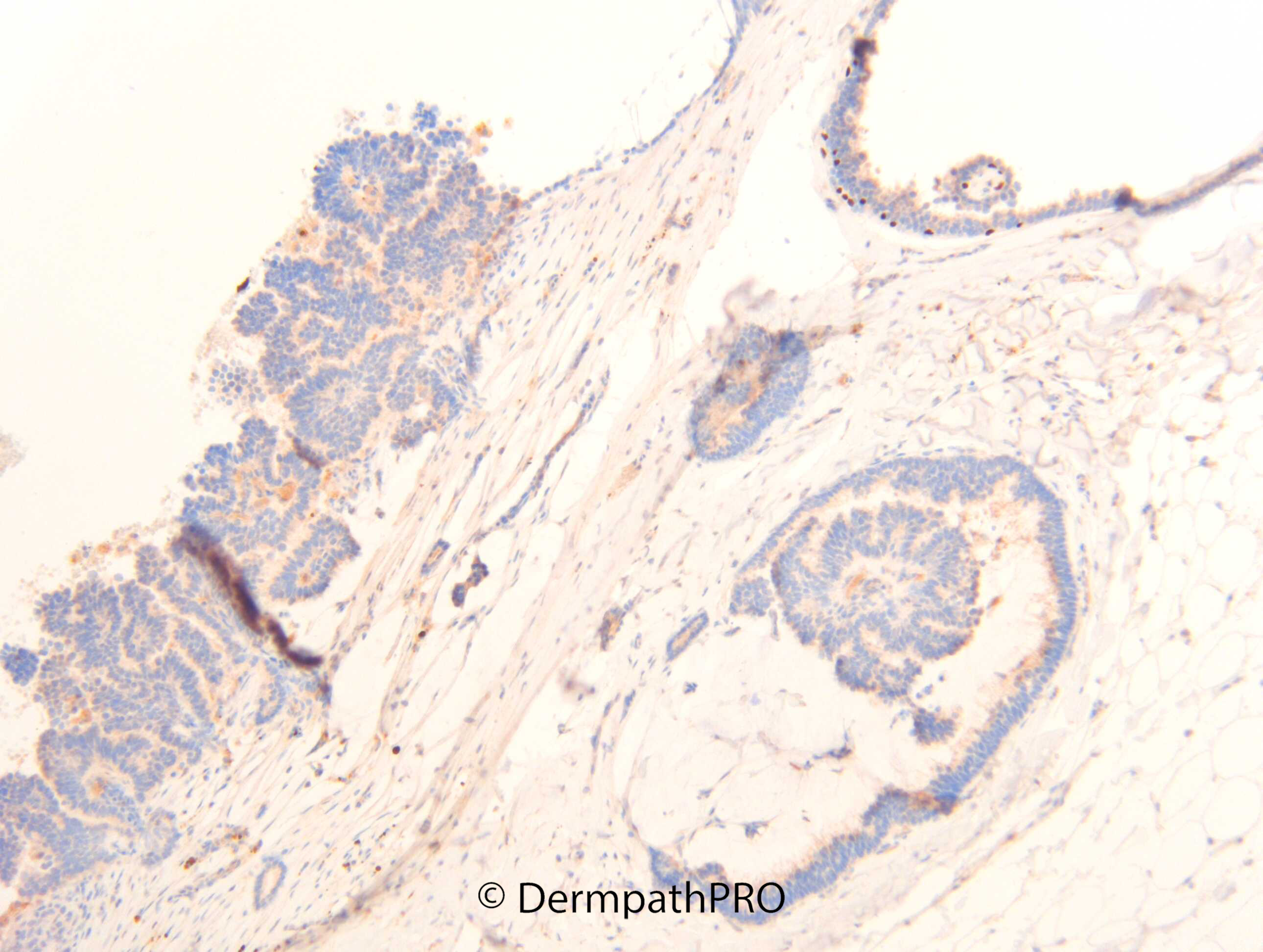
F75. Scalp. 4mm blue papule. ?blue naevus. Punch excision.


  Report Record

User Feedback


Chirag Desai

Posted

Cutaneous metastasis. Neuroendocrine carcinoma??? 

Share this comment


Link to comment
share_externally

Definitive looks like a cystic lesion that's mucinous - an in situ component in the form of EMPSGC, and an invasive mucinous carcinoma? IHC appears to be p63 or some other myoepithelial marker proving invasion vs in situ.

Share this comment


Link to comment
share_externally

Anil Patki

Posted

Apocrine hidrocystoma. Looks benign with nice decapitation secretion. Mucinous secretion is known in apocrine hidrocystomas.

Share this comment


Link to comment
share_externally

?? Unusual PCMC with papillocystic features. 

Share this comment


Link to comment
share_externally

Dr. Richard Carr

Posted

This is a small papillary cystic mucinous carcinoma mimicking a benign cystadenoma. Neuroendocrine markers were negative. In a recent review about 33% of primary cutaneous glandular adnexal carcinomas had a residual myoepithelial cell layer that excludes metastatic carcinoma to the skin. i.e. useful when present but not useful when absent and by far the most likely is a primary cutaneous lesion when this well differentiated. It is also useful here because a benign cystadenoma would have preserved myoepithelial layer throughhout (and no mucin pools). I think 45% of the mucinous carcinomas in the paper below had a partly preserved myoepithelial layer. A small well differentiated lesion like this should have minimal risk for developing metastases. I suggested a modest 5 to 10mm re-excision because mucin pools were abutting margins. In the paper below calponin was considered the best single marker compared with p63 and SMA. I've tend to use p63 as my go to marker.


Plaza, Jose A. MD*; Chung, Catherine MD†; Wick, Mark MD‡; Sangueza, Martin MD§; Gru, Alejandro MD¶ The Utility of Myoepithelial Cell Layer Identification in Adnexal Carcinomas, The American Journal of Dermatopathology: March 2022 - Volume 44 - Issue 3 - p 155-162
doi: 10.1097/DAD.0000000000001844 

Share this comment


Link to comment
share_externally


Join the conversation

You can post now and register later. If you have an account, sign in now to post with your account.

Guest
Add a comment...

×   Pasted as rich text.   Paste as plain text instead

  Only 75 emoji are allowed.

×   Your link has been automatically embedded.   Display as a link instead

×   Your previous content has been restored.   Clear editor

×   You cannot paste images directly. Upload or insert images from URL.

Loading...

×
×
  • Create New...