Jump to content
In this section we have spot diagnoses posted on a daily basis since June 2010, now over 4000! You can review the archived cases and read the suggested diagnoses by users and the final comment by the contributors.
Case are uploaded each week day by 10 am UK time with the correct diagnosis will generally be posted at 8 pm UK time. Why not view the most recent spot diagnosis and proffer a diagnosis?

Case Number : Case 1872 - 1 August - Dr Iskander Chaudhry (Invited) Posted By: Guest

Please read the clinical history and view the images by clicking on them before you proffer your diagnosis.
Submitted Date :
   (0 reviews)

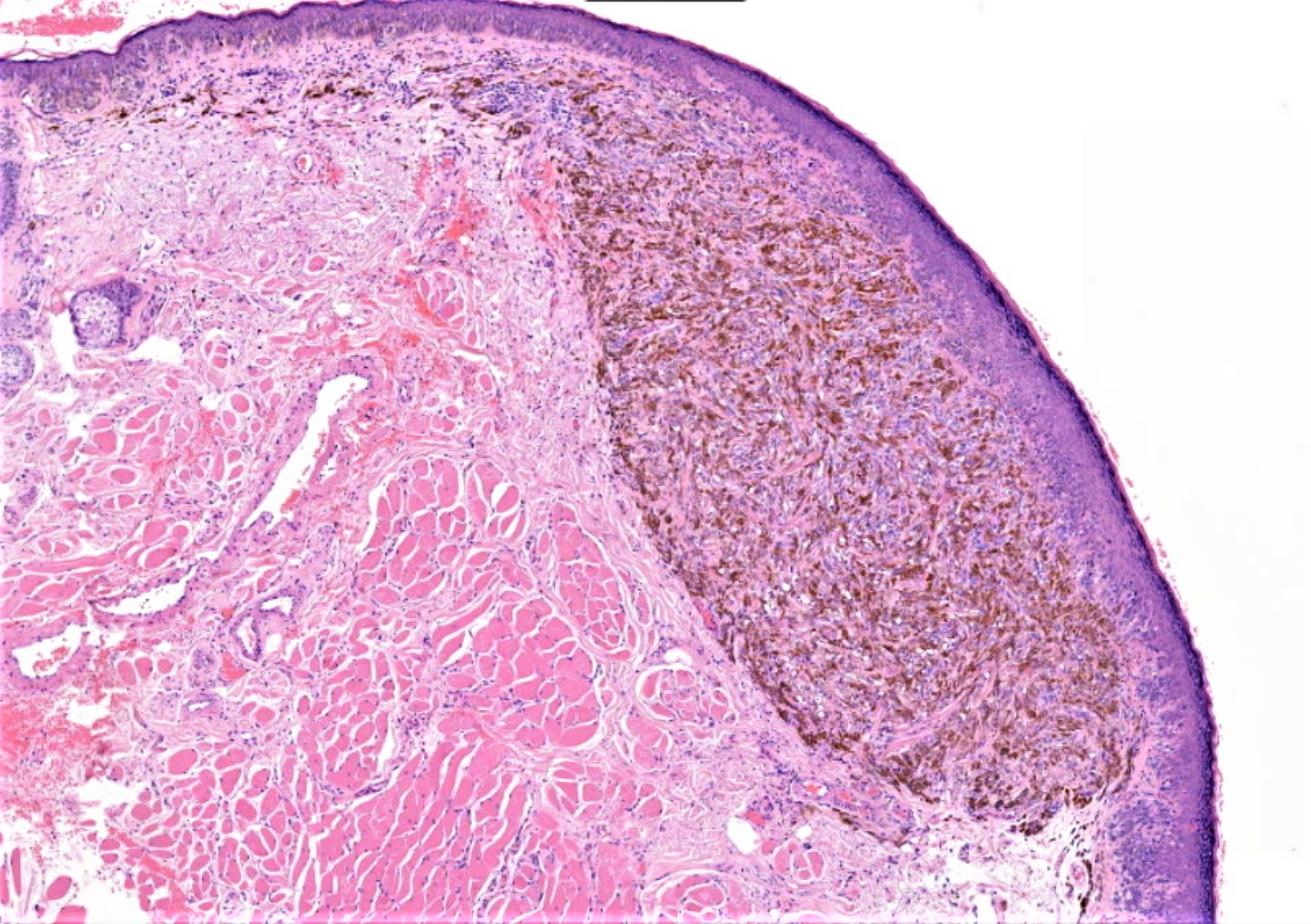
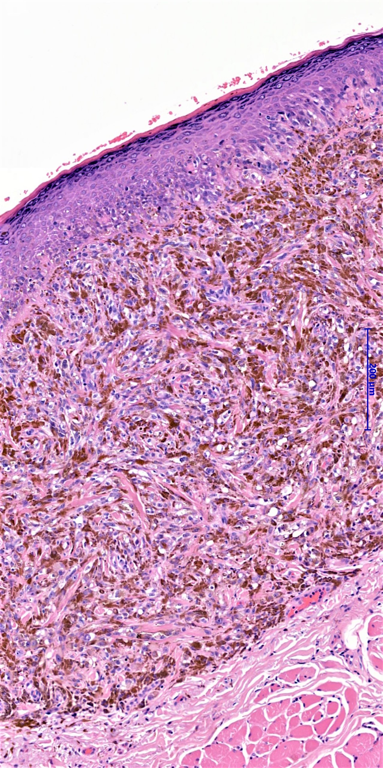
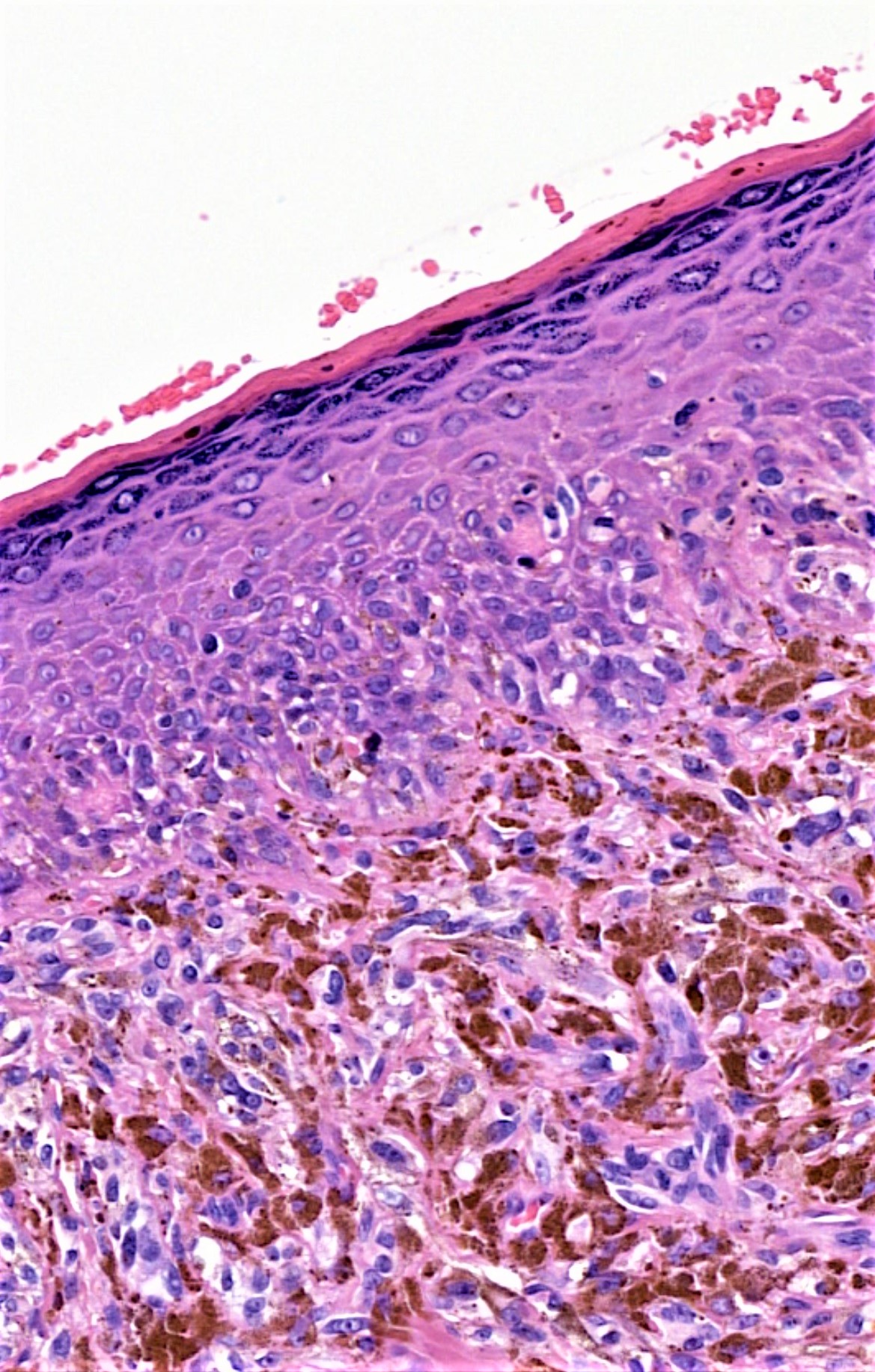
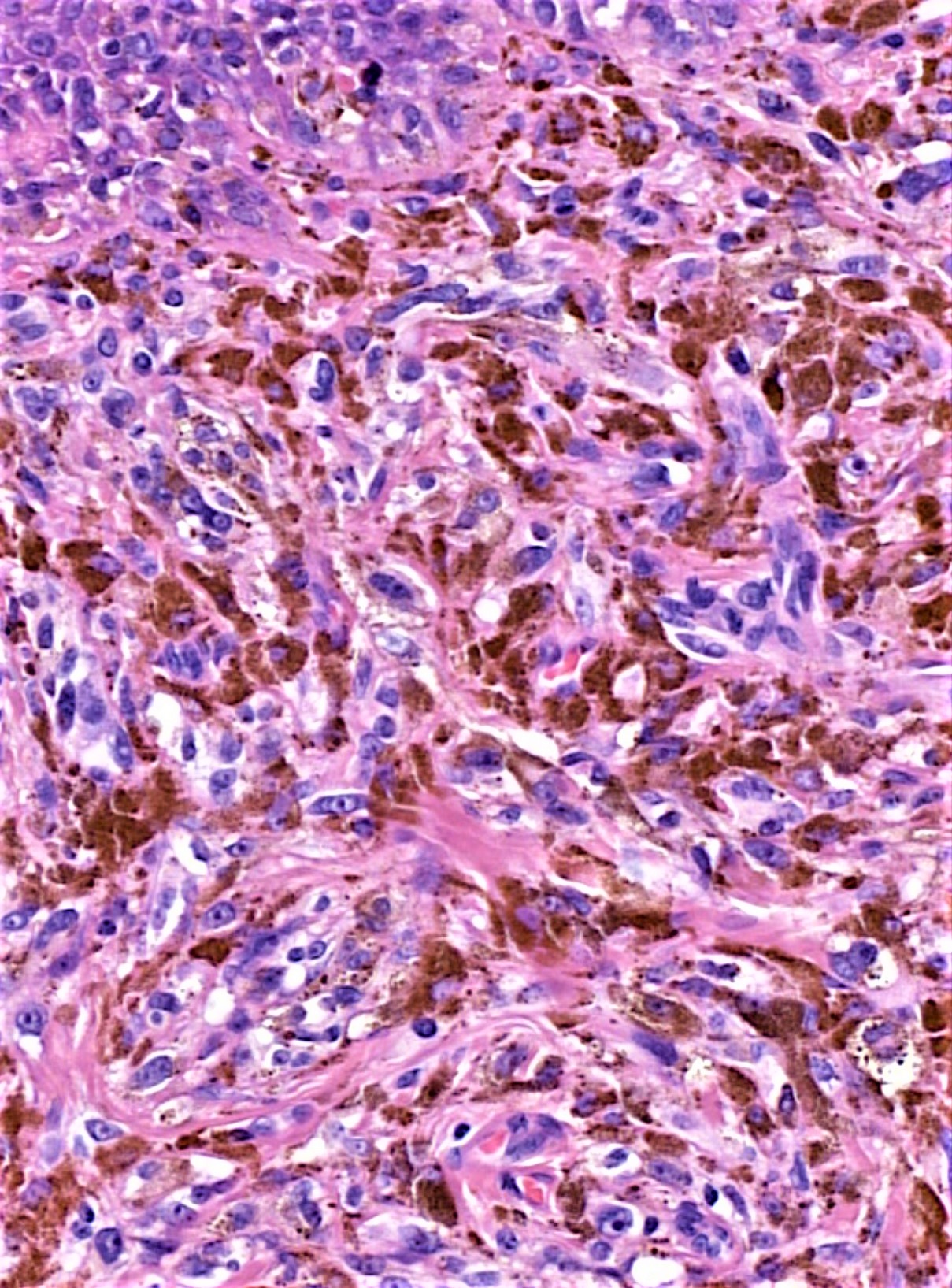
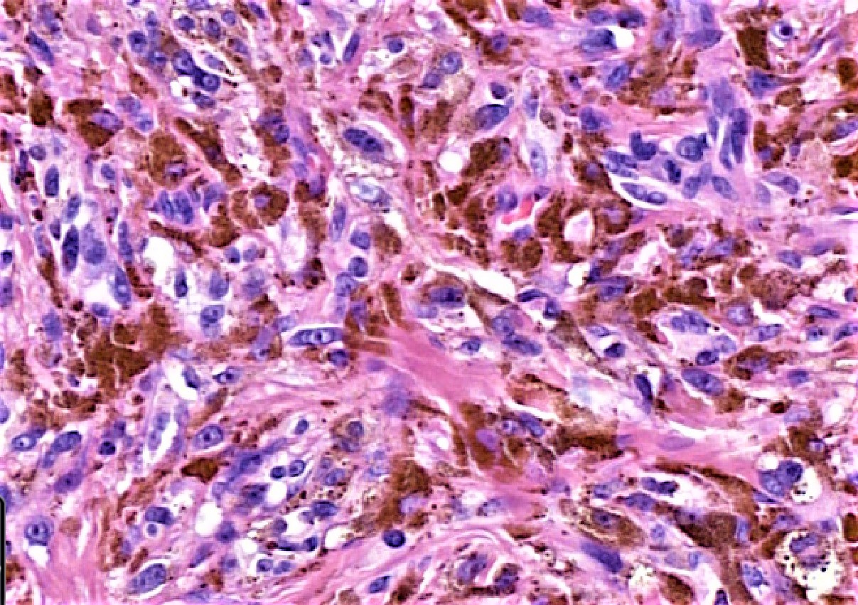
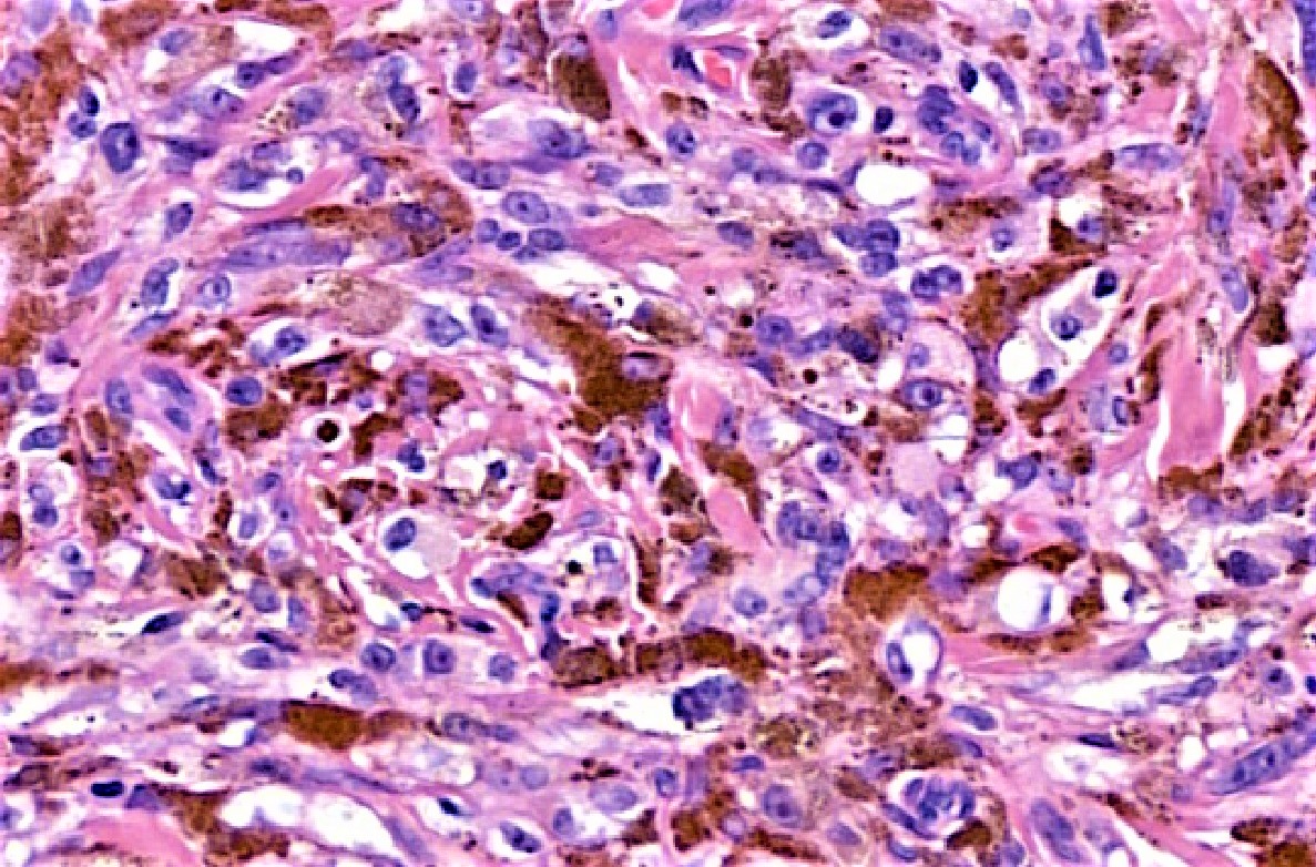
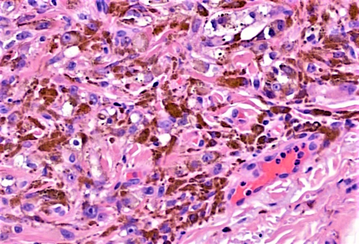
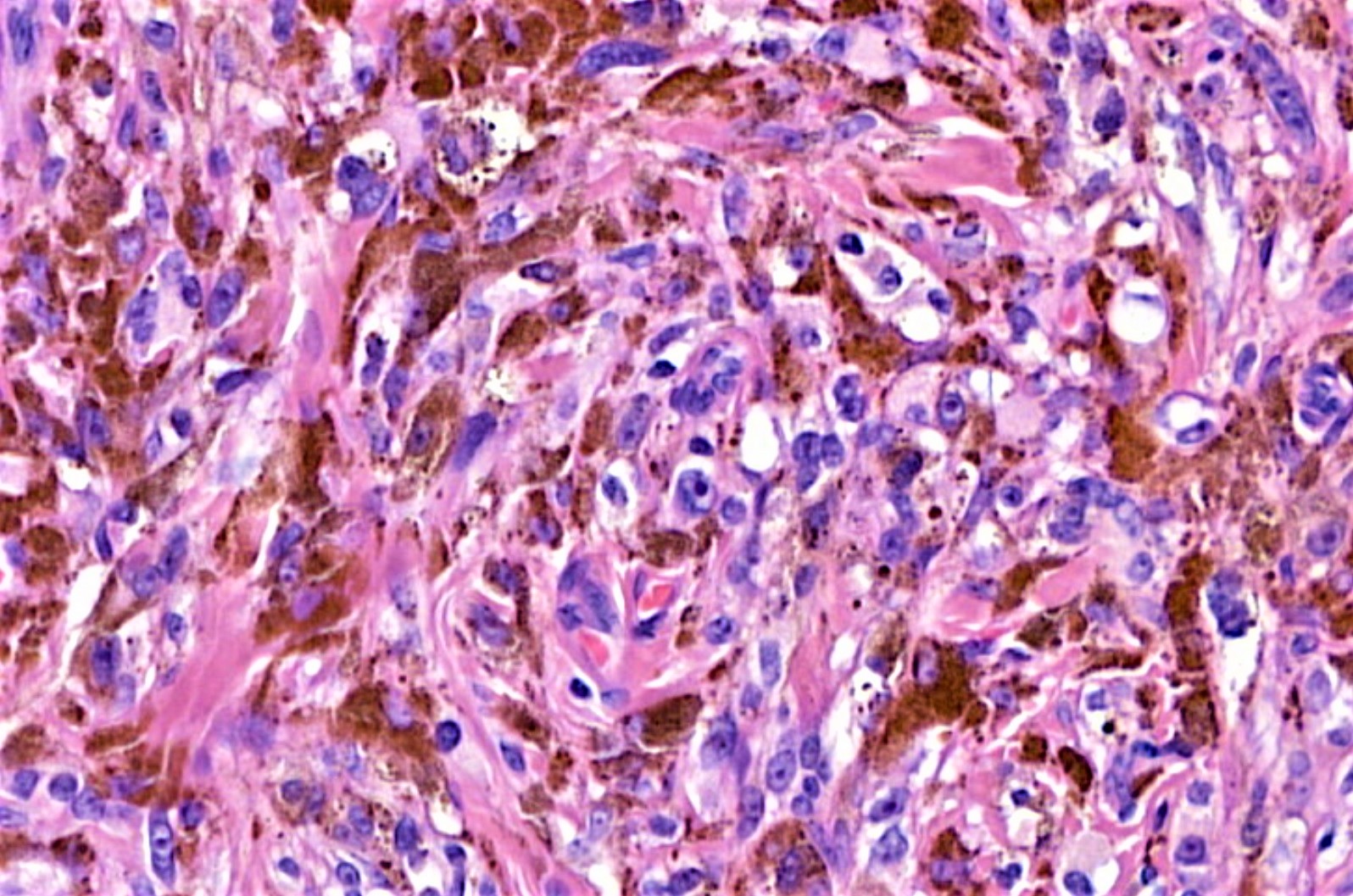
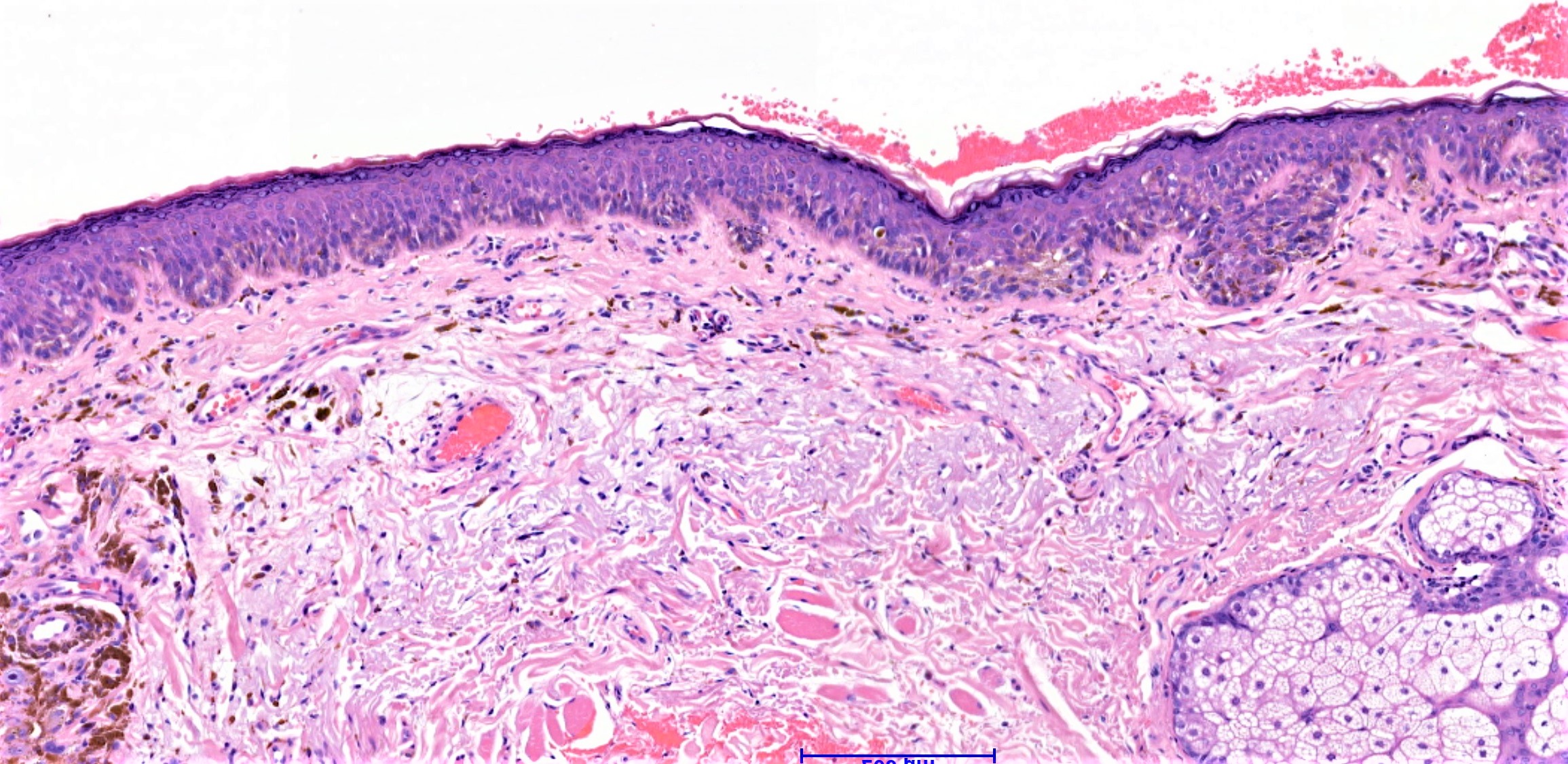
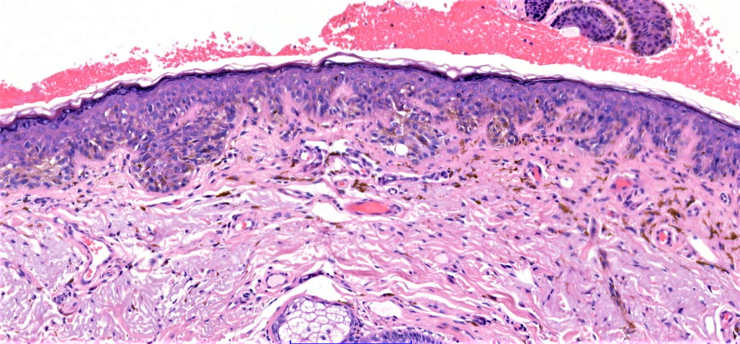
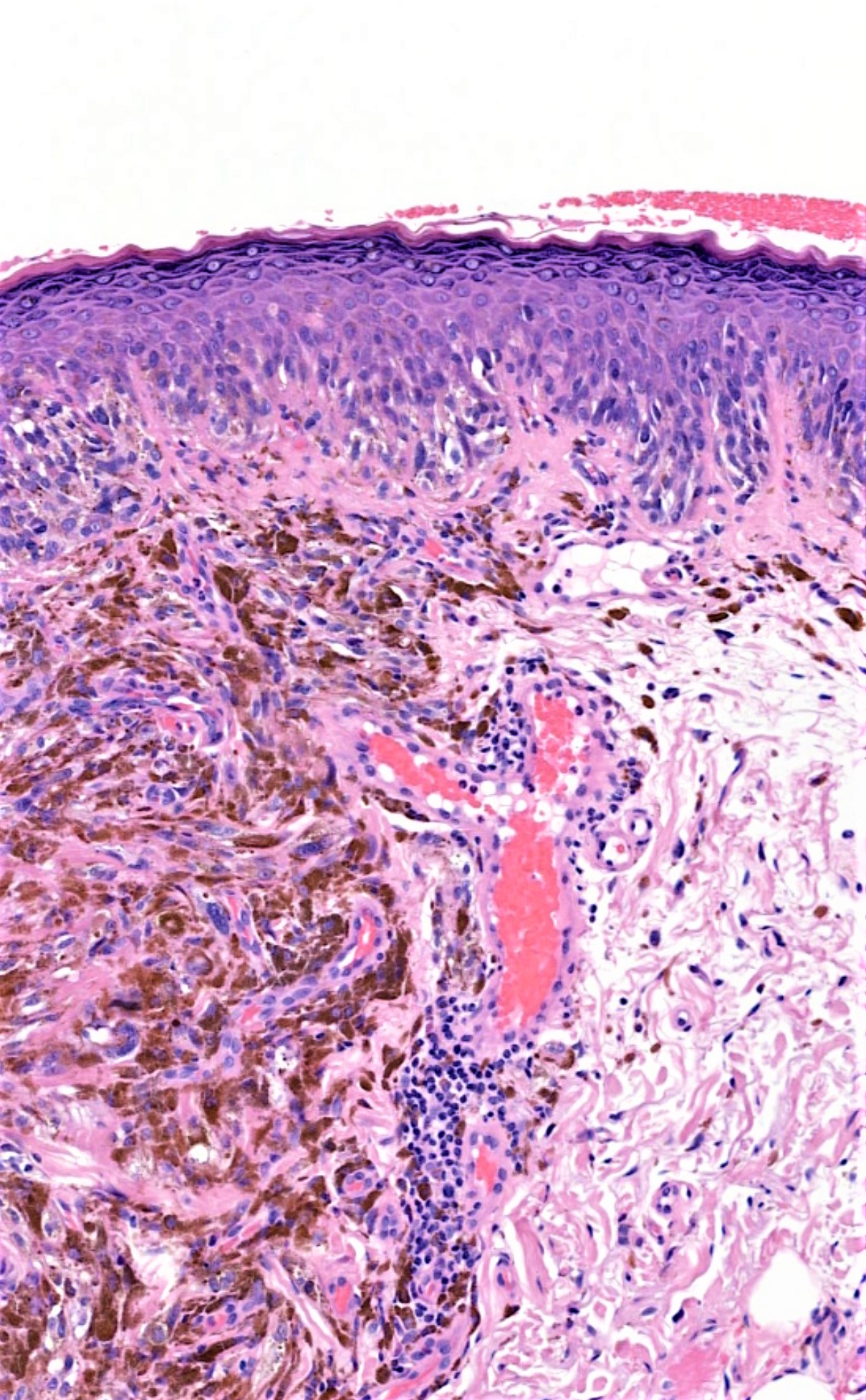
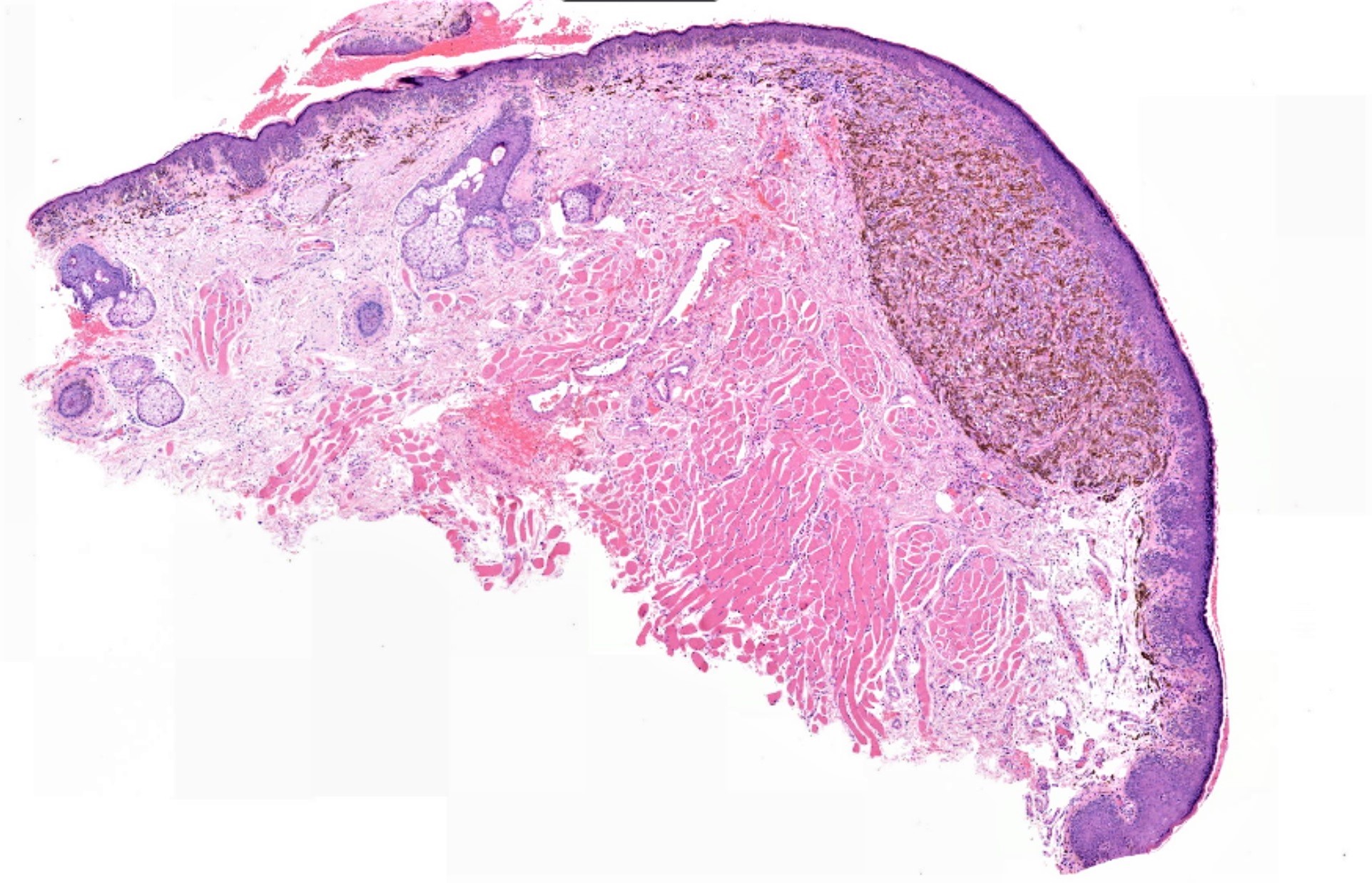
69 year old female. Left upper lip Incisional biopsy. Longstanding pigmented lesion upper lip, has become nodular.

Edited by Admin_Dermpath


  Report Record

User Feedback


Raul Perret

Posted

Im on my phone but excuses appart I thought this case could be compatible with a proliferative nodule on a nevus, due to sharp circumscription, homogeneous distribution of pigment and minimal spitzoid like atypia. PN in acquired nevi are infrequent but reported. In real life I would perform some immunos (ki67, melan A, HMB-45 and bleaching) and probably show the case to someone else as the clinical picture is rather alarming.

Share this comment


Link to comment
share_externally

urmilapandey

Posted

have never seen one before, wonder if a pigmented melanocytoma would be a valid differential on morphology.

Share this comment


Link to comment
share_externally

vincenzo polizzi

Posted

I’m on my iPhone, too, and don’t remember if lip could be a usual site for PEM , but there are many typical large and macro nucleolated cells between the heavily pigmented ones. So Pigmented Epithelioid MelanocytomaI is my favorite spot. 

PS just seen the Urmilapandey comment so am glad we are thinking about the same diagnosis. 

Share this comment


Link to comment
share_externally

Dr. Mona Abdel-Halim

Posted

First impression, pigmented epithelioid melanocytoma too. 

Share this comment


Link to comment
share_externally

Robledo F. Rocha

Posted

Massi and LeBoit, on their absolutely necessary textbook Histological Diagnosis of Nevi and Melanoma, classify melanocytoma as a benign lesion in three circumstances:

  • melanocytoma in the setting of Carney complex;
  • combined melanocytoma; and
  • melanocytoma with maturation in blue nevus, Spitz nevus, or other forms of nevus.

The present lesion has microscopic features of benignity, including small size, confinement to the superficial dermis, sharp circumscription, and insignificant mitotic activity. On the other hand, I don't think it can be accommodate in any of the above three scenarios, and I feel particularly concerned facing a melanocytic tumor that arose from a pre-existing nevus (there seems to be a junctional nevus on the left side of the two first images, and the case history report a nodular change on a longstanding pigmented lesion) on a sun-exposed site of a woman on her late sixties. So I'd consider this lesion as a low-grade malignant melanocytoma with metastatic capability to regional lymph nodes.

Share this comment


Link to comment
share_externally

urmilapandey

Posted

6 minutes ago, Robledo F. Rocha said:

Massi and LeBoit, on their absolutely necessary textbook Histological Diagnosis of Nevi and Melanoma, classify melanocytoma as a benign lesion in three circumstances:

  • melanocytoma in the setting of Carney complex;
  • combined melanocytoma; and
  • melanocytoma with maturation in blue nevus, Spitz nevus, or other forms of nevus.

The present lesion has microscopic features of benignity, including small size, confinement to the superficial dermis, sharp circumscription, and insignificant mitotic activity. On the other hand, I don't think it can be accommodate in any of the above three scenarios, and I feel particularly concerned facing a melanocytic tumor that arose from a pre-existing nevus (there seems to be a junctional nevus on the left side of the two first images, and the case history report a nodular change on a longstanding pigmented lesion) on a sun-exposed site of a woman on her late sixties. So I'd consider this lesion as a low-grade malignant melanocytoma with metastatic capability to regional lymph nodes.

thanks for the information Robeldo

Share this comment


Link to comment
share_externally

Admin_Dermpath

Posted

Dear All 


Dr Chaudhry added 3 more insitu images 


DermpathPRO Admin

Share this comment


Link to comment
share_externally

Raul Perret

Posted

Like it less now, it looks like melanoma in situ with important elastosis and areas suspicious for regression. 

Share this comment


Link to comment
share_externally

Raul Perret

Posted

So agree that this should be up-graded, I was mistaken before.

Share this comment


Link to comment
share_externally

Nitin Khirwadkar

Posted

Don't really like it due to some of the reasons cited by Robledo. There is a junctional run of cells, a nodule with abnormal pigment network, possible fine scarring in the last image and atypia. Going for a melanoma. Would have looked at a bleached section, Mel-A and Ki67 .

Share this comment


Link to comment
share_externally

Admin_Dermpath

Posted

Dear All 

Thank you for your comments. I have now had a few of these cases and call them 'pigment synthesising' melanomas after the name suggested by Eduardo et al. The proliferation index is often low but I have found clinicians manage them in the same way as conventional melanomas. Is this the experience of others ? 

This lesion has a worry atypical in situ component which you don't always see

Best Regards

Iskander 

Share this comment


Link to comment
share_externally

Dr. Richard Carr

Posted

Agree with you Iskander. I expect a low risk for metastasis of this "well differentiated" indolent appearing melanoma but obviously hard to predict.

Share this comment


Link to comment
share_externally

Mariantonieta Tirado

Posted (edited)

Age, sun damaged skin (FACE!) the junctional component seems to trickle to the edge, in addition some dermal atypia: melanoma. But would like to see melanA (red) to evaluate better: symmetry, delimitation and pagetoid spread

Edited by Mariantonieta Tirado

Share this comment


Link to comment
share_externally


Join the conversation

You can post now and register later. If you have an account, sign in now to post with your account.

Guest
Add a comment...

×   Pasted as rich text.   Paste as plain text instead

  Only 75 emoji are allowed.

×   Your link has been automatically embedded.   Display as a link instead

×   Your previous content has been restored.   Clear editor

×   You cannot paste images directly. Upload or insert images from URL.

Loading...

×
×
  • Create New...