Jump to content
In this section we have spot diagnoses posted on a daily basis since June 2010, now over 4000! You can review the archived cases and read the suggested diagnoses by users and the final comment by the contributors.
Case are uploaded each week day by 10 am UK time with the correct diagnosis will generally be posted at 8 pm UK time. Why not view the most recent spot diagnosis and proffer a diagnosis?

Case Number : Case 1165- 10th December Posted By: Guest

Please read the clinical history and view the images by clicking on them before you proffer your diagnosis.
Submitted Date :
   (0 reviews)

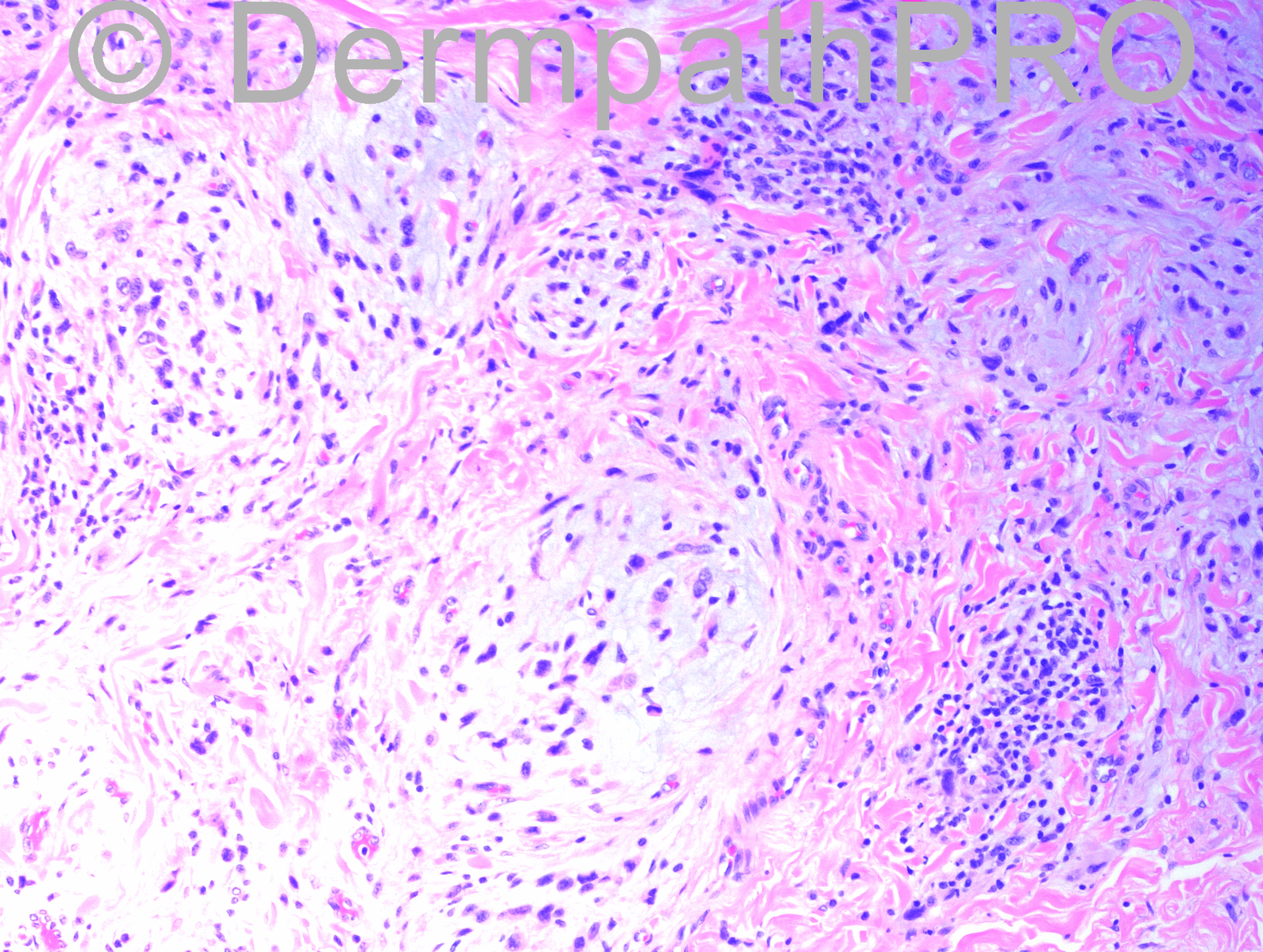
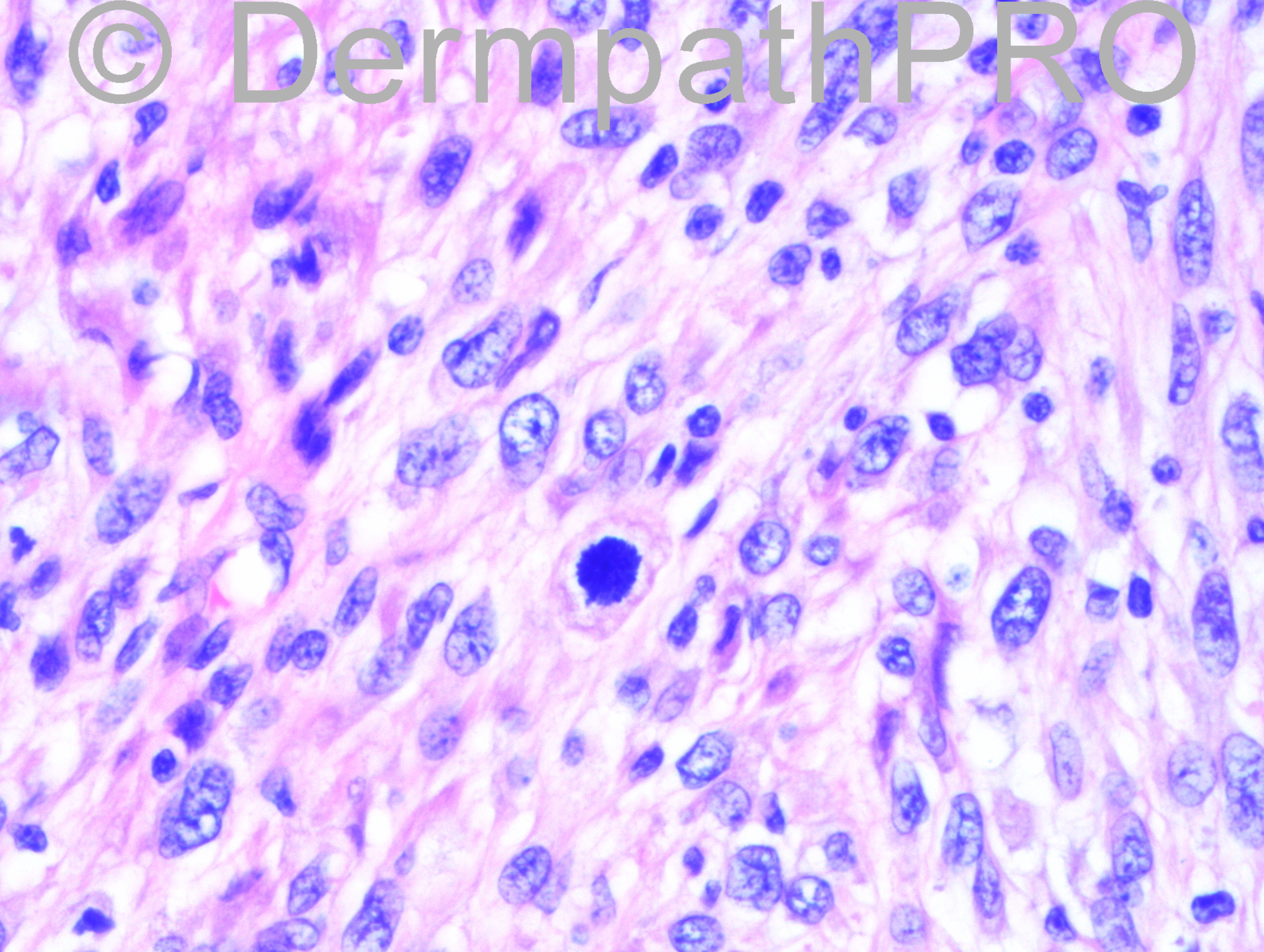
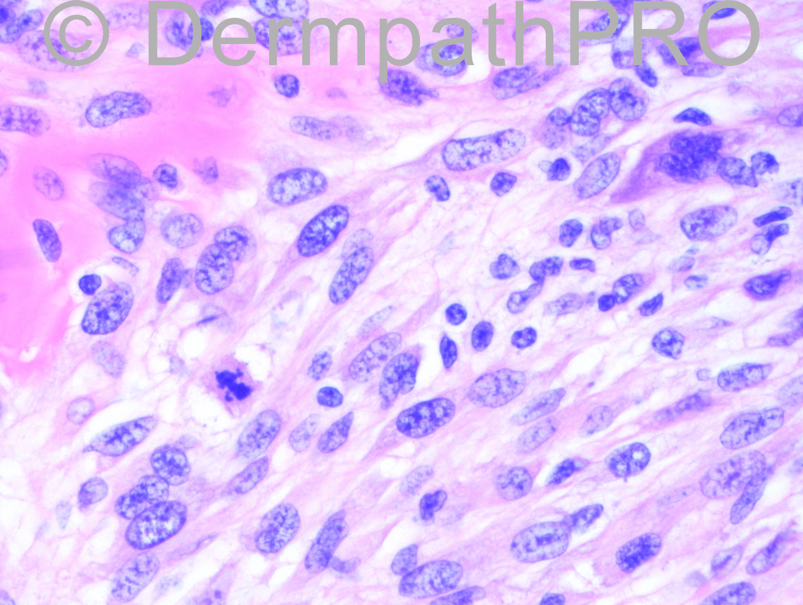
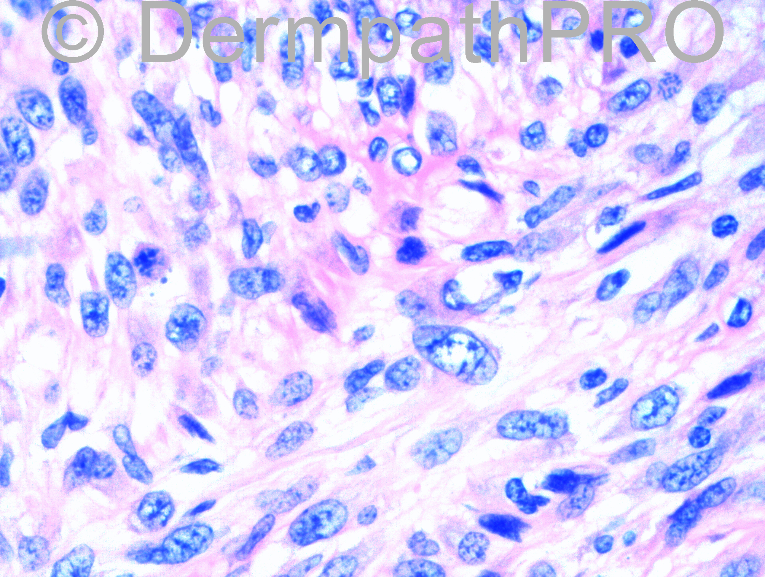
11 year-old female with neck mass. This has been growing slowly for the a year.

Case posted by Dr. Hafeez Diwan


  Report Record

User Feedback


Guest Tiberiu Tebeica

Posted

I also think this is a cellular neurothekeoma.

Share this comment


Link to comment
share_externally

Arti Bakshi

Posted

Cells look too atypical and mitotically active for a cellular neurothekeoma. Also myxoid change is not usually a feature.
Not sure about the diagnosis though, need immunos!

Share this comment


Link to comment
share_externally

Dr. Richard Carr

Posted

The first image, cellular neurothekeoma, was the first thought. I am a little concerned it lacks the typical packeting by hyaline collagen. Most of the CNs I have seen were also slightly more plump epithelioid cells dominating. The cellular atypia and atypical mitotic figures can be seen in cellular neurothekeomas without adverse outcome. I would want to see the IHC of course. My differentials included MPNST and an atypical myoepithelial lesion (before looking at the age which fits better with neurothekeoma).

Share this comment


Link to comment
share_externally

Guest Jim Davie MD

Posted

Cellular neurothekeoma most likely, vs. Type I NF related MPNST.

Share this comment


Link to comment
share_externally

Dr. Hafeez Diwan

Posted

I ended up calling this (after showing the case around to many of my friends and colleagues in the area) atypical cellular neurothekeoma, but it is certainly the most atypical one I have seen. It was negative for S100, MelanA, desmin, CK5/6 and Sox-10. It was positive for CD10 and focally for S100A6. I advised complete excision.

Share this comment


Link to comment
share_externally


Join the conversation

You can post now and register later. If you have an account, sign in now to post with your account.

Guest
Add a comment...

×   Pasted as rich text.   Paste as plain text instead

  Only 75 emoji are allowed.

×   Your link has been automatically embedded.   Display as a link instead

×   Your previous content has been restored.   Clear editor

×   You cannot paste images directly. Upload or insert images from URL.

Loading...

×
×
  • Create New...