Jump to content
In this section we have spot diagnoses posted on a daily basis since June 2010, now over 4000! You can review the archived cases and read the suggested diagnoses by users and the final comment by the contributors.
Case are uploaded each week day by 10 am UK time with the correct diagnosis will generally be posted at 8 pm UK time. Why not view the most recent spot diagnosis and proffer a diagnosis?

Case Number : Case 1959 - 1 Dec 2017 Posted By: Dr. Richard Carr

Please read the clinical history and view the images by clicking on them before you proffer your diagnosis.
Submitted Date :
   (0 reviews)

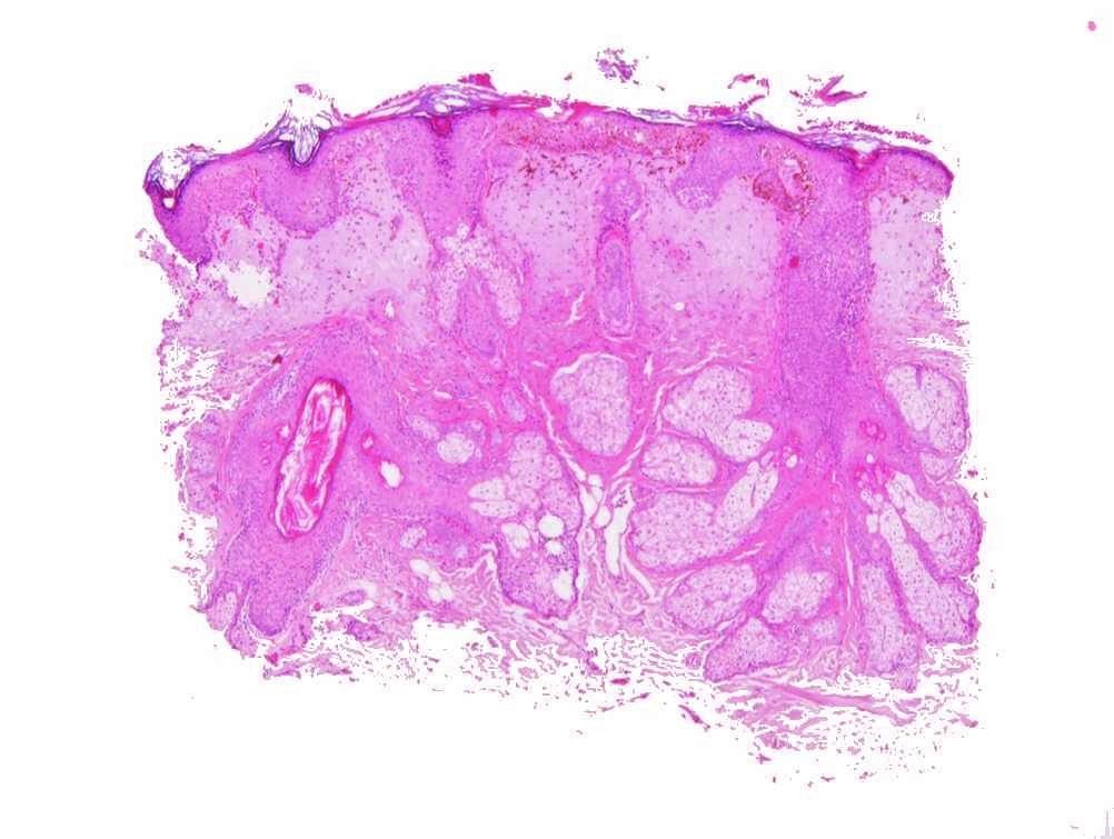
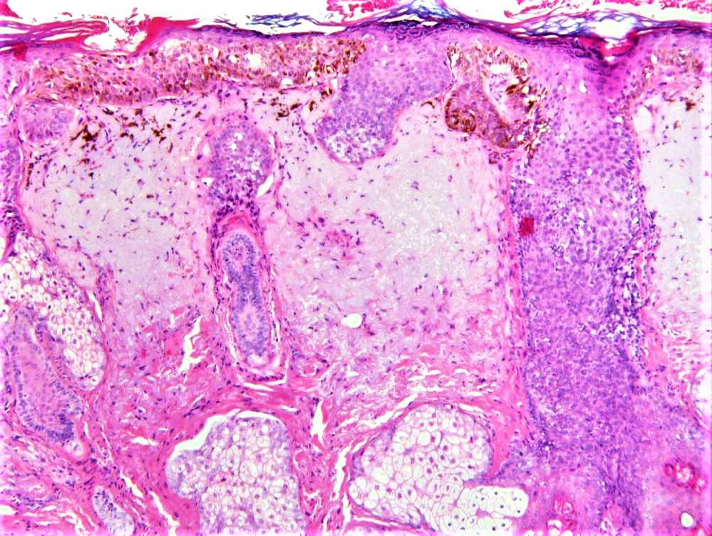
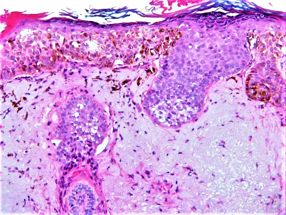
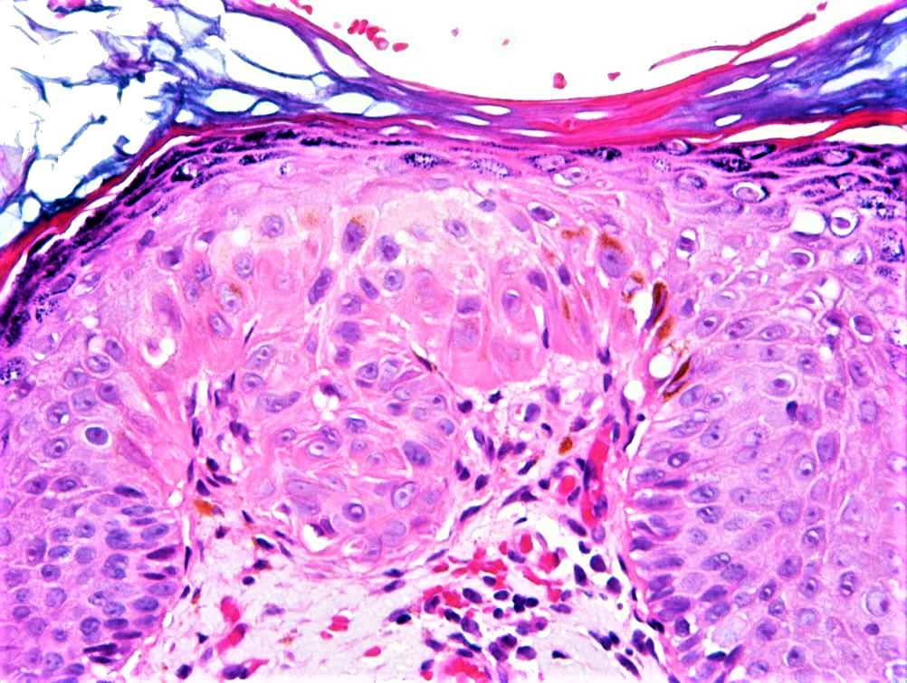
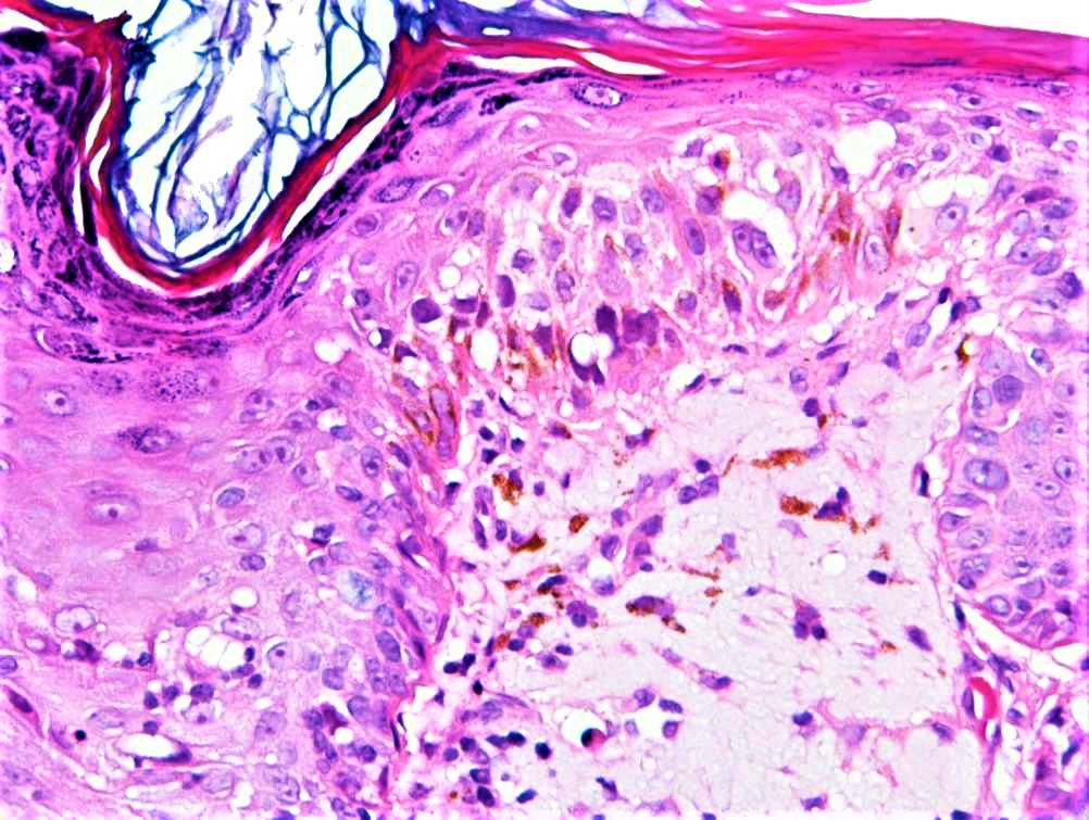
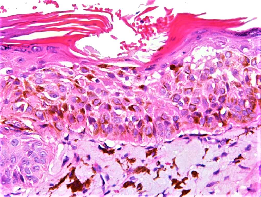
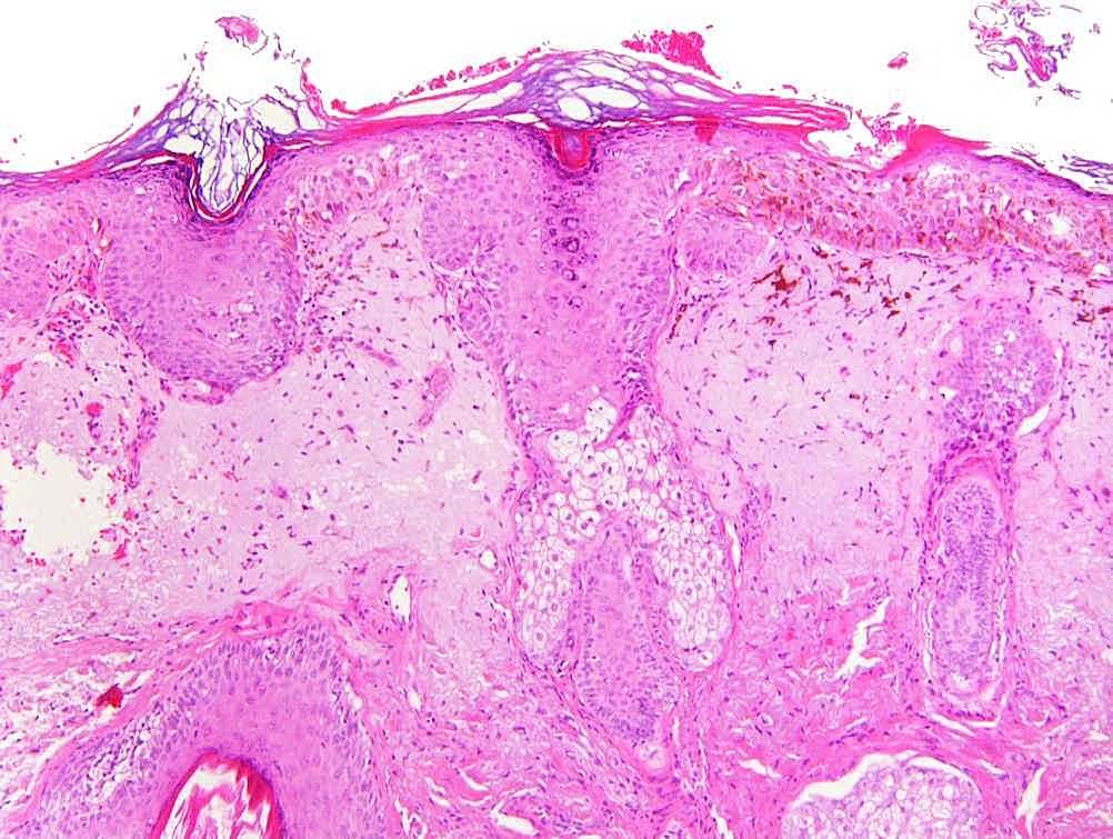
F70. Nose. Lentigo v Lentigo maligna
Edited by Admin_Dermpath


  Report Record

User Feedback


Looks more like a keratinocytic lesion than a melanocytic one, particularly Figures 5 and 6. So would favor a pigmented squamous cell carcinoma in situ.

Share this comment


Link to comment
share_externally

urmilapandey

Posted

does seem to be keratinocyte atypia. not sure if the same cells are pigmented and/or ballooned. so can't exclude a dual pathology.

Share this comment


Link to comment
share_externally

Raul Perret

Posted (edited)

For me there is keratinocyte atypia, and the lesion is discontinuous. I would go for a pigmented variant of actinic keratosis as for me atypia and architectural changes are not enough for SCC in situ. I think though that a nuclear melanocytic marker would be interesting in this case for ruling out coalescent neoplasms.


Edited by Raul Perret

Share this comment


Link to comment
share_externally

vincenzo polizzi

Posted

Agree with Raul. Pigmented actinic keratosis is the best spot. 

Share this comment


Link to comment
share_externally

Uma Sundram

Posted

I am not too sure myself but would probably not sign out without a nuclear melanocytic marker like MITF or SOX10. If it's pigmented AK, it's a pretty good fooler.

Share this comment


Link to comment
share_externally

Nitin Khirwadkar

Posted

Looks like pigmented actinic keratosis to me. However, would not sign out until I get Melan-A and MITF-1.

Share this comment


Link to comment
share_externally

5 hours ago, Nitin Khirwadkar said:

Looks like pigmented actinic keratosis to me. However, would not sign out until I get Melan-A and MITF-1.

just for my own learning experience:

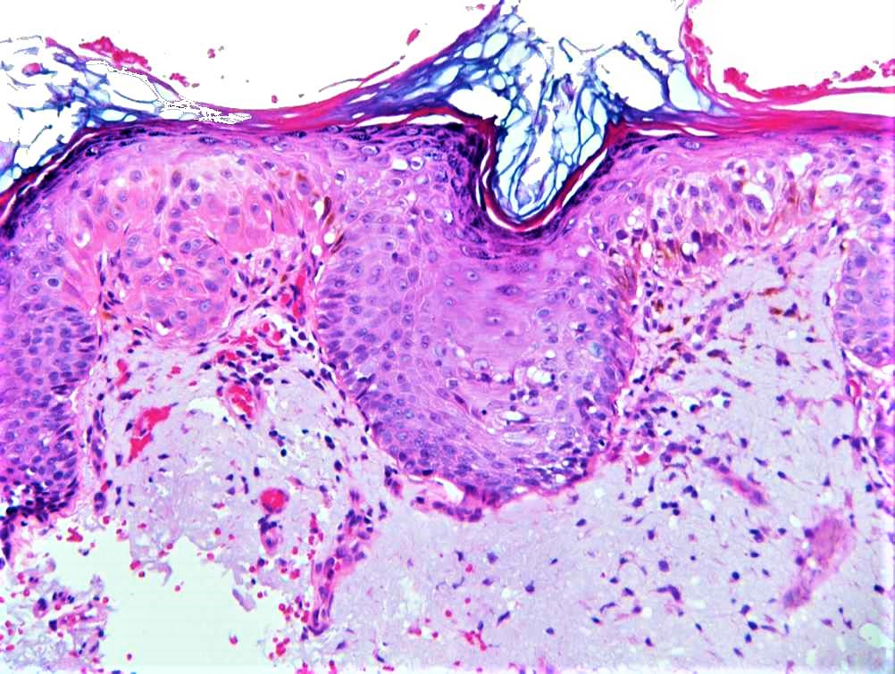
How do you guys differentiate SCC in situ from actinic keratosis in skin. If we agree this is a keratinocytic lesion, the last figure shows atypical cells involving almost the full thickness of the epidermis. Atypical keratinocytes involving less than 1/3 of the epidermis, 2/3 or full to differentiate SCC in situ and AK?


In addition, I almost never see block-like full-thickness involvement of the epidermis like by a melanocytic lesion . Usually we see nests and single cells in malignant melanoma spreading in the epidermis in a pagetoid pattern. But in the last figure, a segment of the epidermis in completely replaced / involved by the atypical cells. Does that favor keratinocytic over melanocytic?

Share this comment


Link to comment
share_externally

In fig 6, cells with clear cytoplasm seem to form a nest and these cells don't display clear cut intercellular bridges.

Share this comment


Link to comment
share_externally

Would think based on the H&E photos the most favoured diagnosis would be pigmented AK, although I would perform immunos (with a non-brown coloured chromogen) to exclude the possibility of a melanocytic lesion.

Share this comment


Link to comment
share_externally

vincenzo polizzi

Posted

13 hours ago, anh said:

just for my own learning experience:

How do you guys differentiate SCC in situ from actinic keratosis in skin. If we agree this is a keratinocytic lesion, the last figure shows atypical cells involving almost the full thickness of the epidermis. Atypical keratinocytes involving less than 1/3 of the epidermis, 2/3 or full to differentiate SCC in situ and AK?


In addition, I almost never see block-like full-thickness involvement of the epidermis like by a melanocytic lesion . Usually we see nests and single cells in malignant melanoma spreading in the epidermis in a pagetoid pattern. But in the last figure, a segment of the epidermis in completely replaced / involved by the atypical cells. Does that favor keratinocytic over melanocytic?

My histopathological differential diagnostic knowledge between SCC and AK is: territorial not adnexal epidermis atypical changes alternating with normal adnexal epidermis appearance    

Share this comment


Link to comment
share_externally

Dr. Richard Carr

Posted

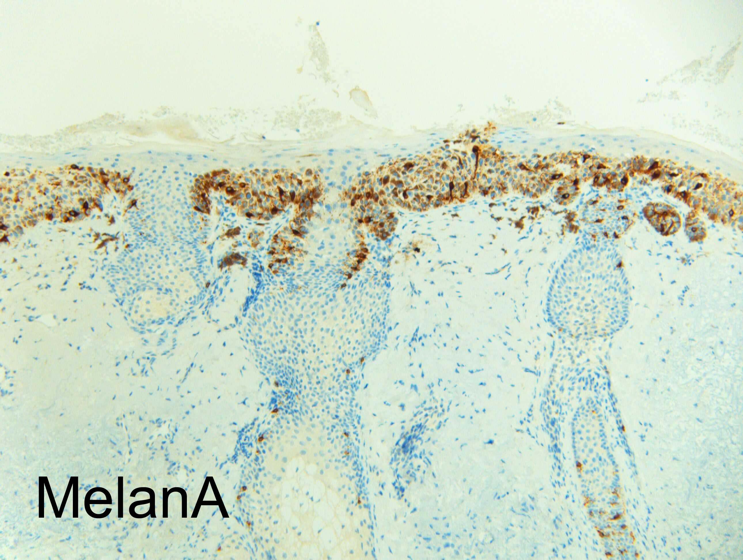
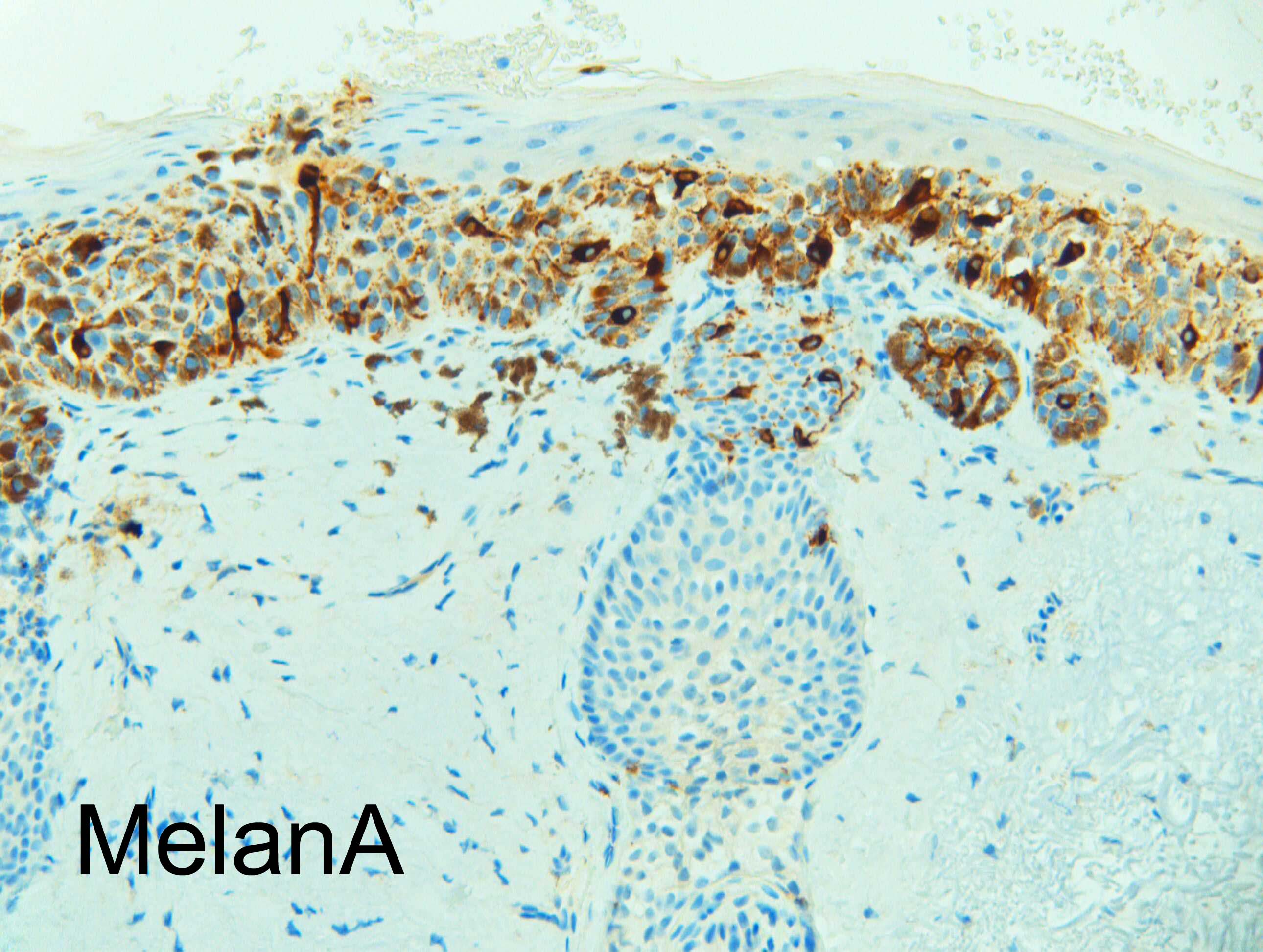
I was reviewing about 30 cases for our MDM meeting (in 20 minutes) and quickly flashed this one on low power diagnosing florid lentigo maligna only to be surprised at the report that it was actually a pigmented actinic keratosis!  MelanA will be posted shortly I hope. I think a low threshold for MelanA (when distinguishing solar lentigo & pigmented actinic keratosis from lentigo maligna) when you're a busy / speed reporter is a good idea and have always taught this locally - this case nicely illustrated the point!

I think you will get different views on grading actinic keratosis (I use mild, moderate and bowenoid) - I have my own ideas but I thought this case quite a subtle, mildly dysplastic, lesion. I avoid "intra-epithelial carcinoma" and "in situ carcinoma" personally. Bowenoid actinic keratosis can be full-thickness (pleomorphic +/- subtle undifferentiated +/- basaloid, thick or thin) or just show markedly atypical basal third with maturation (analagous to differentiated type VIN).

Share this comment


Link to comment
share_externally

Uma Sundram

Posted

I agree w Richard. This is a tough case. A caution about Melan A as it can over represent melanocytes. A non brown chromogen is also a good idea.


Share this comment


Link to comment
share_externally


Join the conversation

You can post now and register later. If you have an account, sign in now to post with your account.

Guest
Add a comment...

×   Pasted as rich text.   Paste as plain text instead

  Only 75 emoji are allowed.

×   Your link has been automatically embedded.   Display as a link instead

×   Your previous content has been restored.   Clear editor

×   You cannot paste images directly. Upload or insert images from URL.

Loading...

×
×
  • Create New...