Jump to content
In this section we have spot diagnoses posted on a daily basis since June 2010, now over 4000! You can review the archived cases and read the suggested diagnoses by users and the final comment by the contributors.
Case are uploaded each week day by 10 am UK time with the correct diagnosis will generally be posted at 8 pm UK time. Why not view the most recent spot diagnosis and proffer a diagnosis?

Case Number : Case 1729 - 12 January - Dr Arti Bakshi Posted By: Guest

Please read the clinical history and view the images by clicking on them before you proffer your diagnosis.
Submitted Date :
   (0 reviews)

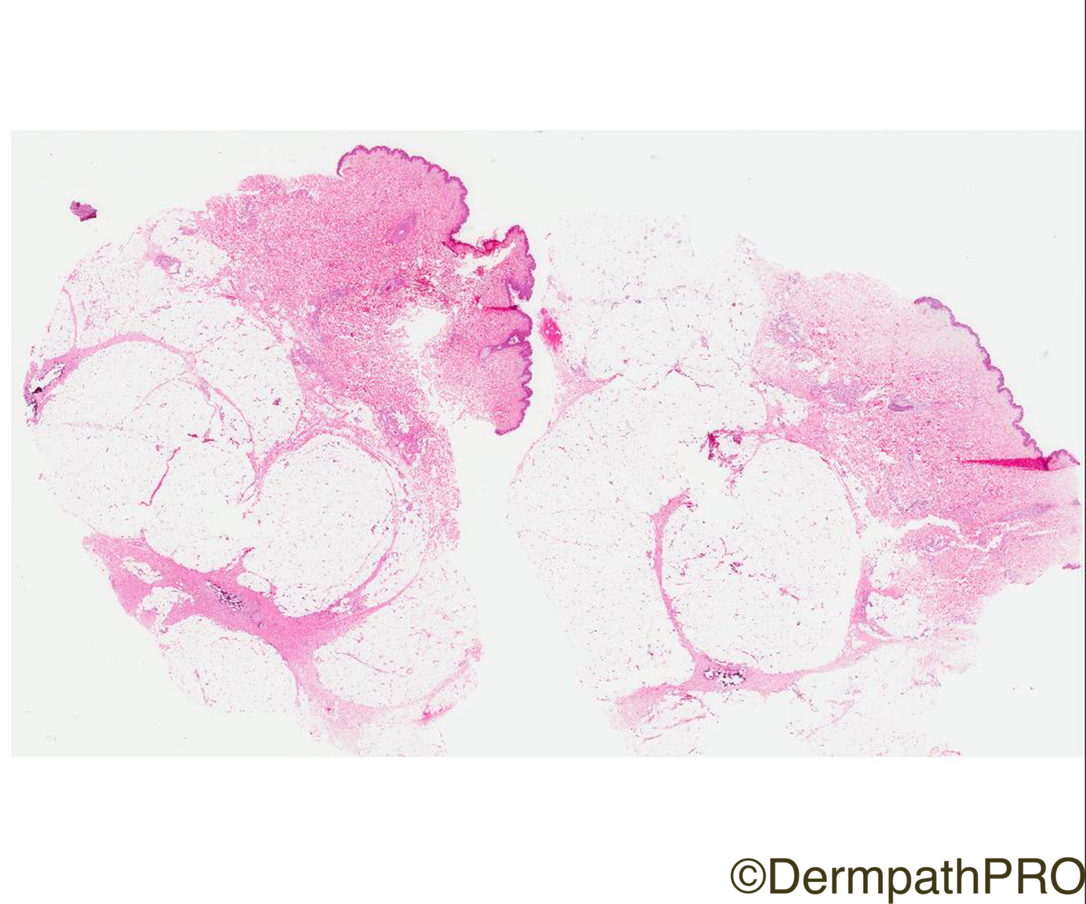
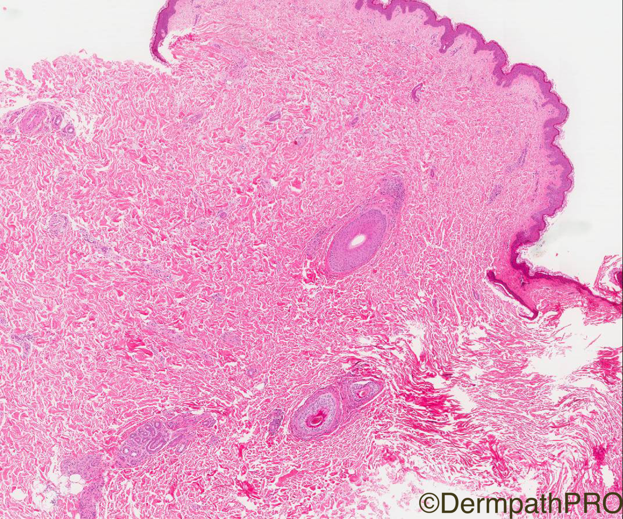
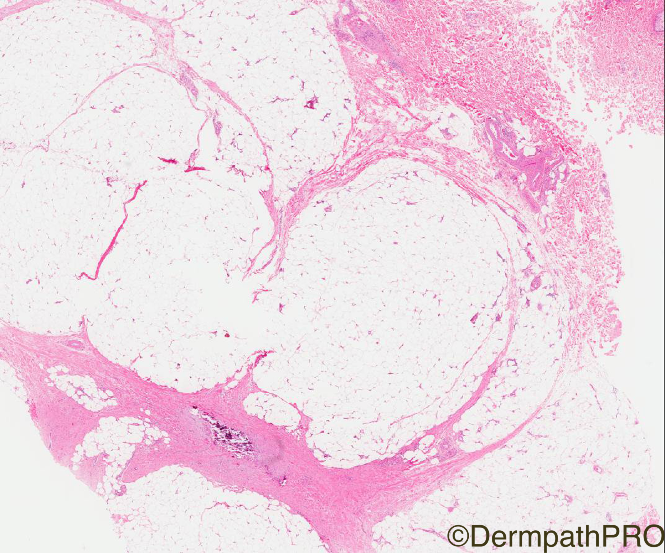
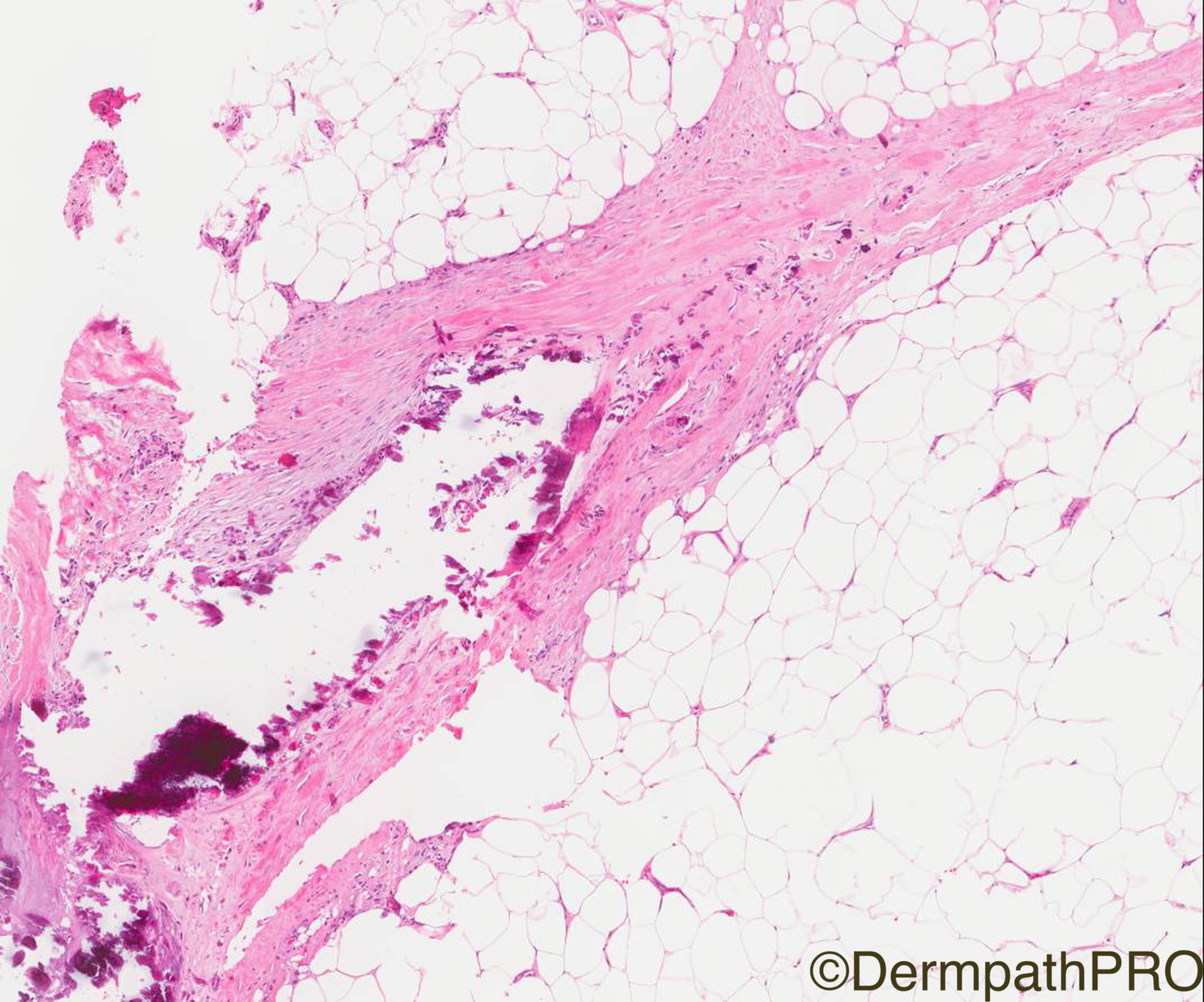
Clinical History: 51/M indurated mass/lump right abdomen?panniculitis ?fat necrosis. Also, has a history of rash for some years face and neck.

Case Posted by Dr Arti Bakshi


  Report Record

User Feedback


Admin_Dermpath

Posted

Straight off her flight back from a well earned vacation Dr Arti Bakshi has a lovely case for you...


Geoff Cross - DermpathPRO Projects

Share this comment


Link to comment
share_externally

vincenzo polizzi

Posted

Calciphylaxis. 

?In a patient with terminal lupus nephropathy

Share this comment


Link to comment
share_externally

Dr. Richard Carr

Posted

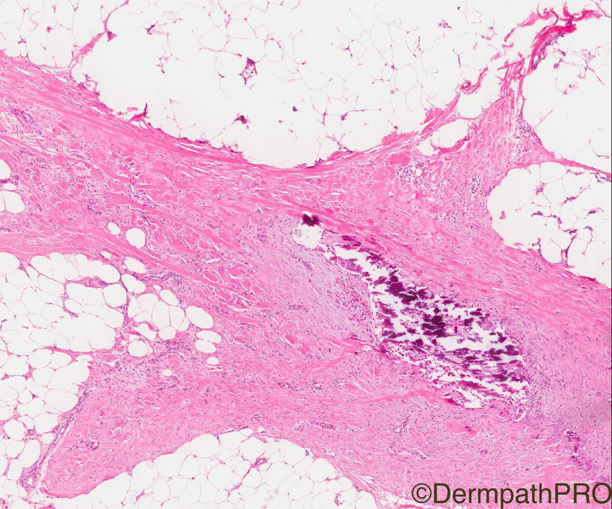
Looks to be dystrophic calcification in the septum with rather morphoeic change to the collagen.  How about the scleroderma spectrum of disorders? I was not sure I could locate the calcification in a vessel. Would need a bit more clinical/laboratory information to consider calciphylaxis.

Share this comment


Link to comment
share_externally

Victor Delgado

Posted

This looks like dystrophic calcification with thickened collagen bundles and a few fibroblasts in the septum. May be Nephrogenic systemic fibrosis or Scleroderma. Clinical information is needed.

Share this comment


Link to comment
share_externally

Robledo F. Rocha

Posted

Thickened sclerotic subcutaneous septa with dystrophic calcification, and adnexal structures that seem to be entrapped favoring morphea/scleroderma spectrum.

Share this comment


Link to comment
share_externally

Nitin Khirwadkar

Posted

Subcutaneous morphoea/morphoea profundus

Share this comment


Link to comment
share_externally

Dr. Mona Abdel-Halim

Posted

My impression is deep morphea with dystrophic calcification. 

Share this comment


Link to comment
share_externally

Raul Perret

Posted

This was for me a difficult case and I took a while but I also thought of deep morphea as favoured diagnosis. The rash on the face and neck bothered me (not frequent in morphea to my knowledge, maybe they were early erythematous lesions of plaque morphea in this case?). In the cases of calciphylaxis that I have seen you find mainly small vessel calcification but a compromise of the whole panniculus and a richer inflammatory infiltrate.

Share this comment


Link to comment
share_externally

vincenzo polizzi

Posted

My impression was that some small, intralobular, periseptal vessels were calcified. But maybe I was wrong...Waiting for the final diagnosis.

Share this comment


Link to comment
share_externally

Arti Bakshi

Posted

My initial impression (and report) was of deep morphoea too.

However, on further discussion with clinicians regarding the rash on face, I was told that the patient had been diagnosed as dermatomyositis and was on treatment for some years for this. (ofcourse all this was conveniently omitted on the histo request card!!). This raised the question whether the changes seen represent subcutaneous dystrophic calcification in association with dermatomyosistis. The patient does not have any other lesions or changes of scleroderma and this is an isolated solitary lump on the abdomen.  In order to have a single unifying diagnosis, the consensus was to regard this as subcutaneous dystrophic calcification secondary to DM.

I do agree that there is septal fibrosis in addition to calcification, which is difficult to explain (unless a secondary process). Although a coexistent solitary lesion of morphoea profunda remains a possibility, on checking the followup information today (the case is 2 yrs old), there has been no progression and no other lesions of morphoea have appeared.  


Share this comment


Link to comment
share_externally


Join the conversation

You can post now and register later. If you have an account, sign in now to post with your account.

Guest
Add a comment...

×   Pasted as rich text.   Paste as plain text instead

  Only 75 emoji are allowed.

×   Your link has been automatically embedded.   Display as a link instead

×   Your previous content has been restored.   Clear editor

×   You cannot paste images directly. Upload or insert images from URL.

Loading...

×
×
  • Create New...