Jump to content
In this section we have spot diagnoses posted on a daily basis since June 2010, now over 4000! You can review the archived cases and read the suggested diagnoses by users and the final comment by the contributors.
Case are uploaded each week day by 10 am UK time with the correct diagnosis will generally be posted at 8 pm UK time. Why not view the most recent spot diagnosis and proffer a diagnosis?

Case Number : Case 1742 - 31 January - Dr Uma Sundram Posted By: Guest

Please read the clinical history and view the images by clicking on them before you proffer your diagnosis.
Submitted Date :
   (0 reviews)

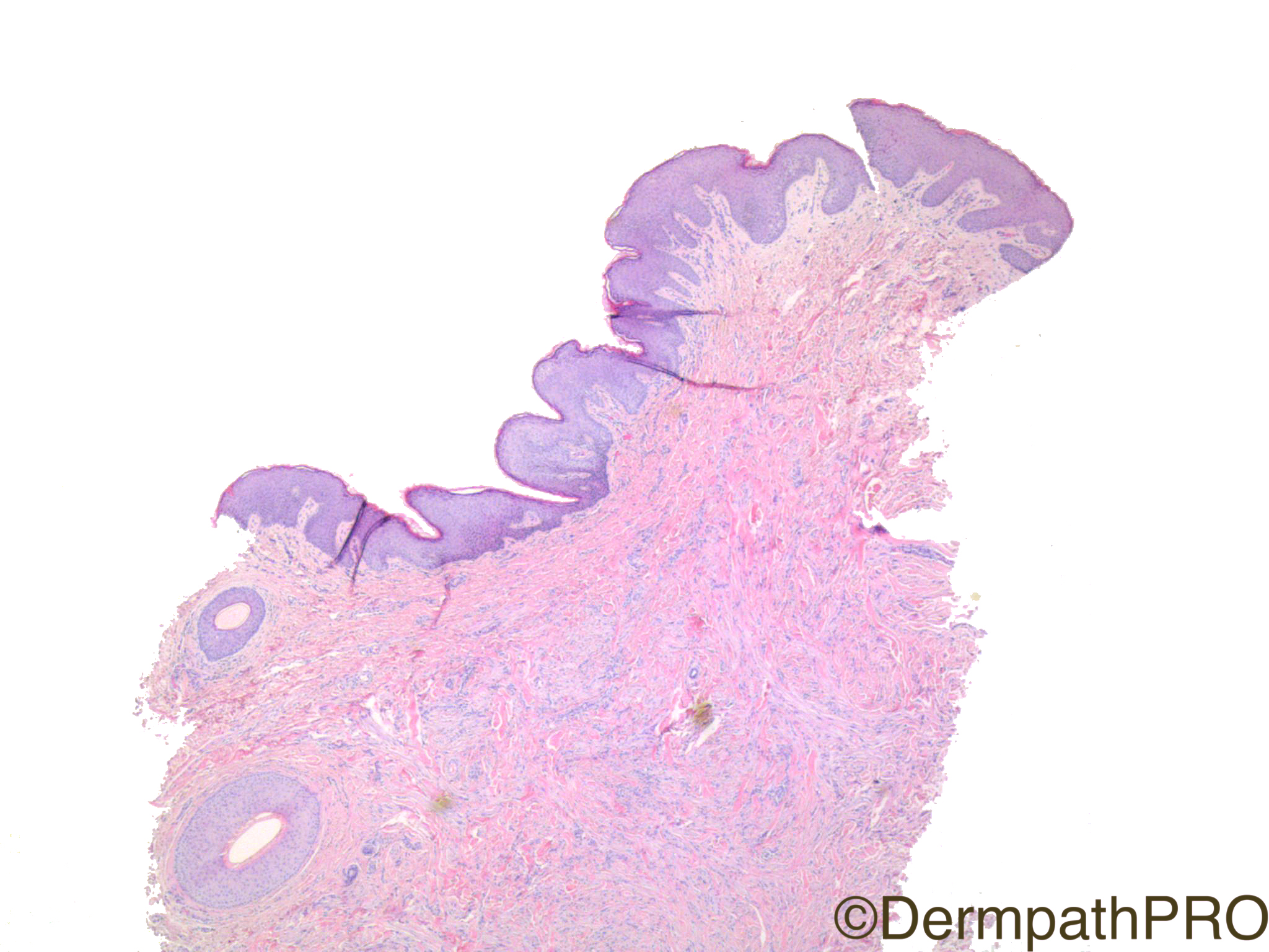
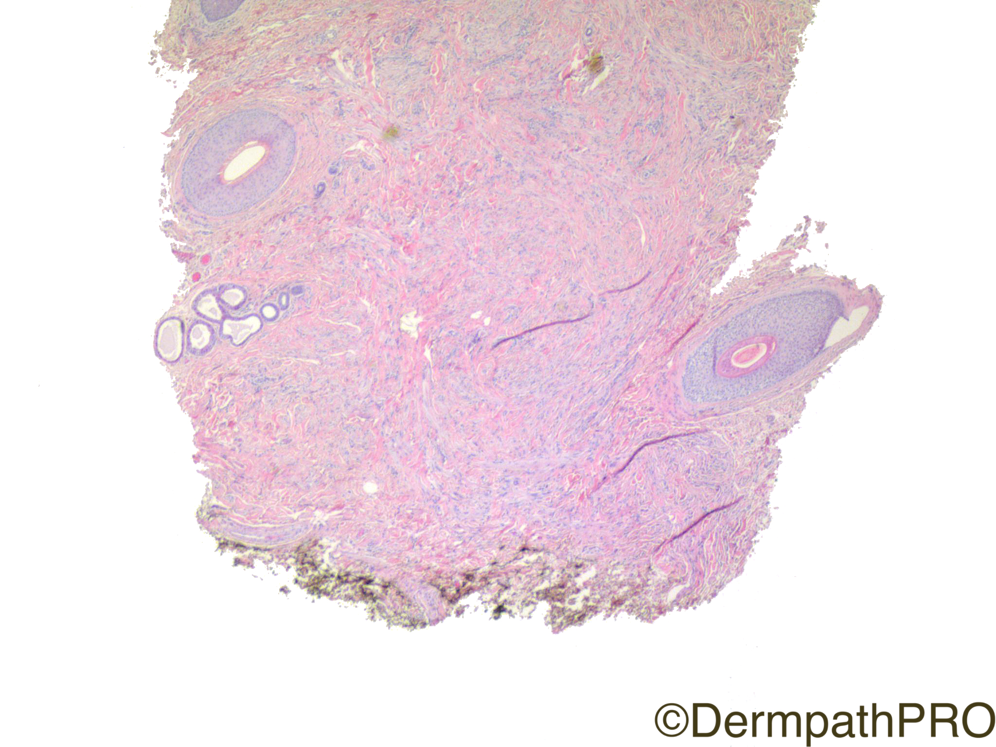
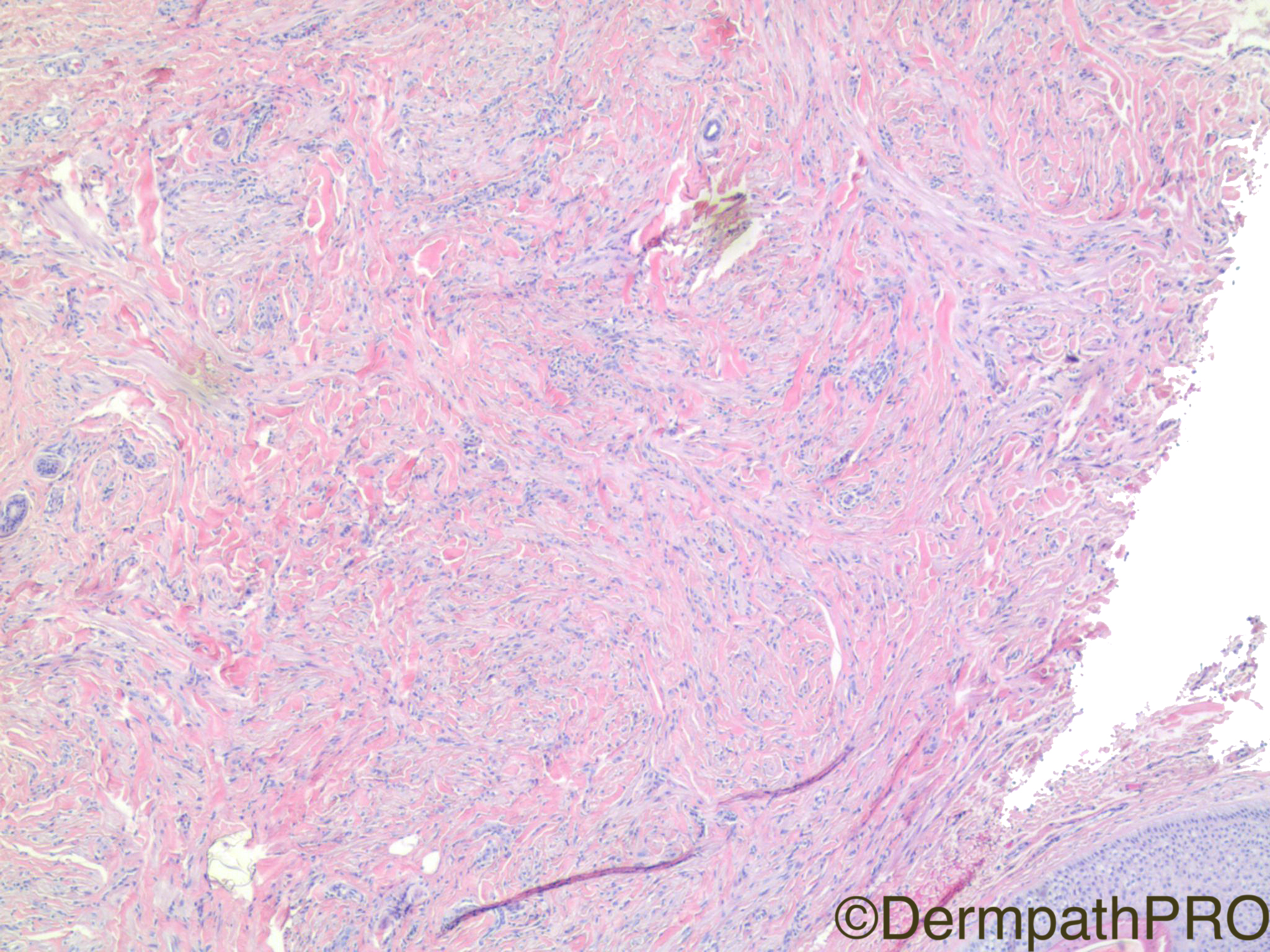
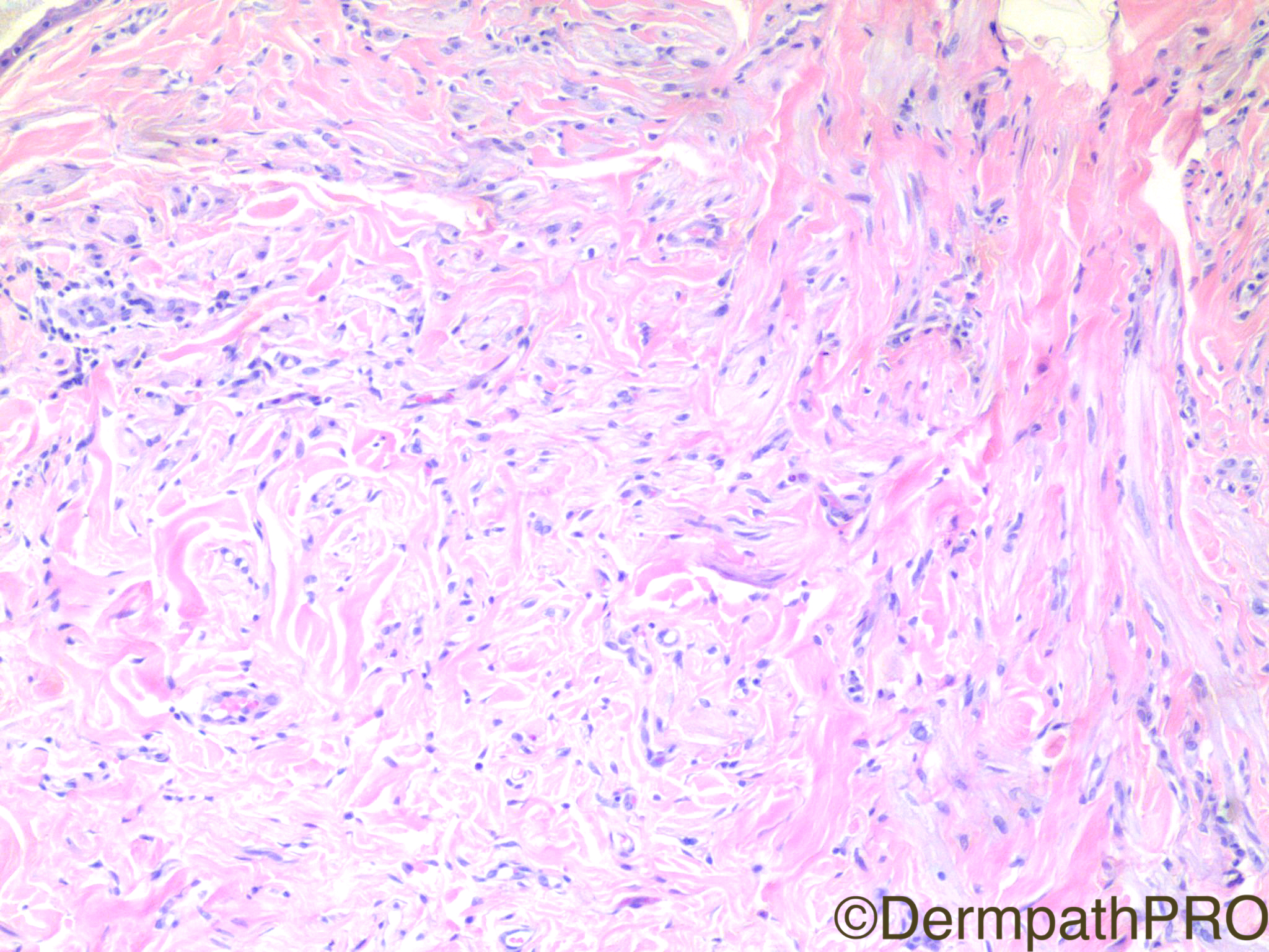
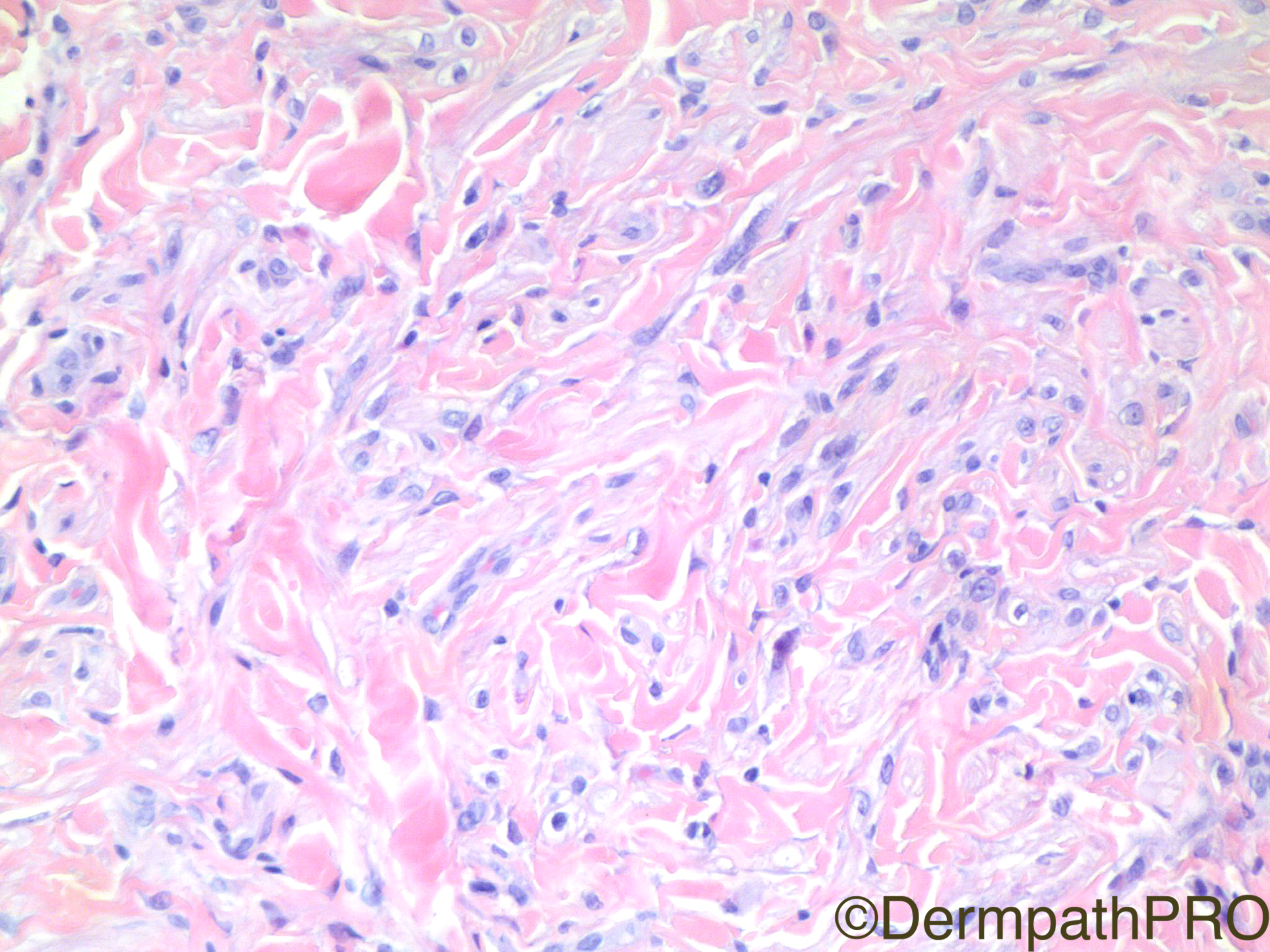
Clinical History: 44 year old woman with right labial ‘cyst’.

Case Posted by Dr Uma Sundram


  Report Record

User Feedback


Admin_Dermpath

Posted

To close off January's Spot Diagnosis Cases we have a nice set of images from Dr Uma Sundram.


Geoff Cross - DermpathPRO Projects

Share this comment


Link to comment
share_externally

vincenzo polizzi

Posted

Just to break the ice, I go with vulvar leiomyoma ( but I thought a lot of vulvar dermatofibroma )...

Share this comment


Link to comment
share_externally

vincenzo polizzi

Posted

Vulvar Leiomyoma is often clinically diagnosed as Bartholin gland cyst, so this pathological diagnosis fits well on the patient's history. The epithelioid and spindle features of the cells in this case are ill demarcated and organised in dispersed fascicles between abundant collagen bands ( this pattern made me think of a dermatofibroma ). But this is the description of VL in the pertinent literature ( Requena-Kutzner Book "Cutaneous Soft Tissue Tumors" ).

So what do you guys think about?

Share this comment


Link to comment
share_externally

Raul Perret

Posted

Hey Vincenzo in this case I thought it could be an hypopigmented blue nevus / neurotized dermal nevus VS dermatofibroma as you suggested. The cells did not struck me as smooth muscle on morphology alone

Share this comment


Link to comment
share_externally

Nitin Khirwadkar

Posted

I think Raul's suggestion of a hypo pigmented blue/neurotised naevus is a good one. Doesn't particularly look smooth muscle. Needs a small battery of IHC.

Share this comment


Link to comment
share_externally

Dr. Mona Abdel-Halim

Posted

Thinking in the line of a nevus also, hypopigmented blue/ desmoplastic nevus

Share this comment


Link to comment
share_externally

Robledo F. Rocha

Posted

I must confess that my first thought was some myxoid fibrohistiocytic tumor or some peripheral nerve sheath tumor, but Raul’s comment on hypopigmented blue nevus convinced me. I’d echo the words of Klaus J. Busam and Raymond L. Barnhill: “Amelanotic melanocytic proliferations are more difficult to be recognized as being melanocytic in origin.”

Share this comment


Link to comment
share_externally

Uma Sundram

Posted

melanocytic markers are negative and desmin is positive

Share this comment


Link to comment
share_externally


Join the conversation

You can post now and register later. If you have an account, sign in now to post with your account.

Guest
Add a comment...

×   Pasted as rich text.   Paste as plain text instead

  Only 75 emoji are allowed.

×   Your link has been automatically embedded.   Display as a link instead

×   Your previous content has been restored.   Clear editor

×   You cannot paste images directly. Upload or insert images from URL.

Loading...

×
×
  • Create New...