Jump to content
In this section we have spot diagnoses posted on a daily basis since June 2010, now over 4000! You can review the archived cases and read the suggested diagnoses by users and the final comment by the contributors.
Case are uploaded each week day by 10 am UK time with the correct diagnosis will generally be posted at 8 pm UK time. Why not view the most recent spot diagnosis and proffer a diagnosis?

Case Number : Case 1865 - 21 July - Dr Iskander Chaudhry (Invited) Posted By: Guest

Please read the clinical history and view the images by clicking on them before you proffer your diagnosis.
Submitted Date :
   (0 reviews)

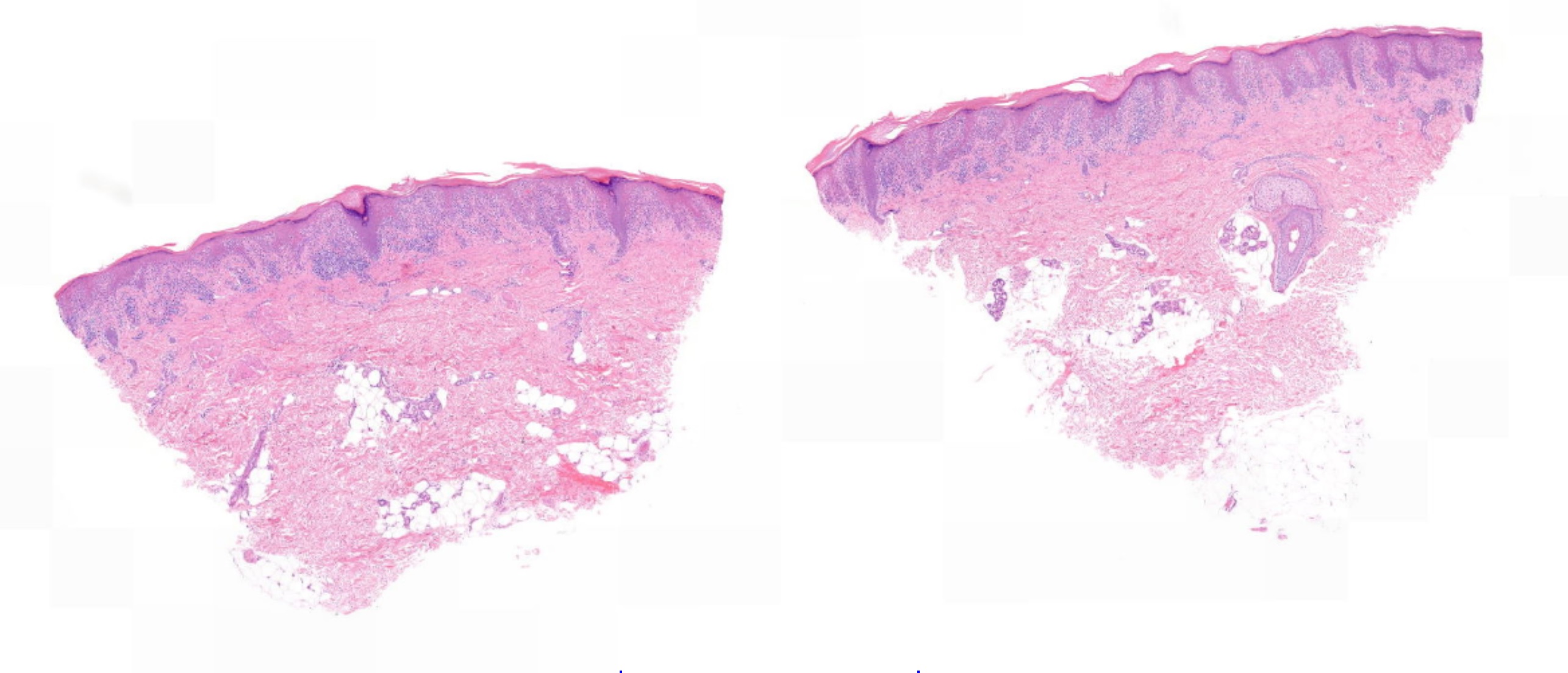
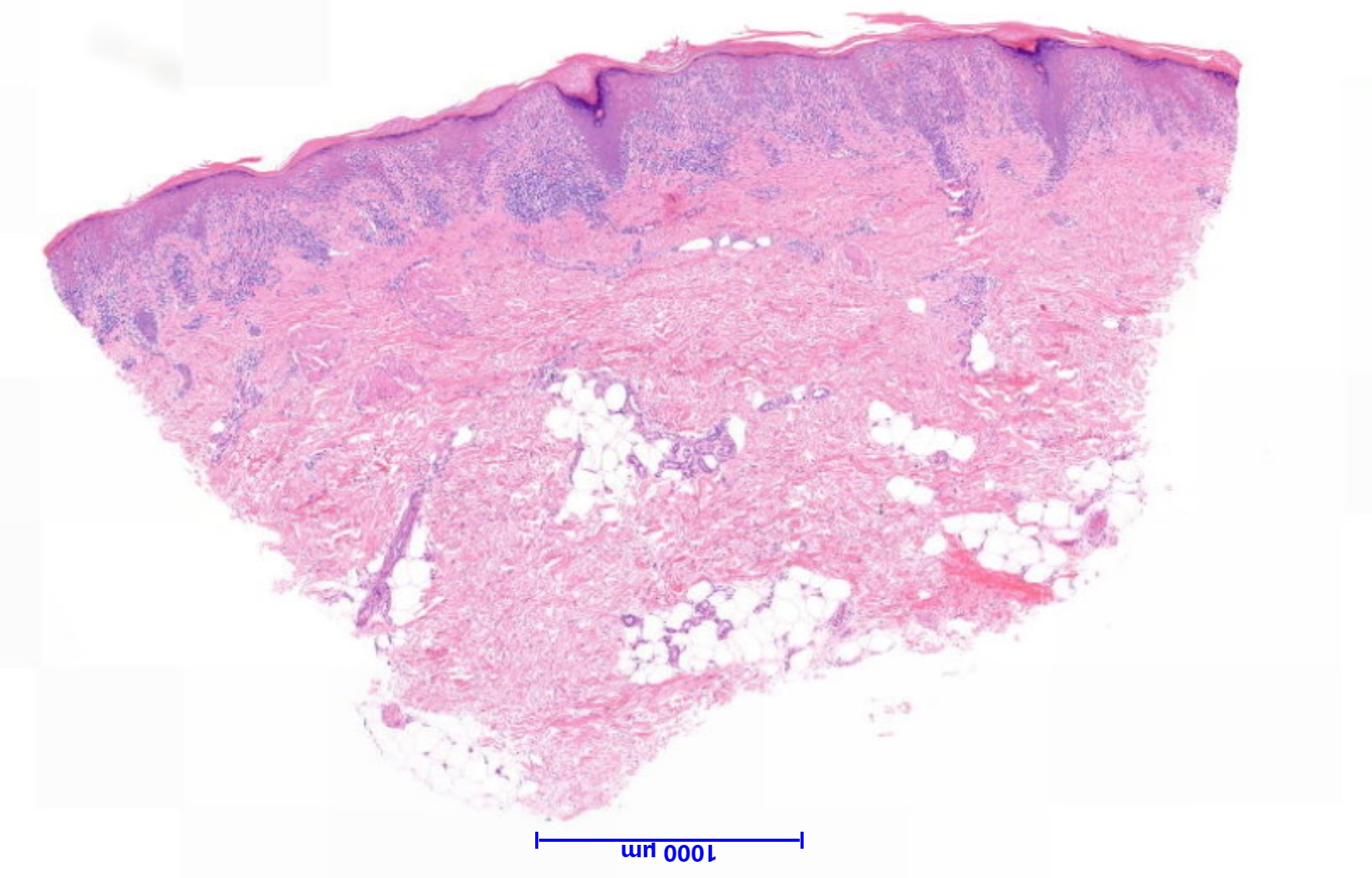
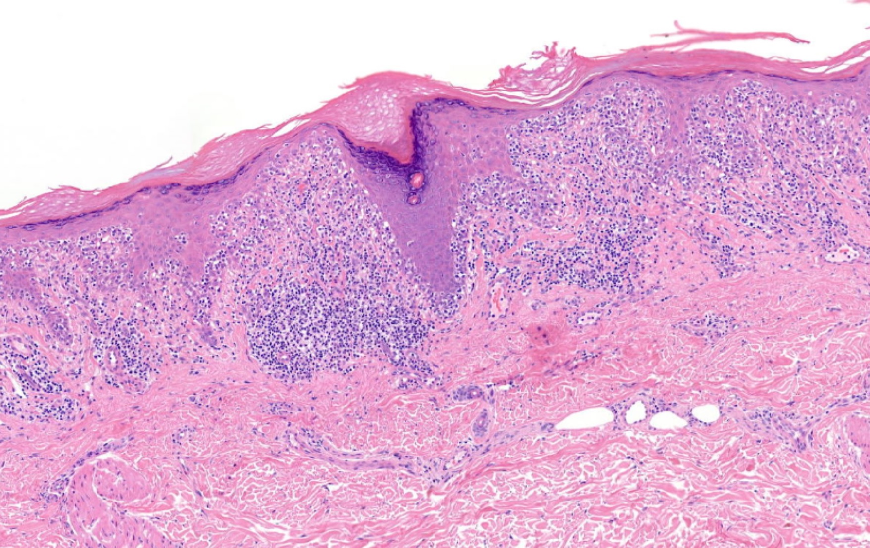
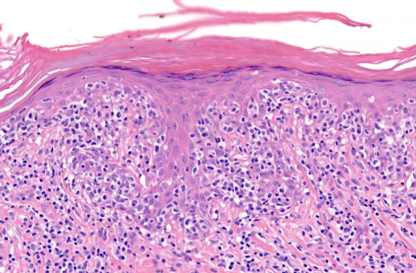
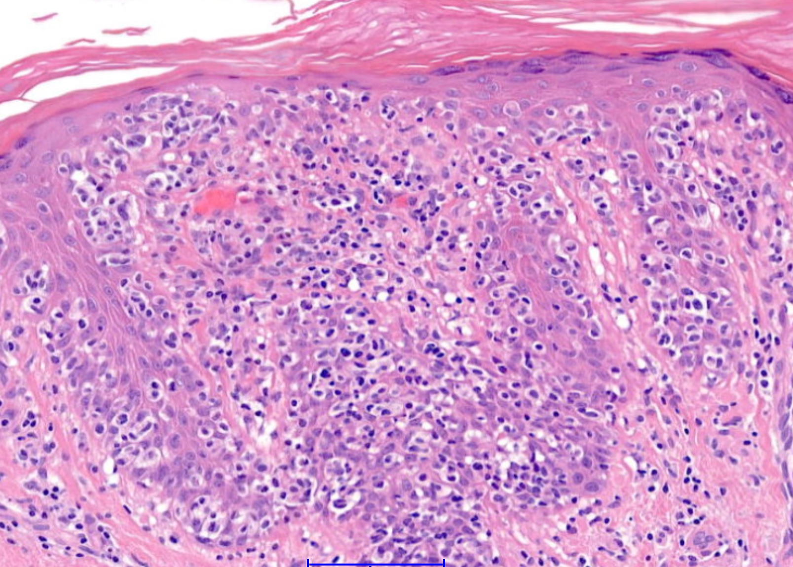
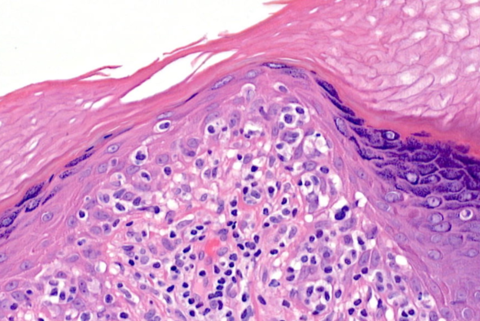
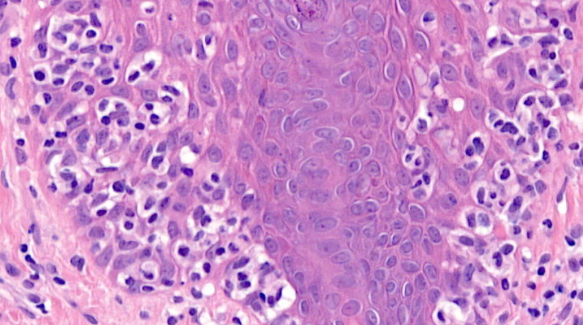
85 year old female. Punch left upper arm. ?Bowen's. ?AK.


  Report Record

User Feedback


vincenzo polizzi

Posted

Very nice case of PagetoidReticulosis/Worringer/Kolopp variant of MF.  Solitary lesion on arm: clinically fit well  

Share this comment


Link to comment
share_externally

Dr. Mona Abdel-Halim

Posted

Would like also to point to the possibility of the variant of LPLK that mimics MF. 

Share this comment


Link to comment
share_externally

Pagetoid reticulosis, melanoma,LPLK

Share this comment


Link to comment
share_externally

Robledo F. Rocha

Posted

One of the clinical hypotheses is actinic keratosis, so the lesion is likely to be a papule. Lymphomatoid papulosis type D is my spot.

Share this comment


Link to comment
share_externally

Raul Perret

Posted

Agree with most of the differentials, the clinical picture will be the ultimate judge in this particular case. Nevertheless, I had the impression that this is pagetoid reticulosis as Vincenzo mentioned.

Share this comment


Link to comment
share_externally

Arti Bakshi

Posted

Agree with pagetoid reticulosis, if this is solitary. Although LyP type D cant be distinguished histologically, it would be odd for LyP to present as a solitary leison.

Share this comment


Link to comment
share_externally

Eman Ahmad

Posted

Favor Pagetoid reticulosis over LPLK (MF like).

Share this comment


Link to comment
share_externally

amiratawdy

Posted

My first thought is LPLK  based on clinicopathological correlation

Share this comment


Link to comment
share_externally

Admin_Dermpath

Posted

Dear All - Many thanks for your comments. The infiltrate was mixed T and B cells.

Our final diagnosis was:

Lymphomatoid Keratosis. 

A case of lymphomatoid keratosis. Choi MJ, Kim HS, Kim HO, Song KY, Park YM. Ann Dermatol. 2010 May;22(2):219-22.

The final report stated:

I do not believe this is cutaneous lymphoma. However, if the patient develops lesions that are clinically concerning for mycosis fungoides, those should be biopsied; or if this lesion returns, there should be a low threshold for re-biopsy.

To date there has been no recurrence or development of additional lesions. 

Acknowledgement to Dr Uma Sundrum who I consulted on this case. 

Share this comment


Link to comment
share_externally


Join the conversation

You can post now and register later. If you have an account, sign in now to post with your account.

Guest
Add a comment...

×   Pasted as rich text.   Paste as plain text instead

  Only 75 emoji are allowed.

×   Your link has been automatically embedded.   Display as a link instead

×   Your previous content has been restored.   Clear editor

×   You cannot paste images directly. Upload or insert images from URL.

Loading...

×
×
  • Create New...