Jump to content
In this section we have spot diagnoses posted on a daily basis since June 2010, now over 4000! You can review the archived cases and read the suggested diagnoses by users and the final comment by the contributors.
Case are uploaded each week day by 10 am UK time with the correct diagnosis will generally be posted at 8 pm UK time. Why not view the most recent spot diagnosis and proffer a diagnosis?

Case Number : Case 1816 - 15 May - Dr Limin Yu Posted By: Guest

Please read the clinical history and view the images by clicking on them before you proffer your diagnosis.
Submitted Date :
   (0 reviews)

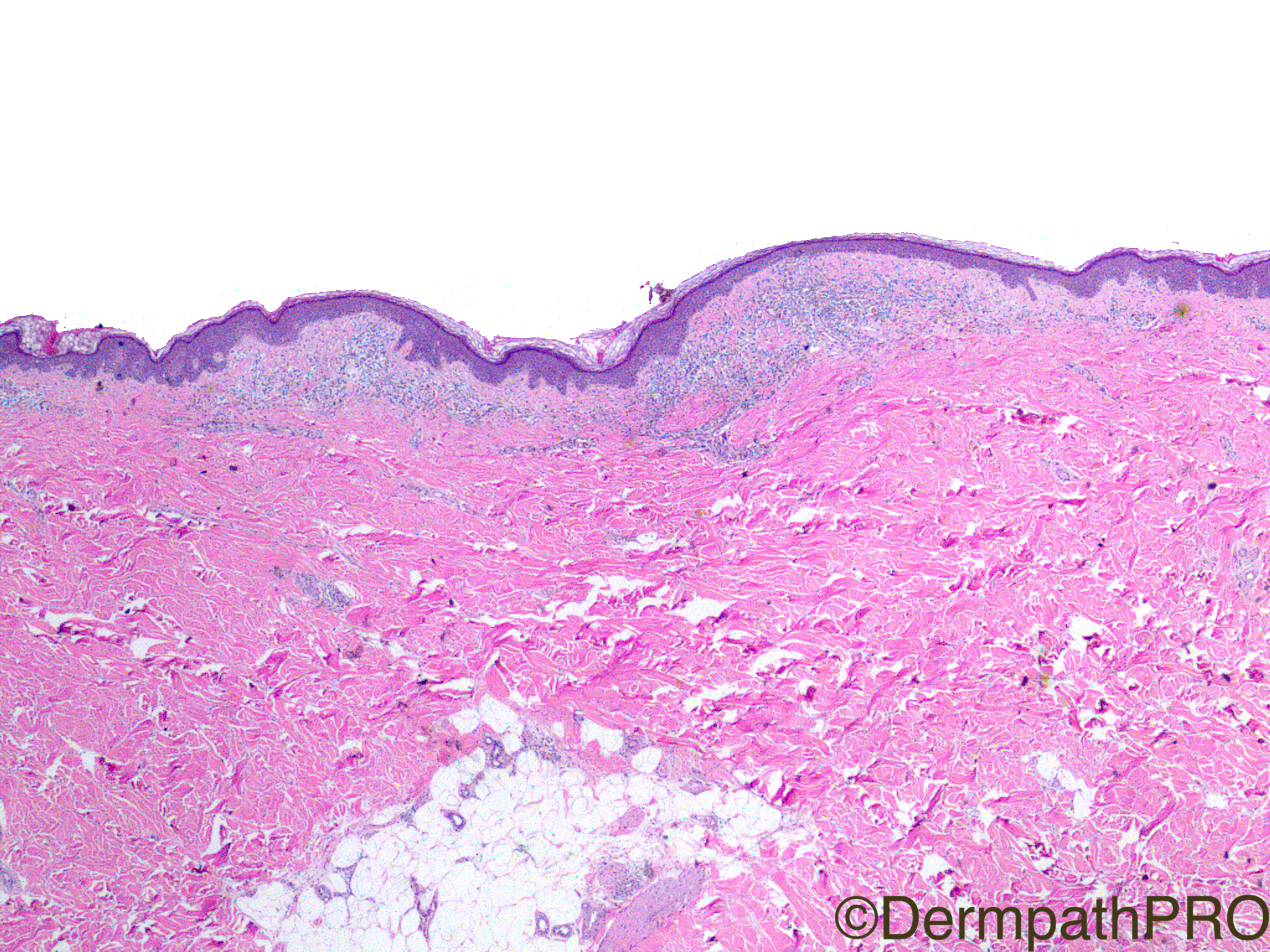
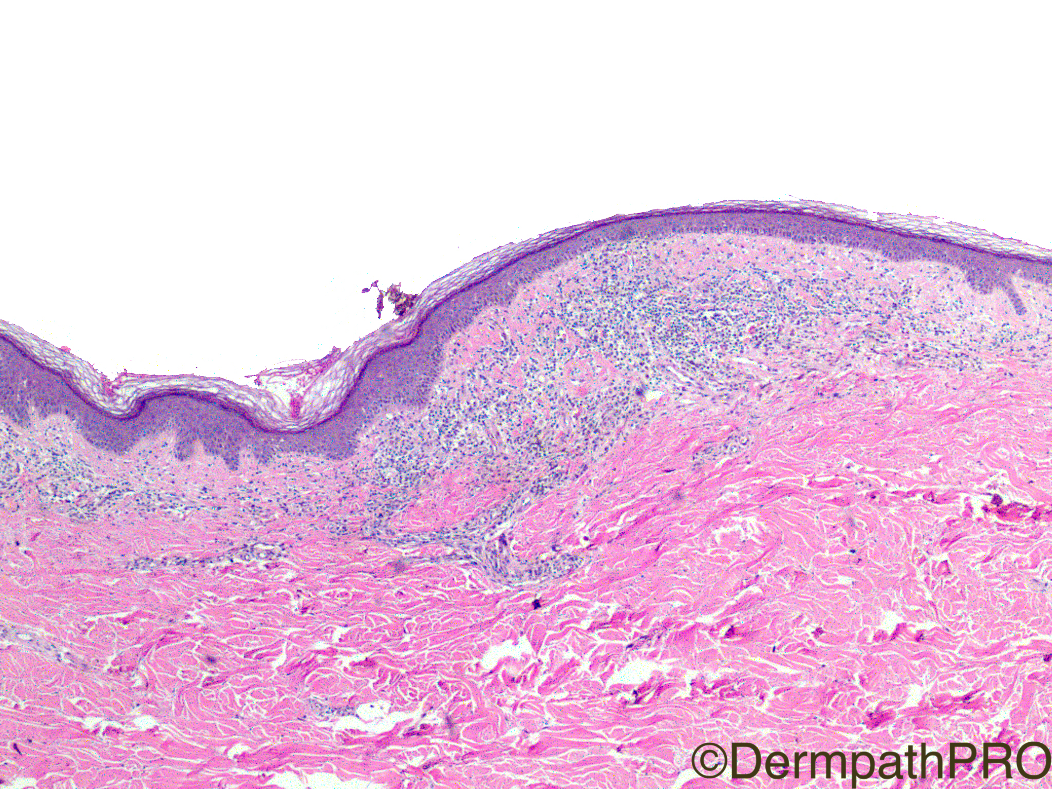
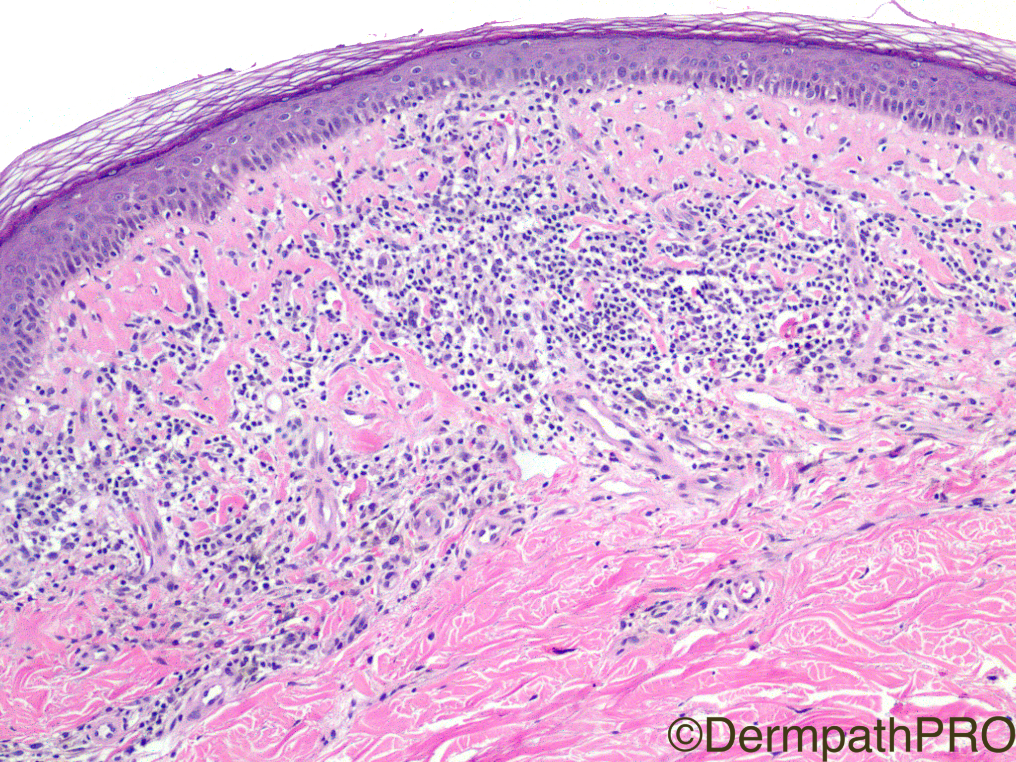
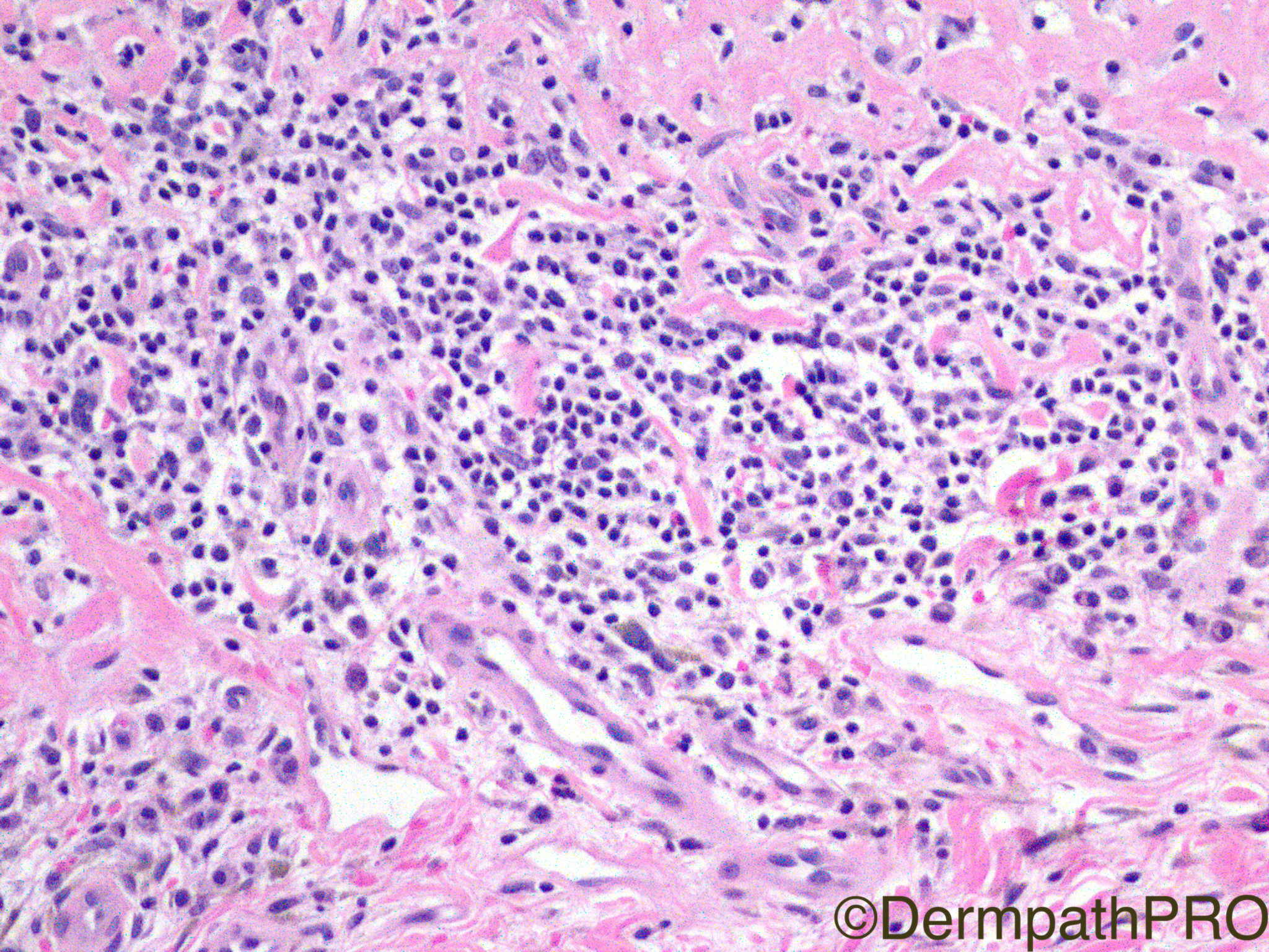
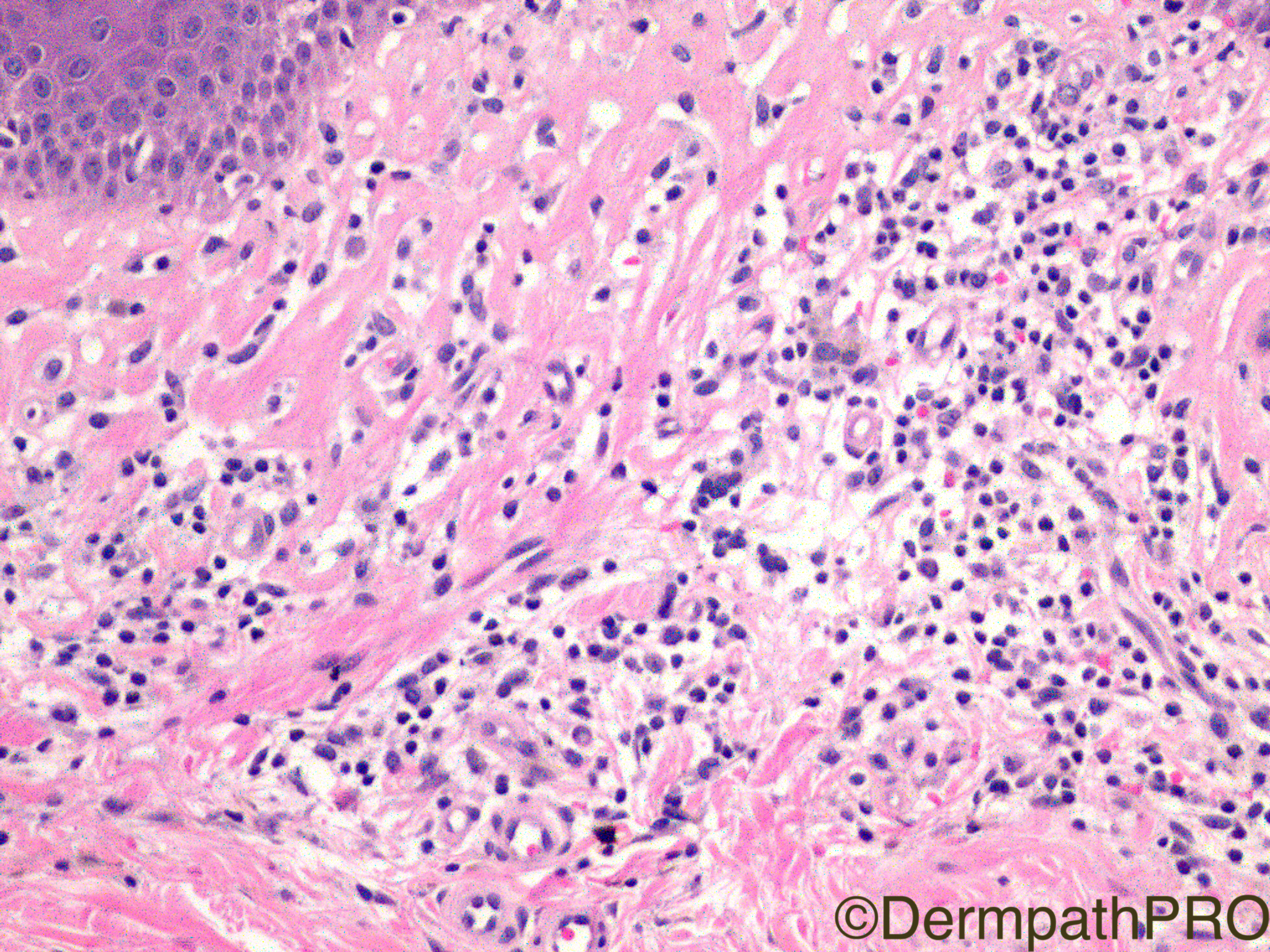
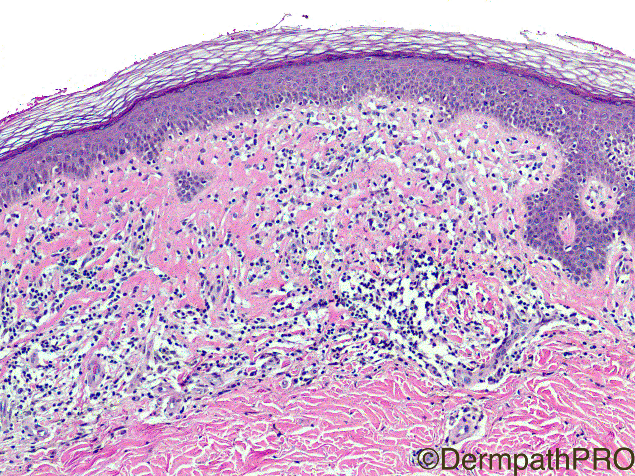
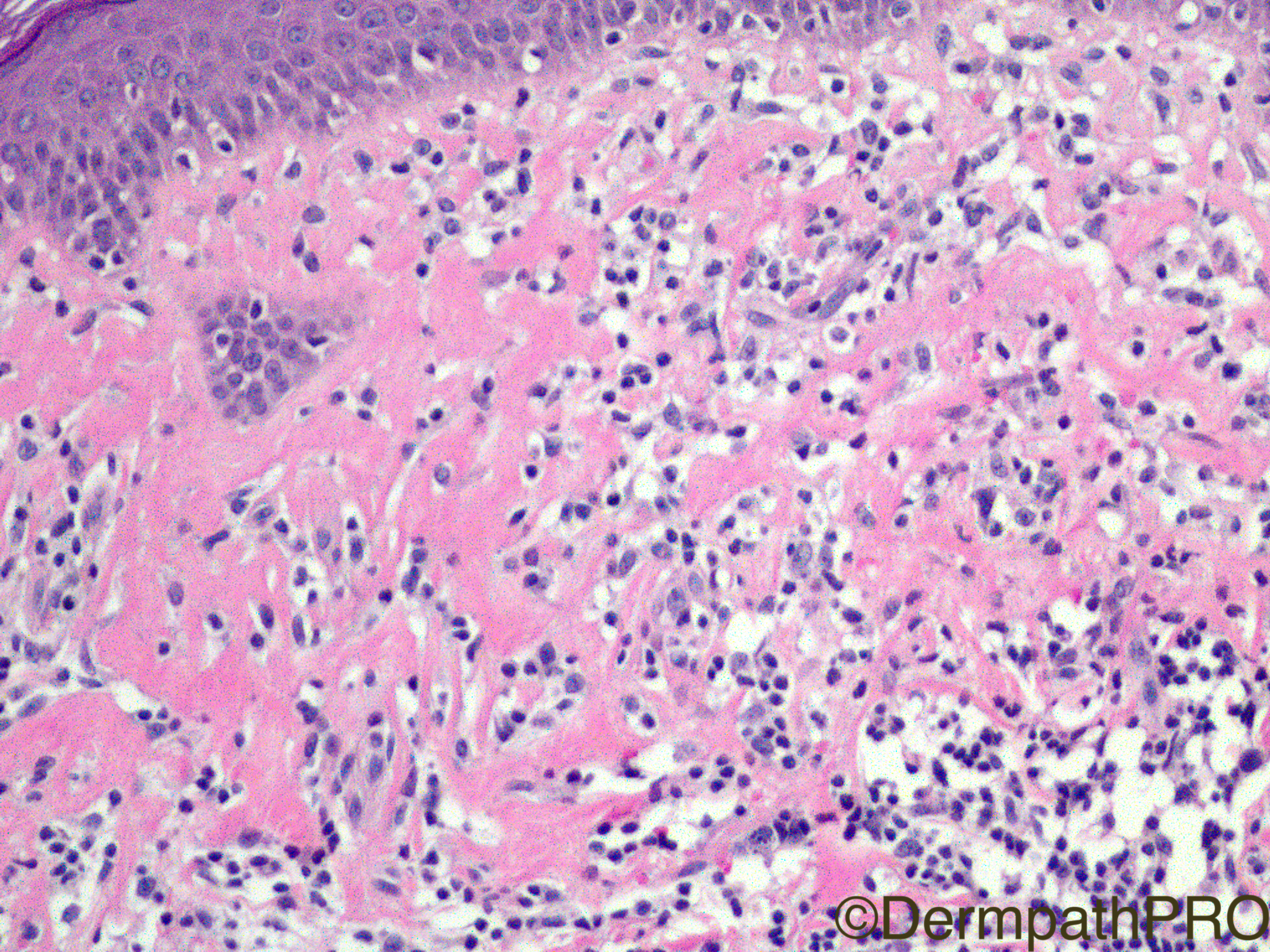
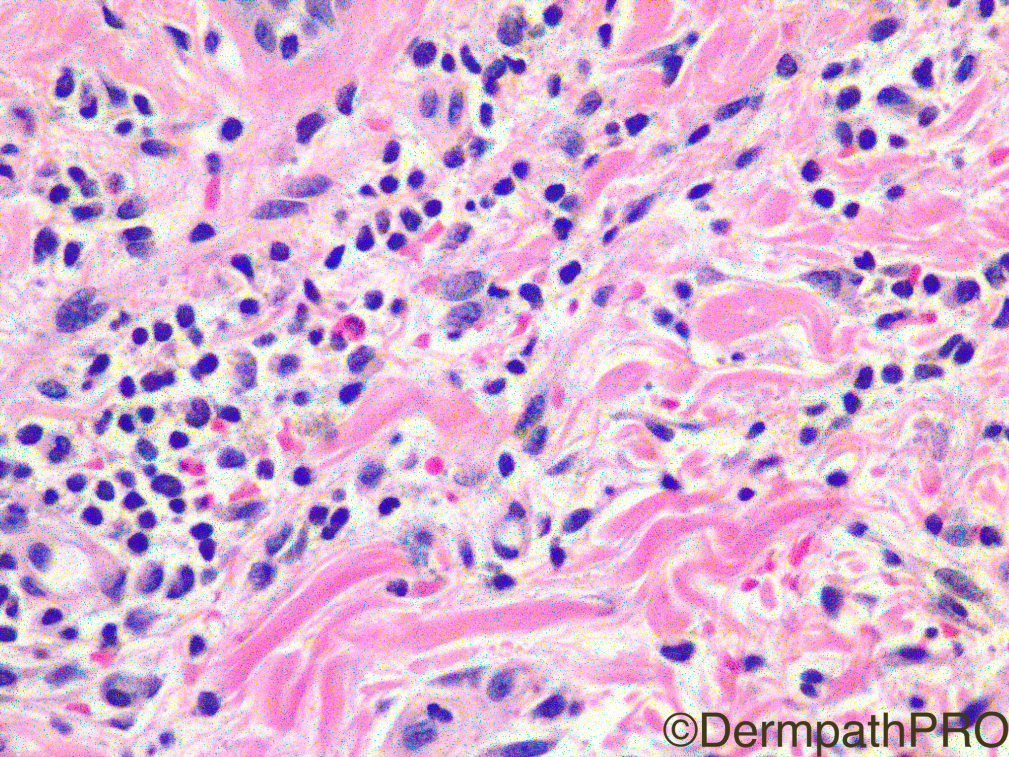
Clinical History: 57 year-old female, right arm rash.

Case Posted by Dr Limin Yu


  Report Record

User Feedback


Admin_Dermpath

Posted

Start your week off with a lovely case from Dr Limin Yu.

Geoff Cross - DermpathPRO Projects

Share this comment


Link to comment
share_externally

vincenzo polizzi

Posted

A PPD-like rash, with eosinophils and chronic-like dermal fibrosis, but with acute-like epidermal features ( no parakeratosis/hyperkeratosis ) makes me think of fixed drug eruption. 

Share this comment


Link to comment
share_externally

Raul Perret

Posted

I also thought of drug adverse effect Vincenzo

Share this comment


Link to comment
share_externally

Dr. Mona Abdel-Halim

Posted

Would like to R/O MF first and check drug history.. could be T cell pseudolymphoma (Lymphomatoid drug reaction) or MF

Share this comment


Link to comment
share_externally

Robledo F. Rocha

Posted

A superficial perivascular and interstitial inflammation of lymphocytes and plasma cells, with rare eosinophils, associated to wiry bundles of collagen and some extravasated erythrocytes in the papillary dermis. There's also lymphocyte tagging along dermoepidermal junction in the absence of spongiosis, disturbance of the stratum corneum, or another epidermal reaction pattern. I favor some sort of drug eruption, perhaps the fixed type, although prominent vacuolar change with apoptotic keratinocytes, deeper extension of the infiltrate, and neutrophils scattered interstitially are lacking.

Share this comment


Link to comment
share_externally

Saman Fatah

Posted

Could this be an early changes of extragenital lichen sclerosus mimicking a CTCL histologically. There is epidermal atrophy with changes of the collagen bundles at the most superficial part of papillary dermis. Clinically such plaques are highly distinctive and a simple genital examination can be rewarding rather than attempting to diagnose CTCL on the histology alone. While lesions of MF can occur at any part of the skin, isolated arm involvement will be slightly unusual as well clinically (if this is the sole site of the lesion).

Share this comment


Link to comment
share_externally

Sasi Attili

Posted

My first thought was inflammatory stage of Lichen sclerosus. Kept quiet as no one else mentioned it! 

11 hours ago, Saman Fatah said:

Could this be an early changes of extragenital lichen sclerosus mimicking a CTCL histologically. There is epidermal atrophy with changes of the collagen bundles at the most superficial part of papillary dermis. Clinically such plaques are highly distinctive and a simple genital examination can be rewarding rather than attempting to diagnose CTCL on the histology alone. While lesions of MF can occur at any part of the skin, isolated arm involvement will be slightly unusual as well clinically (if this is the sole site of the lesion).


Share this comment


Link to comment
share_externally

Dr. Mona Abdel-Halim

Posted

Very good suggestion of inflammatory LS !!

Share this comment


Link to comment
share_externally

Dr. Richard Carr

Posted

LS was my "spot diagnosis" but curious amount of haemosiderin! There are reports of vascuolopathic changes in genital LS.

Share this comment


Link to comment
share_externally

Nitin Khirwadkar

Posted

My differentials are LS and a drug reaction. I have had a case where LS had unusual amounts of hemosiderin.

Share this comment


Link to comment
share_externally

Limin Yu

Posted

I hope you enjoyed this case, the diagnosis is: Pigmented Purpuric Dermatosis

Share this comment


Link to comment
share_externally


Join the conversation

You can post now and register later. If you have an account, sign in now to post with your account.

Guest
Add a comment...

×   Pasted as rich text.   Paste as plain text instead

  Only 75 emoji are allowed.

×   Your link has been automatically embedded.   Display as a link instead

×   Your previous content has been restored.   Clear editor

×   You cannot paste images directly. Upload or insert images from URL.

Loading...

×
×
  • Create New...