Jump to content
In this section we have spot diagnoses posted on a daily basis since June 2010, now over 4000! You can review the archived cases and read the suggested diagnoses by users and the final comment by the contributors.
Case are uploaded each week day by 10 am UK time with the correct diagnosis will generally be posted at 8 pm UK time. Why not view the most recent spot diagnosis and proffer a diagnosis?

Case Number : Case 1917 - 04 Oct - Dr Iskander Chaudhry Posted By: Guest

Please read the clinical history and view the images by clicking on them before you proffer your diagnosis.
Submitted Date :
   (0 reviews)

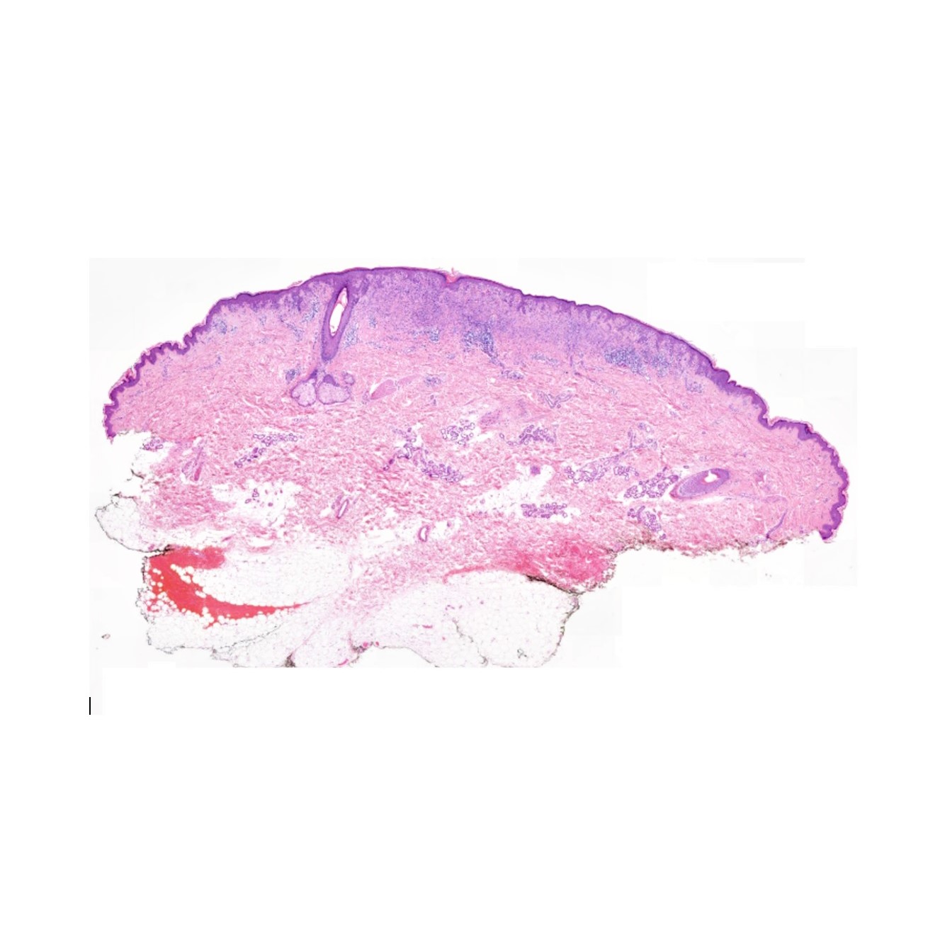
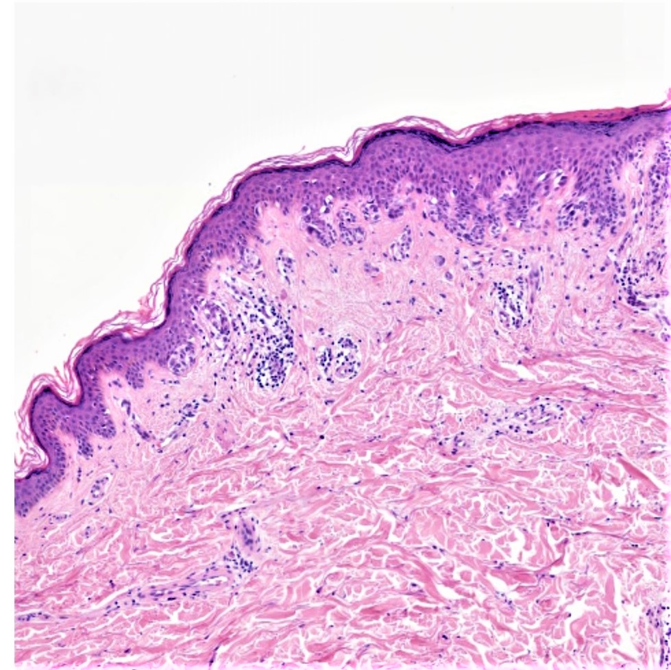
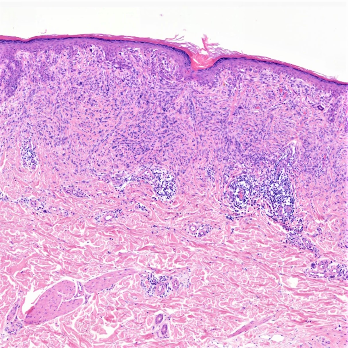
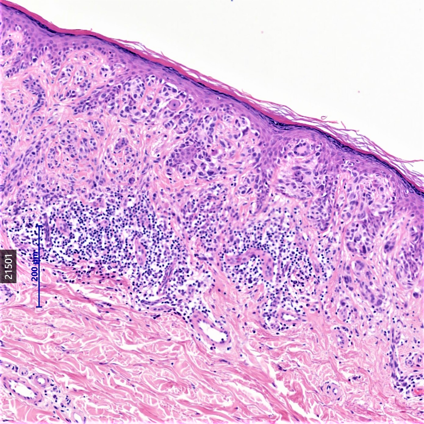
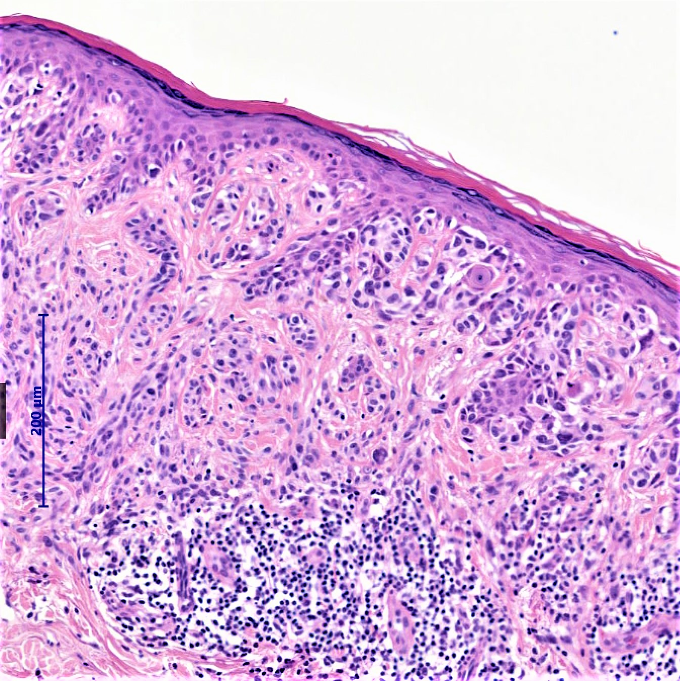
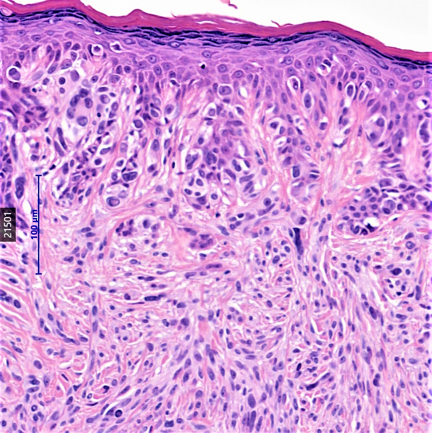
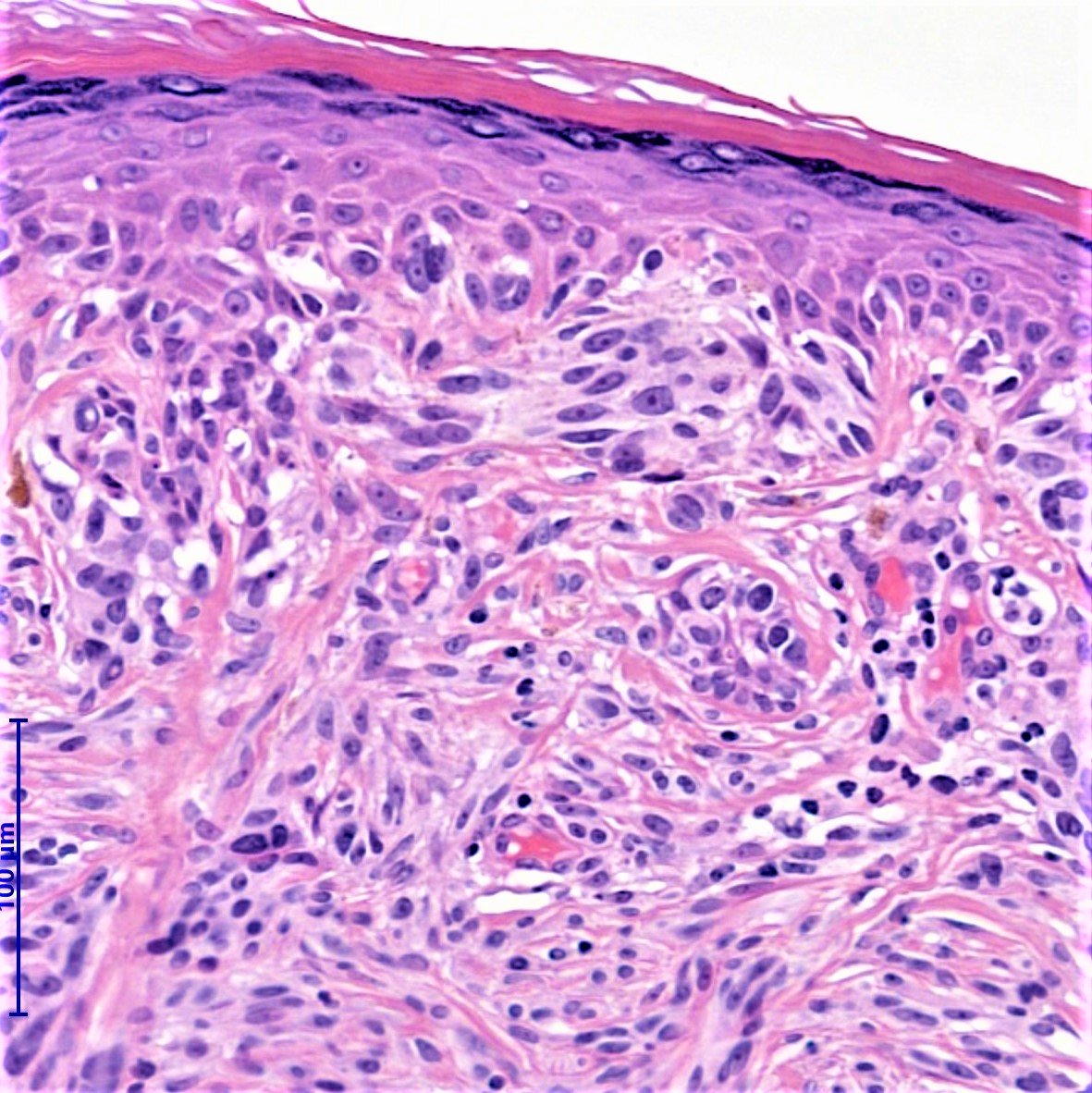
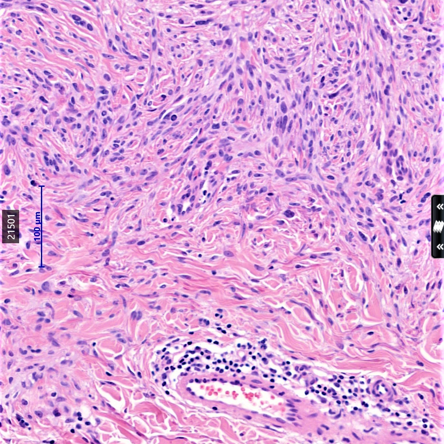
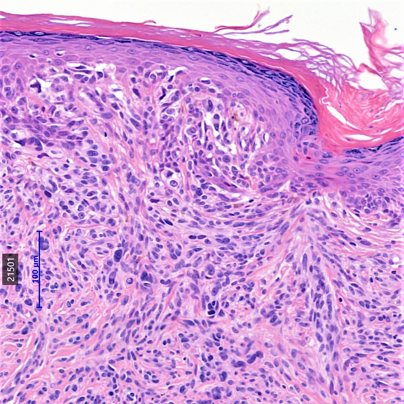
85 year old male. Right forearm excision
Multiple moles. Ugly duckling red flat topped "mole". Dotted vessels ?what


  Report Record

User Feedback


Admin_Dermpath

Posted

Liverpool Update in Dermatopathology

Tues 28th November 2017

Organised by DermpathPRO & Supported by the British Society of Dermatopathology

Time: 9:00 am - 5.00 pm CPD: 6.5 points

Speakers:

  • Arti Bakhsi - Histopathologist, Royal Hospital Liverpool - UK
  • Marijke van Dijk – Dermatopathologist University Medical Centre Ultrecht- Netherlands
  • Robert Phelps - Professor of Dermatologyand Pathology Mount Sinai Medical Center New York - USA
  • Gerald Saldana - ConsultantHistopathologist Leicester Royal Infirmary - UK
  • Alan Evans - Ninewells Hospitaland Medical School Dundee - UK


Programme:

 Chairperson: Leena Joseph

  • 09:00 – 09:45 – Blistering dermatoses - Robert Phelps
  • 0945 – 10:30 – Breslow density is a novel prognostic feature in cutaneous malignant melanoma, Gerald Saldanha
  • 10:45 – 11:35 – ABC of Lymphomatoid Papulosis, Arti Bakhsi
  • 11:35 – 12:30 – Deep penetrating naevus and other less common variants, Dr Alan Evans

Chairperson: Arti Bakshi

  • 13:15 – 13:45 Infectious diseases - what European pathologist need to be aware of. Dr Marijke van Dijk
  • 13:45 – 14:30 Interesting cases from my referral practice, Dr Marijke van Dijk

Chairperson: Nitin Khirwadkar

  • 14:45 – 17:00 Slide Seminar Cases

Register Now!!

HOW TO BOOK

1.  Select the paypal button and select either Consultant or Trainee rates (BSD member or not).

2.  Then email info@dermpathpro.com with your name, your institution and confirmation of your paypal booking, and we will email you back within 24-48 hours (week days) with your confirmed place and further details about the course.

3.  If you're a trainee we would need an email confirmation from your Programme Director or equivalent confirmation (certificate/ other documentation).

 COURSE FEE: Trainees £75 Consultants: £140 (BSD Members), £160 (Non BSD Member)

Become a BSD member:

  • Membership costs £15 annually and members can receive discounted registration rates for meetings organised and supported by the BSD.
  • Click here to Join

Register for Course:

Book Place

 pixel.gif

 Venue: Hilton Liverpool,3 Thomas Steers Way,Liverpool,L1 8LW

Share this comment


Link to comment
share_externally

vincenzo polizzi

Posted

Endocrine Mucin Producing Sweat Gland Carcinoma. 

It’s the same case showed in 6 September.  

Share this comment


Link to comment
share_externally

Admin_Dermpath

Posted

2 hours ago, vincenzo polizzi said:

Endocrine Mucin Producing Sweat Gland Carcinoma. 

It’s the same case showed in 6 September.  


This is now corrected and a new case has been added! 

Share this comment


Link to comment
share_externally

Dr. Richard Carr

Posted

Dear Iskander & Arti. Looks like I was too late to accept your kind invitation! I was a little distracted with the ISDP meeting in Glasgow. Maybe next year. Good luck with the course. Warm regards, Richard

Share this comment


Link to comment
share_externally

Dr. Mona Abdel-Halim

Posted

To me, melanoma arising in a nevus. 

Share this comment


Link to comment
share_externally

Admin_Dermpath

Posted

From Dr Chaudhry:

Thank you I would like to wait for other comments. I would be interested if you both thought the dermal component was benign or malignant - how compelling is the epidermal component towards melanoma in situ? 

Share this comment


Link to comment
share_externally

In situ melanoma, but I´m concerned about the dermal component. Maybe it represents a desmoplastic melanoma. The lymphocytic inflammation is a clue to this diagnosis.

Share this comment


Link to comment
share_externally

Nitin Khirwadkar

Posted

Severely dysplastic at-least. Not entirely sure about the dermal component on these images. Can't see mitotic figures. Ki67 and p16.

Share this comment


Link to comment
share_externally

M A Rahman

Posted

I agree with Nitin, at least dysplastic nevus and I too have not seen any mitoses. But moderate lymphocytic response with areas of fibrosis in the dermis worries me. ?regression

Share this comment


Link to comment
share_externally

vincenzo polizzi

Posted

Although dermal component shows smaller cells then junctional ones, I think this is IV level Melanoma, because my impression is there aren’t two different lesions here.  The spindle cells in dermis have spitzoid features and would like knowing about this same case in a teenager.....?????

Share this comment


Link to comment
share_externally

Arti Bakshi

Posted

Difficult one!.... I would probably go for melanoma in situ for the junctional component mainly because the atypia gets diffuse (as opposed to random) and confluent in the 4th image.

Dermal component more tricky. It does appear to mature, but some large cells at the base. Would look carefully for dermal mitoses (including levels) before going for invasive vertical phase melanoma though.

Share this comment


Link to comment
share_externally

Admin_Dermpath

Posted

Thanks every one: this case was shown to Dr Alistair Robson and his comments are below:


'It is odd, agreed, but I think this is benign. 

 
1. It is symmetrical (in the biological sense).
2. Not only is there no significant pagetoid spread but there is a uniformity to the junctional architecture as you skim over this component at scanning level.
3. Whilst cells do vary, particularly within the junctional portion, there are multinucleate cells (commonly though not, of course, exclusively a benign phenomenon) and there is a spitzoid appearance to many.
4. Atypia is not marked (The (definite) observed variation appears more akin to what I prefer to term anisonucleosis). 
5. There is maturation; indeed, the dermal cells appear bland.
6. The dermal cells sit within the collagen, for the most part, without expansile groups obscuring the dermis.
7. Both junctional and dermal cells vary rather than having the uniformity of a clonal growth pattern (cytological "equivalent" to point "6").
8. No mitoses.
9. I think the dermal element is simply a desmoplastic response.
 
On balance, and I feel reading through the earlier comments on the lesion, the overall impression is that it is an unusual lesion, which naturally worries everyone, but also that there are no hard grounds for malignancy. This, I suggest, is also a good basis for preferring a benign diagnosis whilst acknowledging the unusual nature of the lesion.
 
If desired, FISH will likely be instructive here.'

Share this comment


Link to comment
share_externally

Arti Bakshi

Posted

Melanocytic lesions!!....refuse to get easier with time.

A range of diagnosis as usual but have to say Dr Robson's assessment is pretty comprehensive and probably accurate.

Share this comment


Link to comment
share_externally

Dr. Richard Carr

Posted

Wow we have some very malignant dermatopathologists on this site. My report is brief. Combined dysplastic (Clark) /desmoplastic Spitz (SPARK naevus). A nice combination that makes it stand out clinically and histologically. It needs removing for a blissful nights sleep but surely not a melanoma?

Share this comment


Link to comment
share_externally


Join the conversation

You can post now and register later. If you have an account, sign in now to post with your account.

Guest
Add a comment...

×   Pasted as rich text.   Paste as plain text instead

  Only 75 emoji are allowed.

×   Your link has been automatically embedded.   Display as a link instead

×   Your previous content has been restored.   Clear editor

×   You cannot paste images directly. Upload or insert images from URL.

Loading...

×
×
  • Create New...