Jump to content

Building Blocks of Dermatopathology

BAD DermpathPRO Learning Hub: Basics of Immuno

Dermatopathology
Course Overview
Quizze

Case Number : IM0004 Dr. Hafeez Diwan

Please read the clinical history and view the images by clicking on them before you proffer your diagnosis.
Submitted Date :
   (0 reviews)

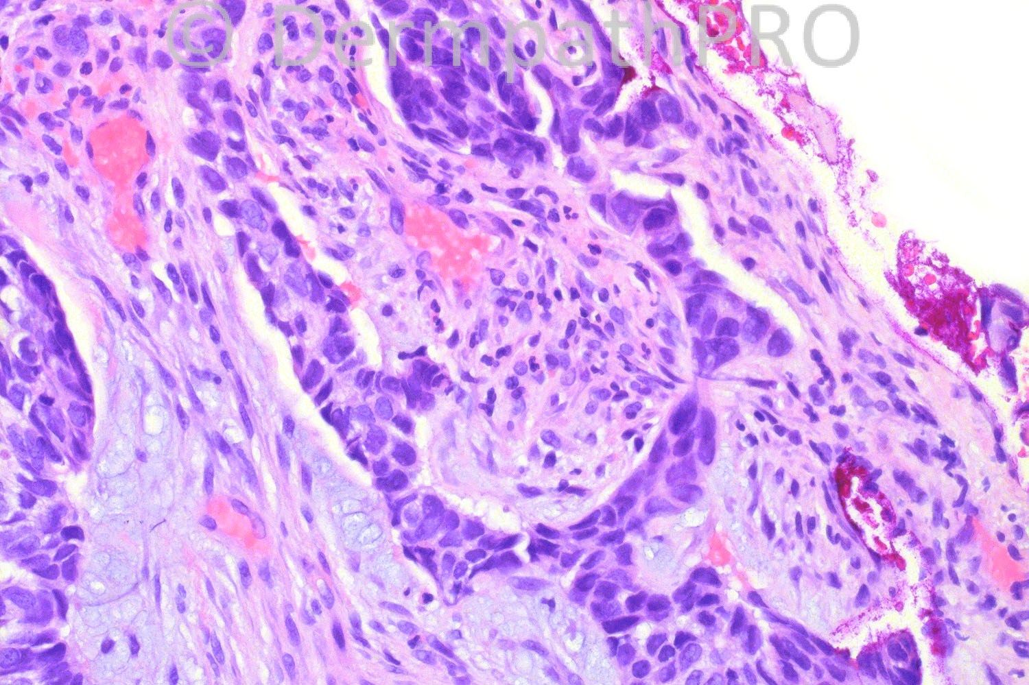
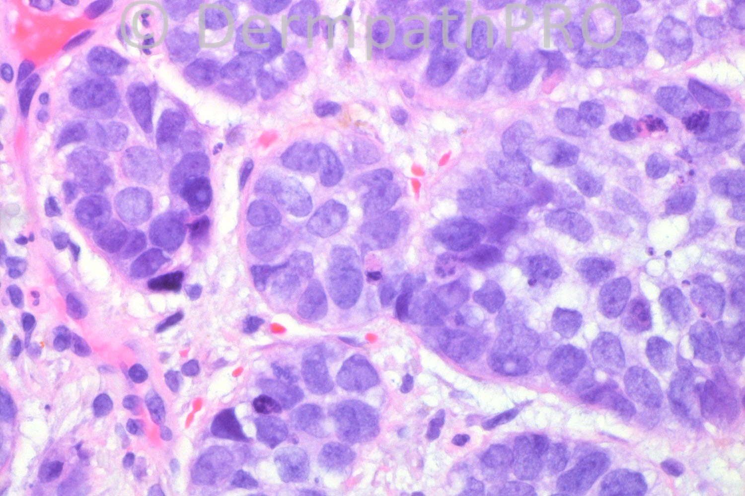
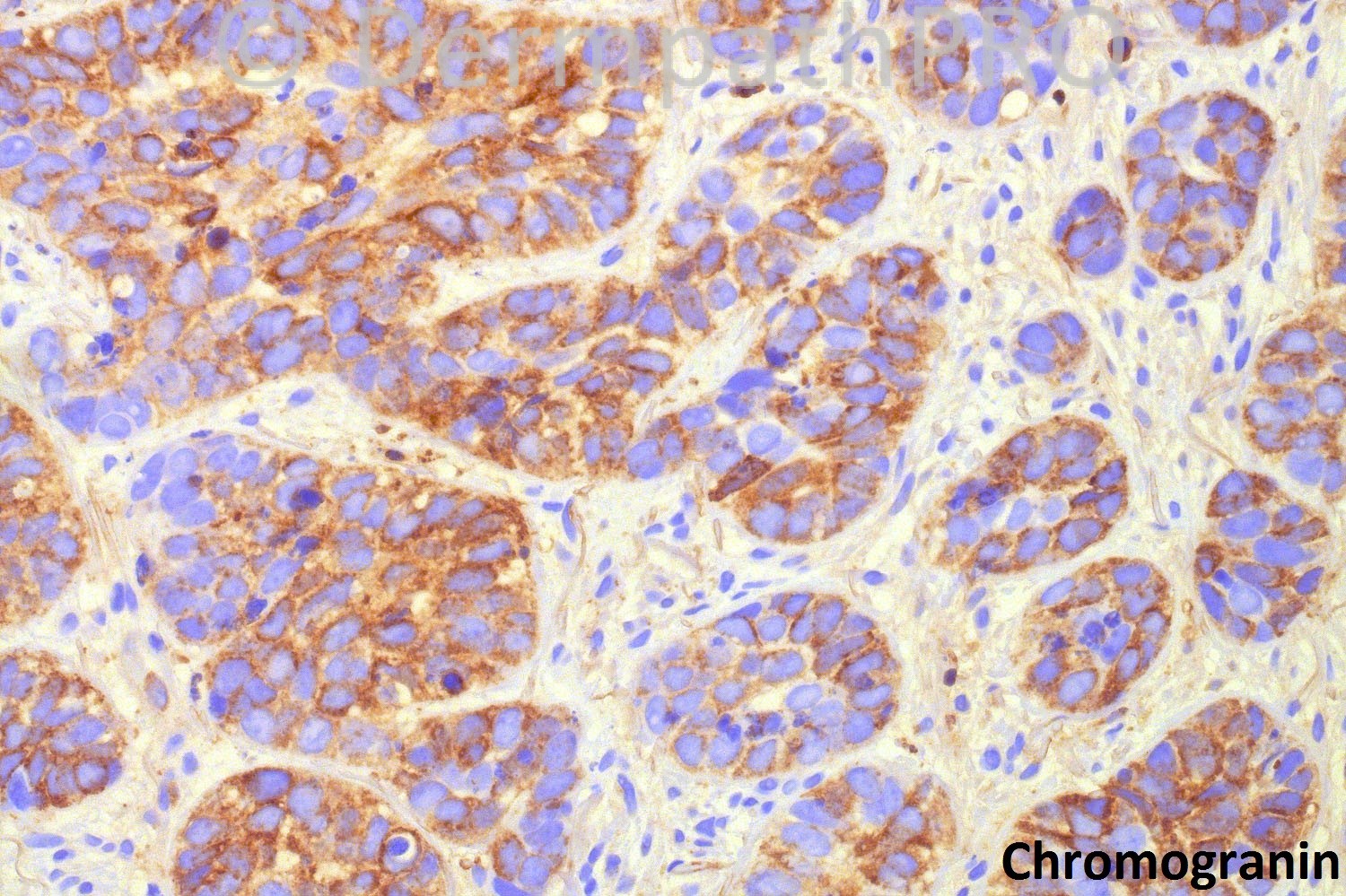
79 years-old male with chronic wound on left medial lower leg. A biopsy was performed to rule out skin cancer.


  Report Record

User Feedback


Operations Pathhub

Posted

Guest Hazem Hamed- Basal cell carcinoma with neuroendocrine differentiation. Clefting and mucin are seen in 2nd image and a hint of peripheral pallisading in 3rd image.
 
Guest - There is connection to the epidermis, mucinous stroma, clefting, vague palisading, moderate quantity of oesinophilic cytoplasm and visible nucleoli. Additionally, the clinical course seems to be indolent ("chronic wound"). All this points towards basal cell carcinoma with neuroendocrine differentiation (and away from Merkel).
 
Azza Esmat - Agree BCC with neuroendocrine differentiation
 
Dr. Mona Abdel-Halim - Yes BCC with neuroendocrine differentiation.
 
Sasi Attili - Agree with all
  
Arif Usmani - Agree. Basal cell carcinoma with neuroendocrine differentiation arising in a scar: A case report
Manuela Krokowski MD1, Josef Hoch MD2, Alfred C Feller MD1, Heinz-Wolfram Bernd MD1, Christoph Thorns MD1, Stefan Krueger MD3 Dermatology Online Journal 15 (10): 4
 
Dr. Hafeez Diwan - Yes. This is a basal cell carcinoma with neuroendocrine differentiation.

Share this comment


Link to comment
share_externally


×
×
  • Create New...