Jump to content

Building Blocks of Dermatopathology

BAD DermpathPRO Learning Hub: Basics of Immuno

Dermatopathology
Course Overview
Quizze

Case Number : IM0005

Please read the clinical history and view the images by clicking on them before you proffer your diagnosis.
Submitted Date :
   (0 reviews)

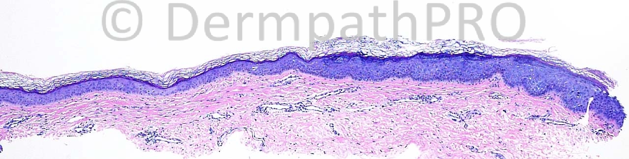
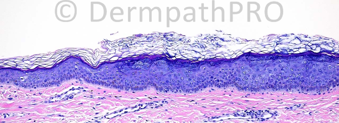
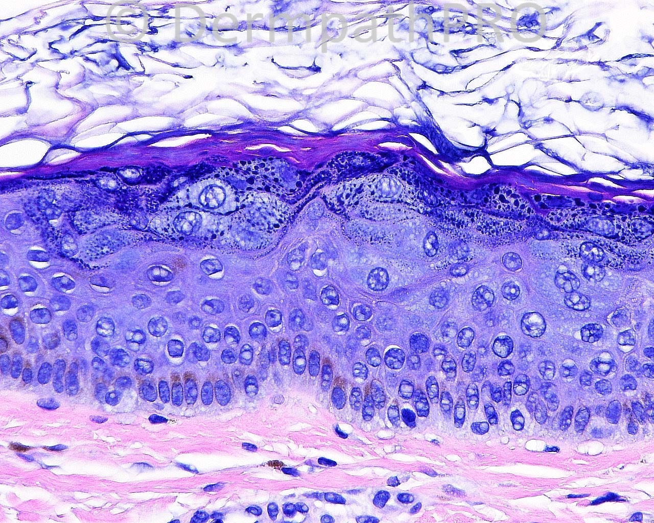
The patient is a 44-year-old white man with excisions taken from A - the left temple by the forehead.


  Report Record

User Feedback


Operations Pathhub

Posted

Guest Romualdo - Agree: epidermodysplasia verruciformis.

Guest Rodrigo Restrepo - Epidermodysplasia verruciformis
 
Guest nwpath - Agree, epidermodysplasia verruciformis.
  
Robledo F. Rocha - A plane wart-like lesion on the head that shows thickened epidermis with striking swollen keratinocytes only in the upper layers is indicative of epidermodysplasia verruciformis.
  
Guest Jim Davie MD - Epidermodysplasia verruciformis. Atypical enlarged keratinocytes with light blue cytoplasm, arranged as wedge-shaped or columnar aggregates in the stratum spinosum.[/size][/font][/color]
 
Dr. Mona Abdel-Halim - EDV
 
Mark A. Hurt MD - My diagnosis:

-- VERRUCA PLANA WITH
EPIDERMODYSPLASIA VERRUCIFORMIS LIKE CHANGES

COMMENT:
These kinds of findings have been identified as focal lesions in what has been termed "EV acanthoma". Further correlation is suggested, as some patients can have lesions like this in a disseminated pattern.

Reference:

Ko CJ, Iftner T, Barr RJ, Binder SW. Changes of epidermodysplasia verruciformis in benign skin lesions: the EV acanthoma. J Cutan Pathol. 2007 Jan;34(1):44-8. PubMed PMID: 17214854.

   
Guest Jim Davie MD - EDV changes (clusters of large keratinocytes with voluminous blue cytoplasm, and atypical keratohyaline granules) are seen as incidental findings in a wide variety of lesions such as SK, verruca, etc.

Share this comment


Link to comment
share_externally


×
×
  • Create New...