Jump to content

Building Blocks of Dermatopathology

BAD DermpathPRO Learning Hub: Basics of Immuno

Dermatopathology
Course Overview
Quizze

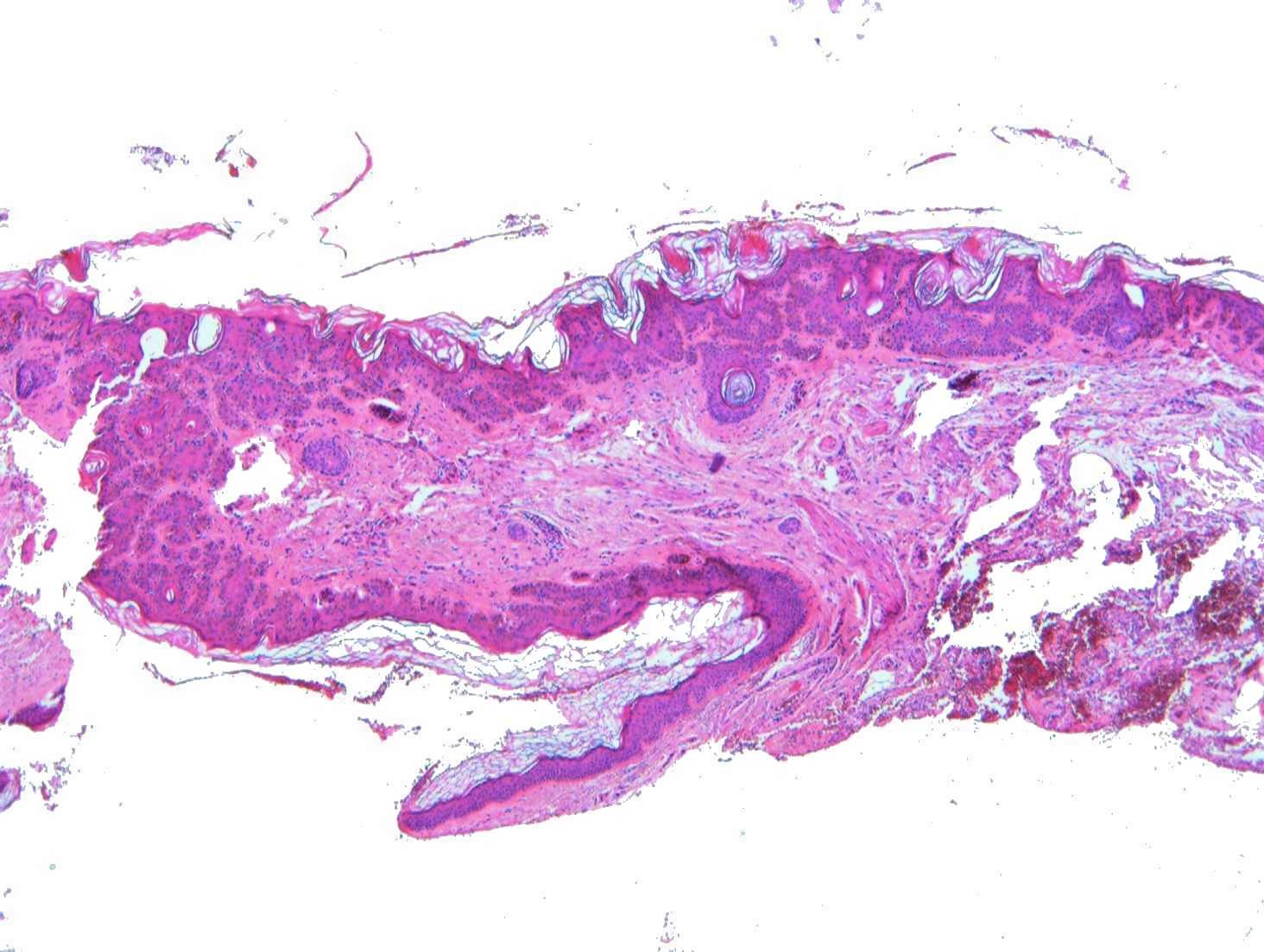
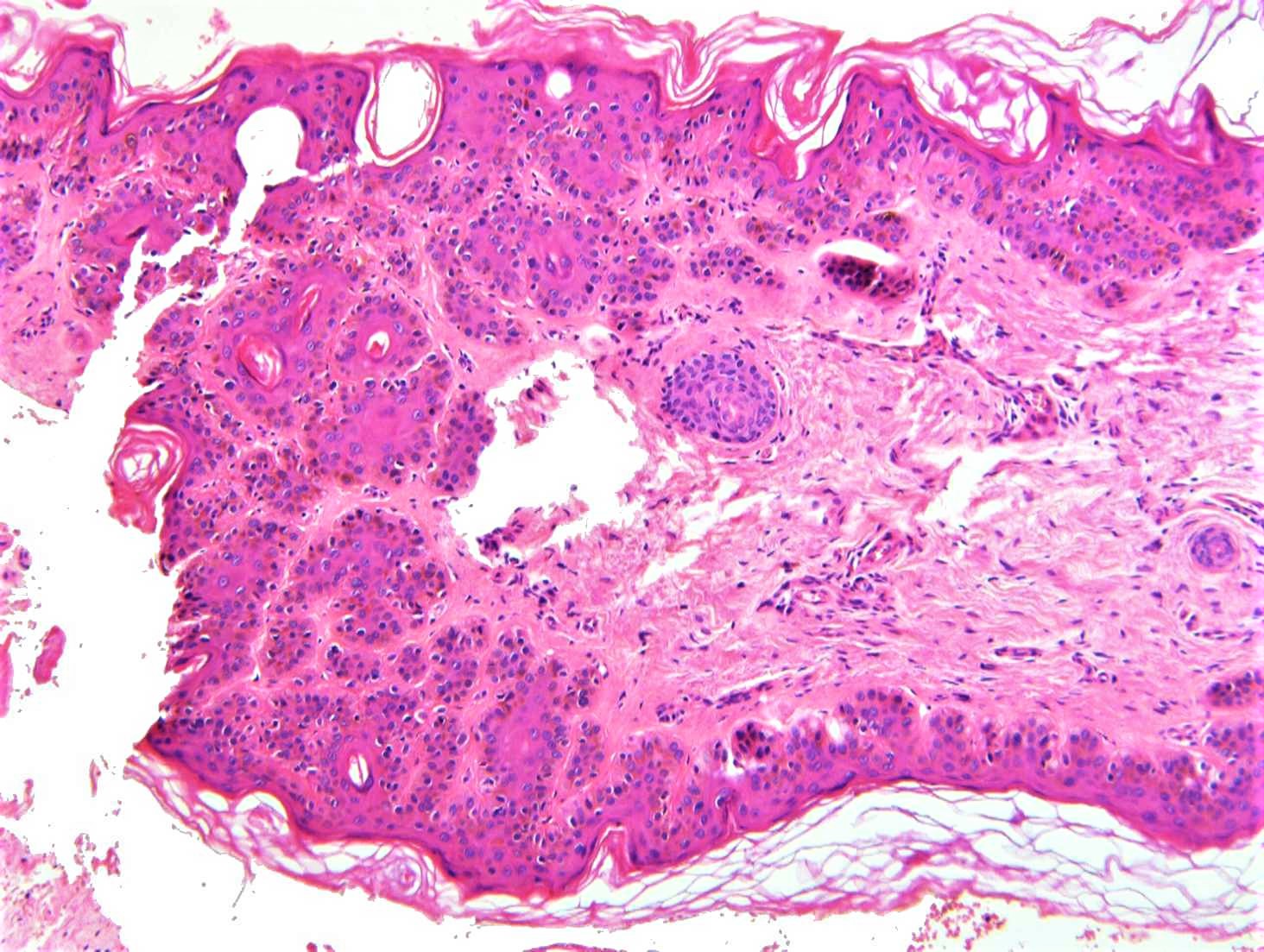
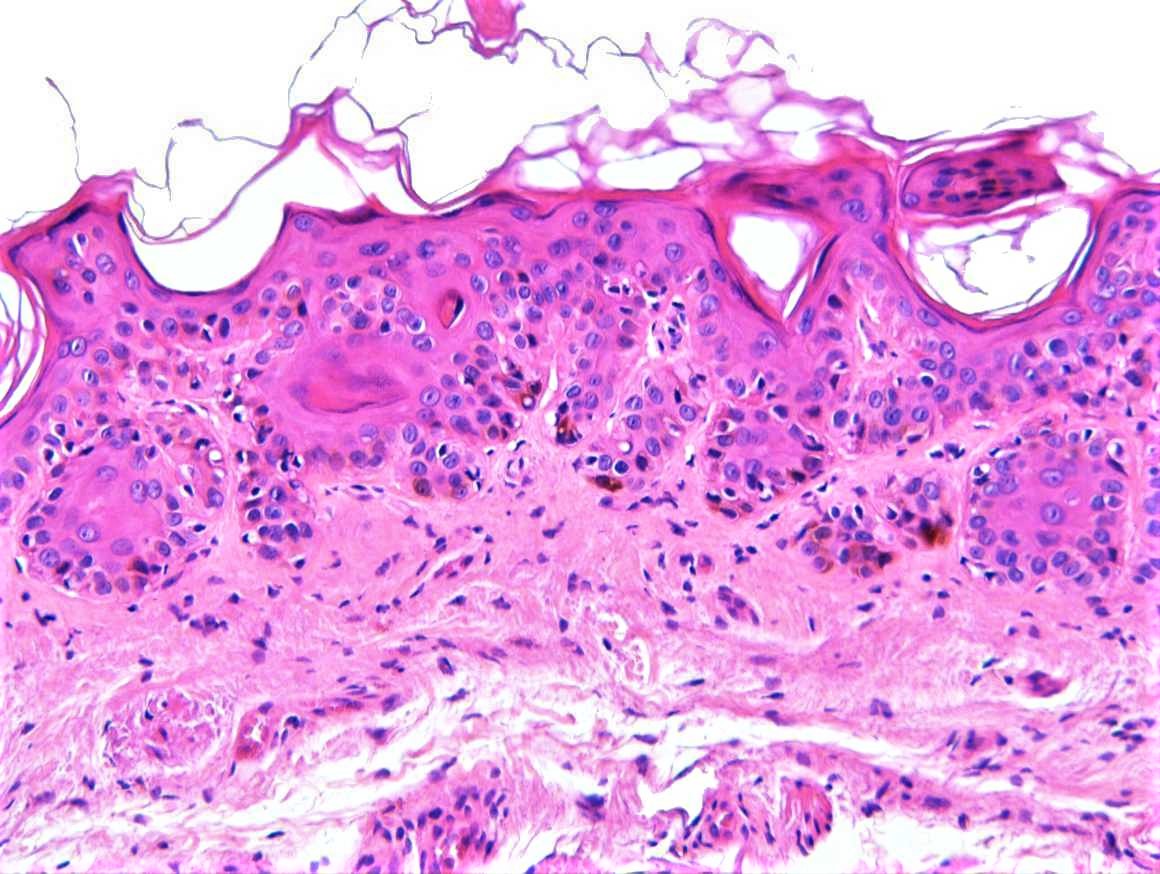
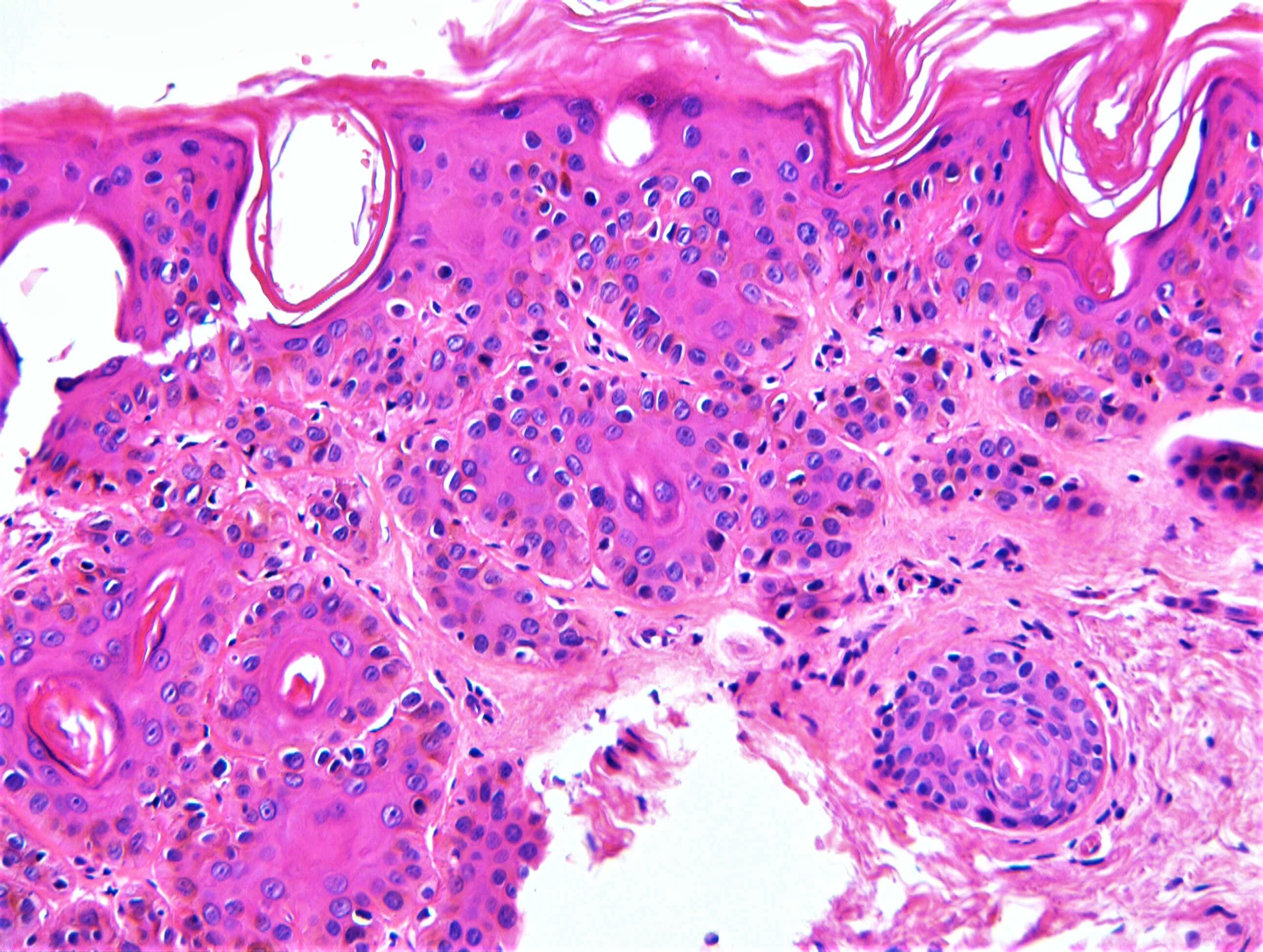
Case Number : IM0008 Dr. Richard Carr

Please read the clinical history and view the images by clicking on them before you proffer your diagnosis.
Submitted Date :
   (0 reviews)

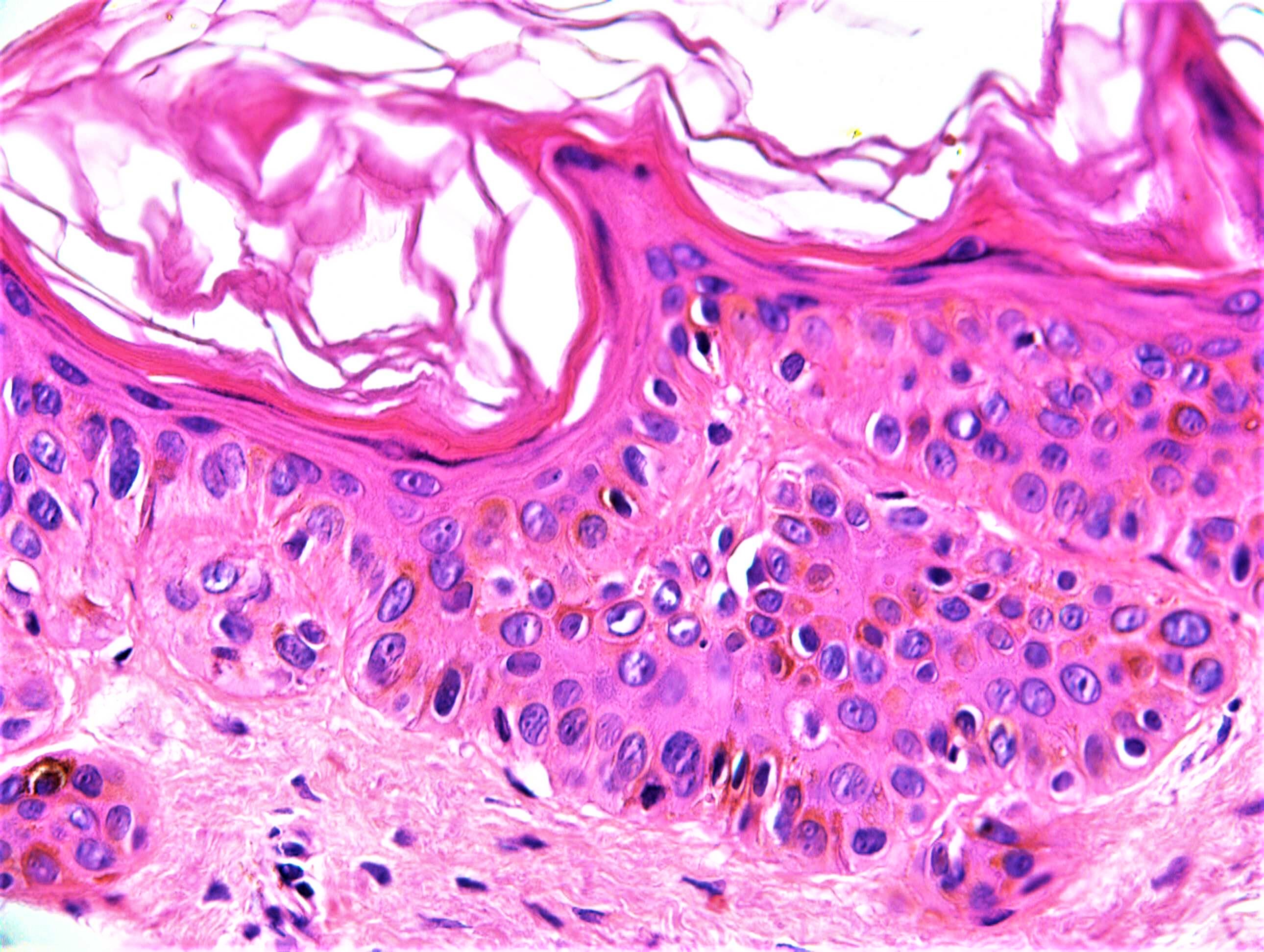
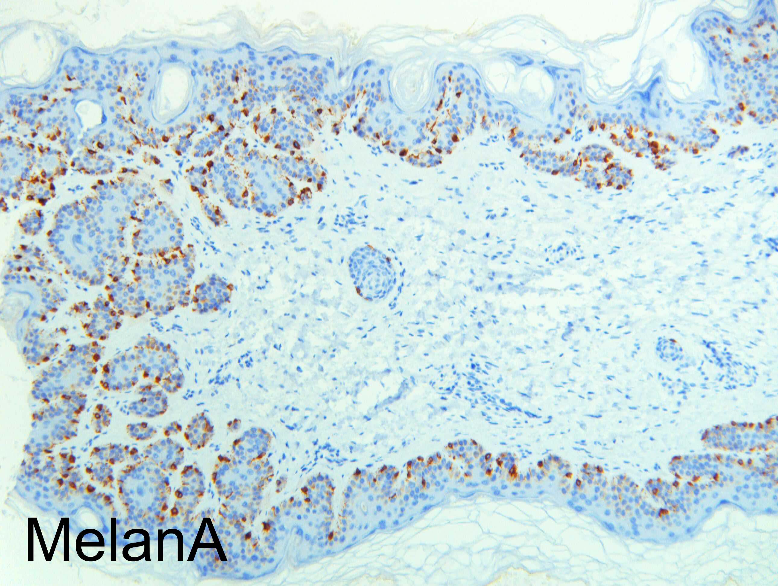
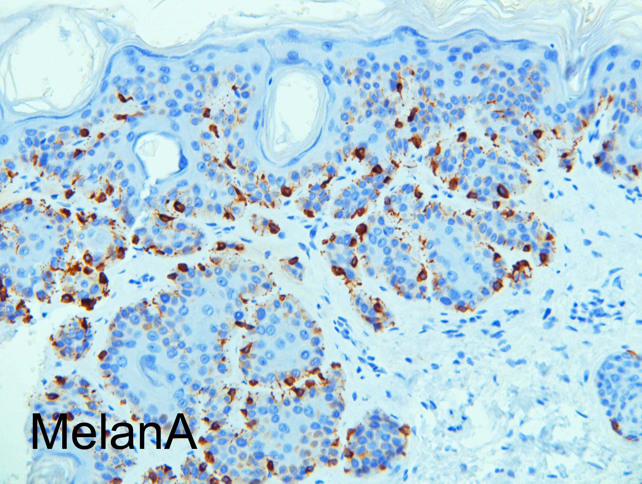
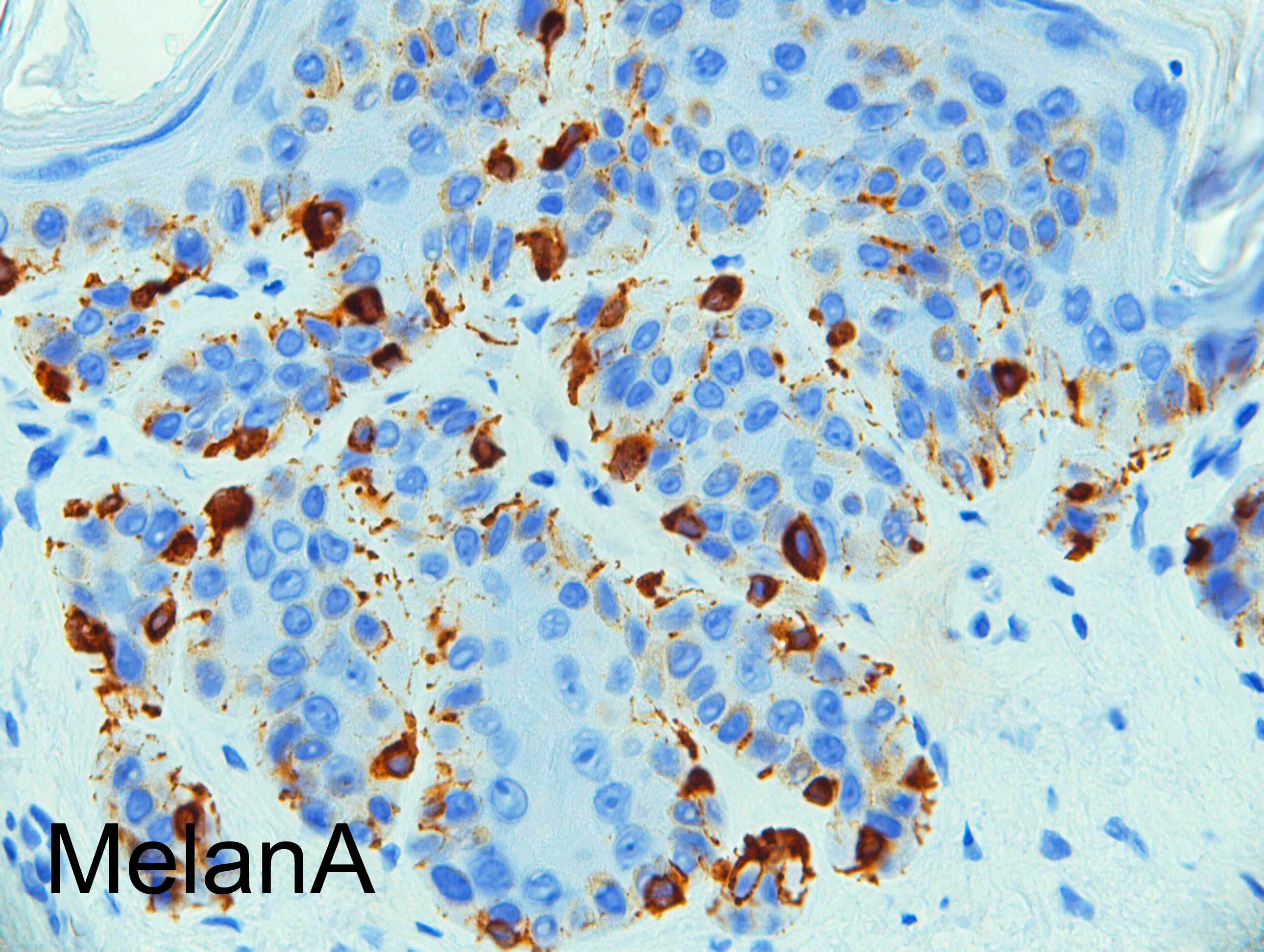
F80. Ayptical mole on the wrist. Curetted.


  Report Record

User Feedback


Operations Pathhub

Posted

Anil Patki - Pigmented seborrhoeic keratosis

Dr. Mona Abdel-Halim - Reticulated seb k, but do not know what is going in the dermis (first image) from this magnification. Otherwise, reticulated seb K. 

Urmilapandey - wondering if there is cystic invagination  in image 1

Vincenzo Polizzi - Yes, SK, reticulated type, agree! A typical example of what may happens in pathology: sometimes the diagnostically easiest lesion become the most difficult one....!

Anil Patki - One may call it acanthotic type rather than reticulated. Reticulum= net and reticulated SK should show a network of thin strands of keratinocytes.

Nitin Khirwadkar - Pigmented Seb K/actinic lentigo. As Mona has pointed out, not sure what is happening in the dermis in the first image ? some calcification

Dr. Richard Carr - Dear All. Diagnosis: Seborrhoeic keratosis with pale basal keratinocytes mimicking atypical basal melanocytic proliferation. Have a great weekend! 

Share this comment


Link to comment
share_externally


×
×
  • Create New...