Jump to content

Building Blocks of Dermatopathology

BAD DermpathPRO Learning Hub: Basics of Immuno

Dermatopathology
Course Overview
Quizze

Case Number : IM0012 Dr. Richard Carr

Please read the clinical history and view the images by clicking on them before you proffer your diagnosis.
Submitted Date :
   (0 reviews)

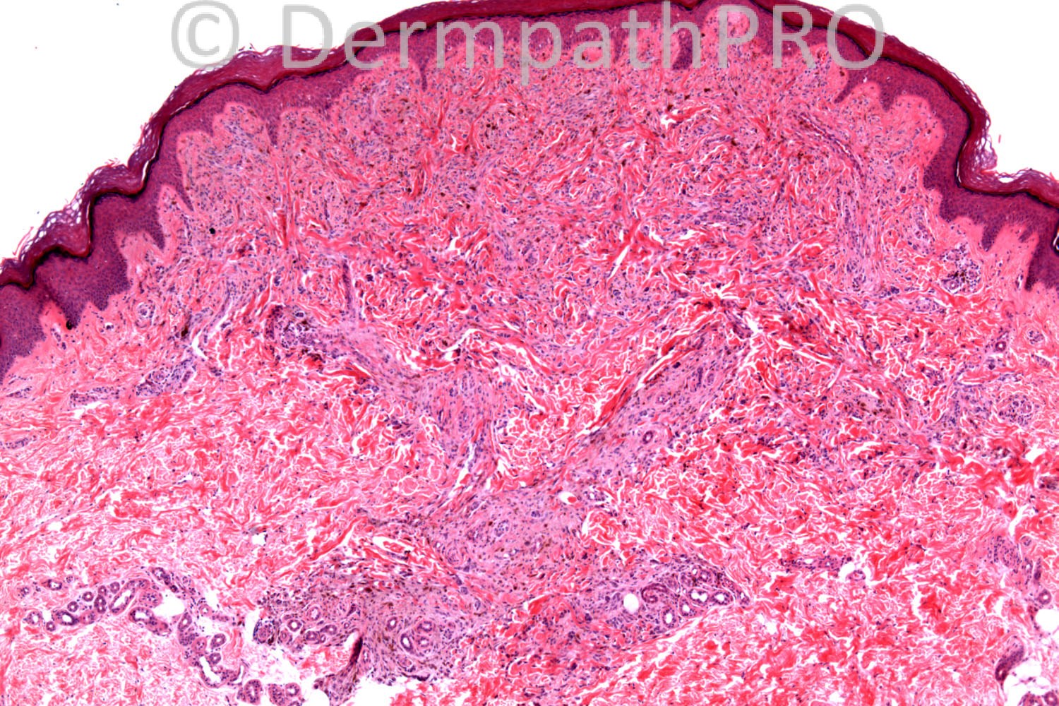
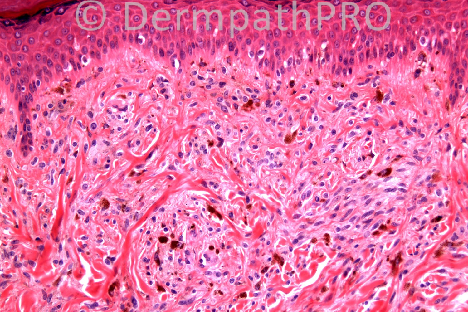
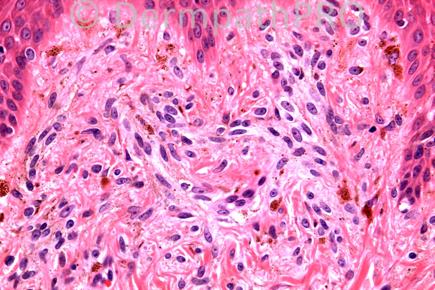
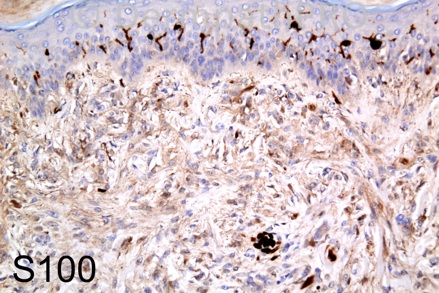
16 years-old female, lesion on foot. ?Spitz naevus


  Report Record

User Feedback


Operations Pathhub

Posted

Guest Maria George - Could it be a nevus which lost s100 staining or cellular neurothekoma which is not staining with s 100.
 
Mark A. Hurt MD - Favor "cellular" neurothekeoma. I interpret the s100 as negative. I would like to know whether the lesion is positive for cd63, d2-40, and NSE. If it is a "C"N, it should be positive for all three, but negative with s100 and Melan-A.
  
Guest Maria George - Just to supplment the IHC suggested by Mark, I did find the paragraphs in this online link useful;
http://emedicine.medscape.com/article/2034472-overview#aw2aab6b6
 
Dr. Mona Abdel-Halim - And what about the melanophages/ melanin granules dispersed among the lesion? I am not sure that S100 here is negative. Thought of hypopigmented type blue nevus or less likely desmoplastic nevus??
 
Guest - Sclerotic hypopigmented blue nevus vs Desmoplastic nevus

IgorSC - Hypopigmented blue nevus. I don´t think that there is sclerosis or a juntional component for a Desmoplastic nevus. Nice case. Waiting for more comments.
 
Robledo F. Rocha - I think this is a desmoplastic Spitz nevus. I found a wedge-shaped intradermal melanocytic proliferation with epithelioid superficial cells and smaller deep ones, all of them embedded in a sclerotic stroma. Typically melanin pigment is sparse and the site is an extremity of a young patient.
S100 is negative, since normal dendritic melanocytes within the epidermis are strongly highlighted by this immunostain. This might mean a loss of s100 expression by lesional cells. Edited November 3, 2013 by Guest
  
Guest Juan Carlos Garcés, Ecuador - Hypopigmented blue nevus
 
Eman El-Nabarawy - Cellular neurothekeoma with melanocytosis.
 
Guest Jim Davie MD - I'd favor Blue nevus with anomalous S100 negative staining, if we are dealing with a situation of anomalous negative S100 staining. (S100-negative blue nevi are uncommon [<3%], and are usually hypopigmented).
 
Guest Romualdo - I agree with the diagnosis of blue nevus with aberrant S100 negative staining.
 
Dr. Richard Carr - This is a "relatively" hypopigmented blue naevus (HBN). One definition for HBN is melanin detected in <5% of the lesional cells and I guess this lesion qualifies if we exclude the melanophages. The clinical mis-diagnosis of Spitz (usually non-pigmented lesions) would support the "hypopigmented" classification. I agree if you are not familiar with hypopigmented BN then you will consider an odd naevocellular or desmoplastic naevus (cells are quite plump in this case and in other similar lesions we have seen) but as correctly mentioned by responders above there are nice dendritic cells on close inspection and the dendritic nature can easily be confirmed with MelanA which is diffusely positive in all cases. What is not in the textbooks and is very much our experience is that blue naevi are usually either only focally or weakly S100 positive or very commonly, as in this case, completely S100 negative. Putting this case in has prompted me to finish off our paper on this topic. It is not novel information as some authors have mentioned it in the literature but it seems to be totally overlooked in most textbooks. It has to be said that some papers do state S100 as being positive in HBN but in our experience S100-/weak/focal; diffuse melanA positive (dendritic) pattern is a diagnostic clue to blue naevus. We have been caught out by near completely melanin negative case and made the wrong diagnosis of a benign fibrohistiocytic lesion based on S100 neg before spotting very sparse melanin on H&E and confirming the correct diagnosis with MelanA. A subsequent case we correctly diagnosed only because of this prior experience. Thanks for mentioning cellular neurothekeoma as this can now go in to our discussion and I was not familiar with cellular neurothekeoma with melanocytosis (do you have a reference?) but melanA will confirmed the correct diagnosis in this case. Jim I would be interested in your reference that <3% of blue naevi are S100 negative (because that is not our experience that includes cases referred in from other pathologists/labs). I interpreted the S100 positive dendritic cells in the epidermis to be Langerhan's rather than melanocytes but there may be a combination here. Remember S100 nuclear staining is helpful in heavily pigmented lesions. If you look at the S100 again you can see the subtle melanin pigmentation of the dendritic processes in this case. Edited June 22, 2013 by Guest
  
Mark A. Hurt MD - Richard, my experience is similar to yours with blue nevi. The usual profile is strong Melan-A, weak to "negative" S100, negative Ki-67. If a case with this histopathology is negative with Melan-A and S100, then consider "cellular" neurothekeoma and do CD63, D2-40, and NSE.

Share this comment


Link to comment
share_externally


×
×
  • Create New...