Jump to content

Building Blocks of Dermatopathology

BAD DermpathPRO Learning Hub: Basics of Immuno

Dermatopathology
Course Overview
Quizze

Case Number : IM0013 Uma Sundram

Please read the clinical history and view the images by clicking on them before you proffer your diagnosis.
Submitted Date :
   (0 reviews)

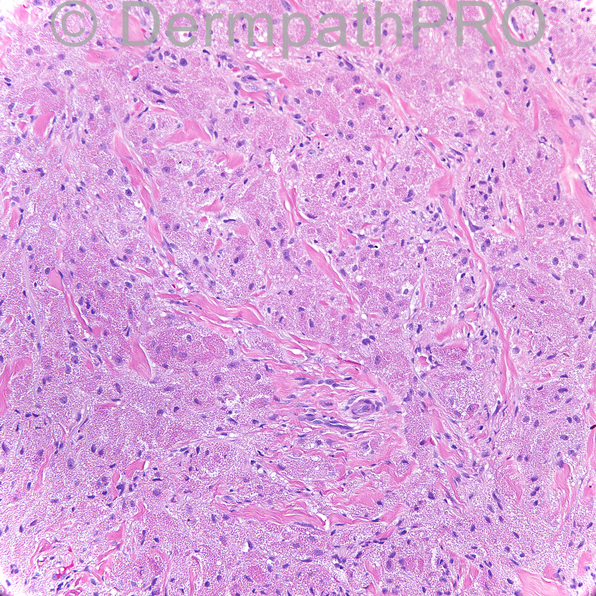
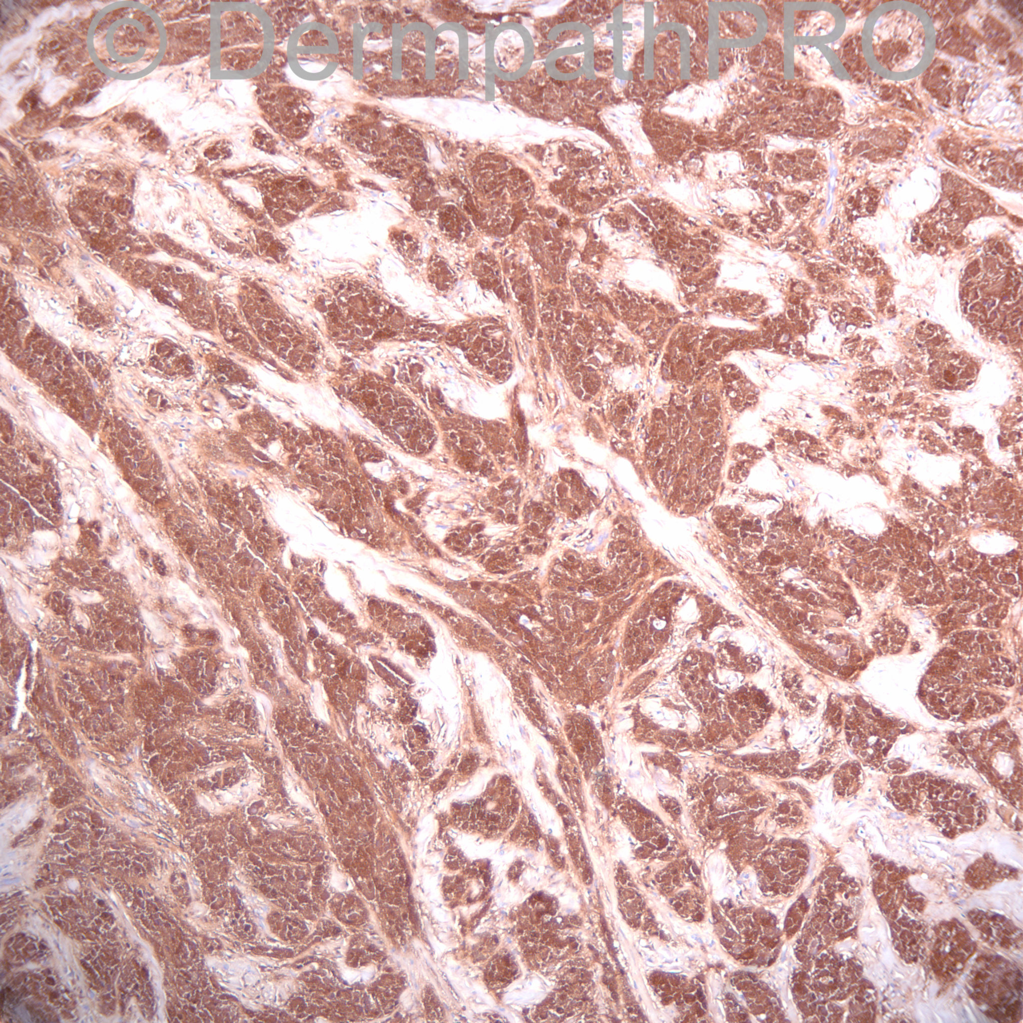
88 year old man with 2.5 cm erythematous very firm nodule on the left abdomen.


  Report Record

User Feedback


Operations Pathhub

Posted

Guest Romualdo - Granular cell tumor.
  
Guest - Agree with granular cell tumour. Coincidentally, I was reading about tumours with granular cells last week, after seeing an undifferentiated pleomorphic sarcoma with granular cells. I came up with this list: Melanoma, hibernoma, rhabdomyoma, leiomyoma and leiomyosarcoma, dermatofibroma and dermatofibrosarcoma and metastatic histiocytoid carcinoma. If anyone has more to add here, please do so.
 
Dr. Mona Abdel-Halim - Very lovely tumor... Have seen five cases this last year ...
   
Dr. Richard Carr - I checked my slide database and reference database with the following results:
Non-neural granular cell tumour; Granular cell BCC; Granular cell trichoblastoma (Battistella, Am J Dermatopathol 2013); case of spindle cell angiosarcoma that Arno Rutten put in the UK Skin EQA educational cases I noted focal granular cell changes; focal granular cell change in a glomangiopericytoma; AFX (very xanthomatous case almost granular cell); congenital granular cell epulis (mouth)
   
Guest - Thank you, Dr Carr. Much appreciated.
   
Eman El-Nabarawy - GCT.
 
Uma Sundram - Dear friends, my apologies for the late post. This is indeed granular cell tumor. The stain is S100 as you probably have already guessed, and the tumor is very strongly positive.

Share this comment


Link to comment
share_externally


×
×
  • Create New...