Jump to content

Building Blocks of Dermatopathology

BAD DermpathPRO Learning Hub: Diagnostic Clues

Diagnostic Challenge
Interesting Case
Pitfalls

Case Number : CT0005 Uma Sundram

Please read the clinical history and view the images by clicking on them before you proffer your diagnosis.
Submitted Date :
   (0 reviews)

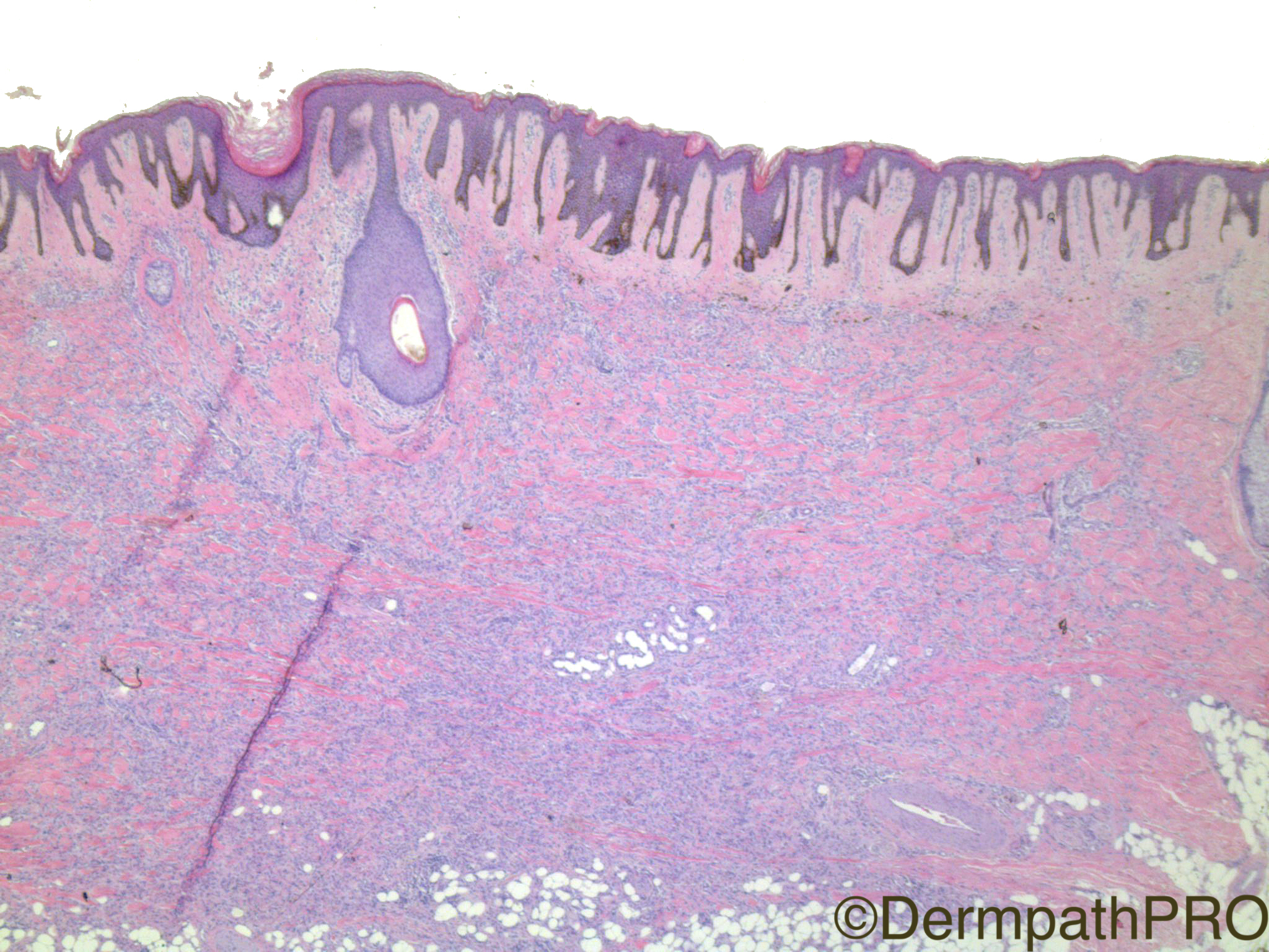
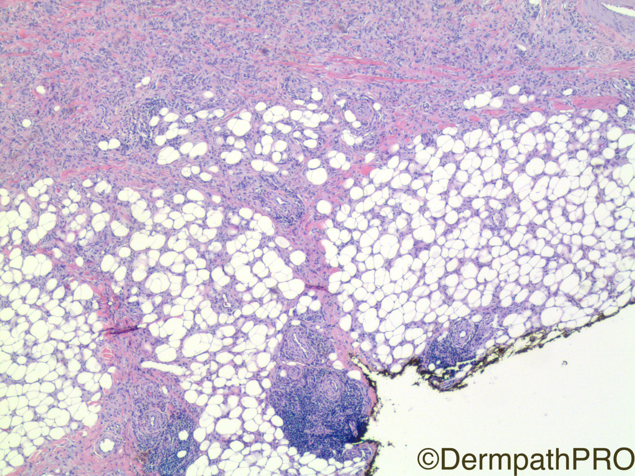
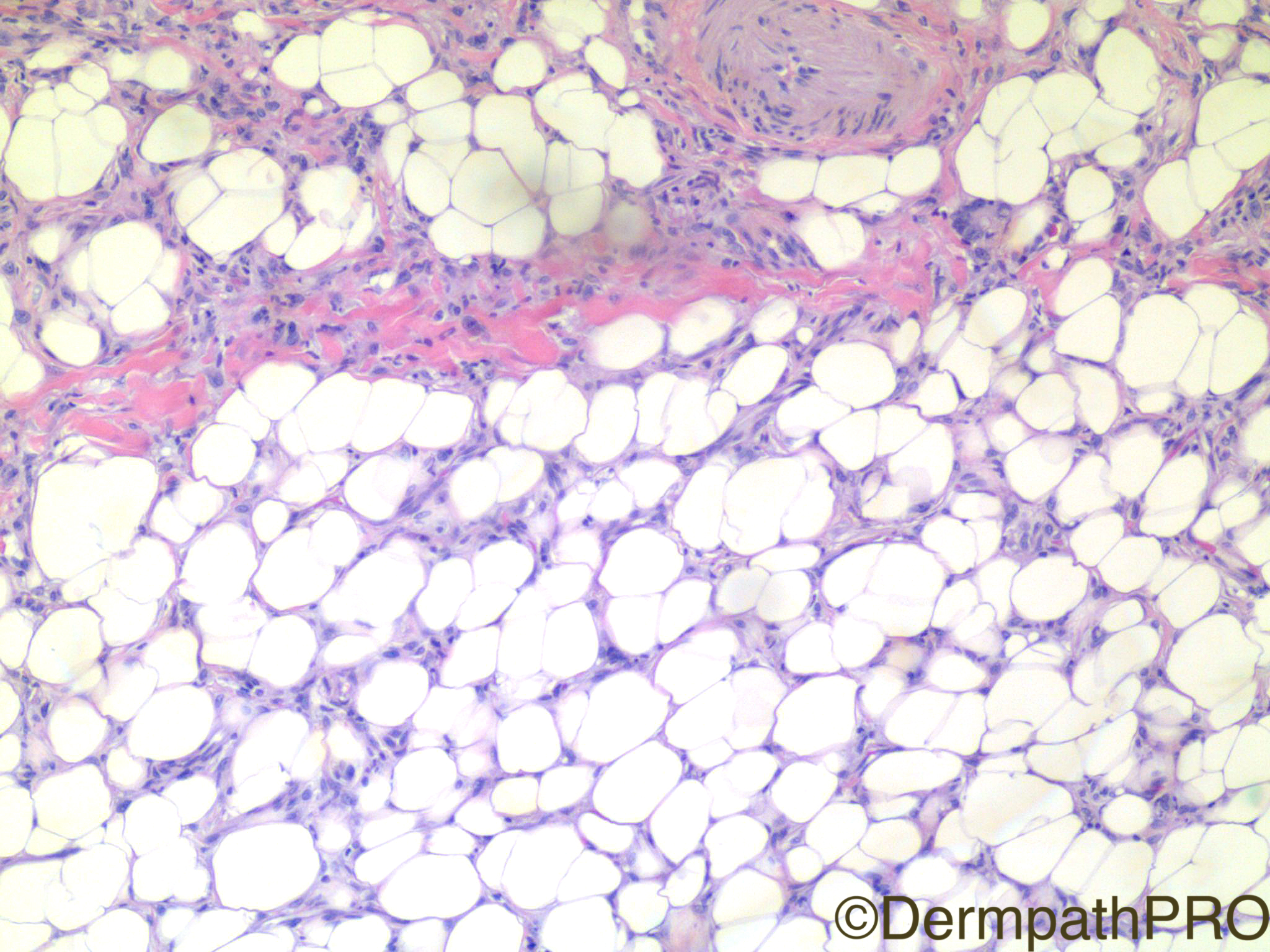
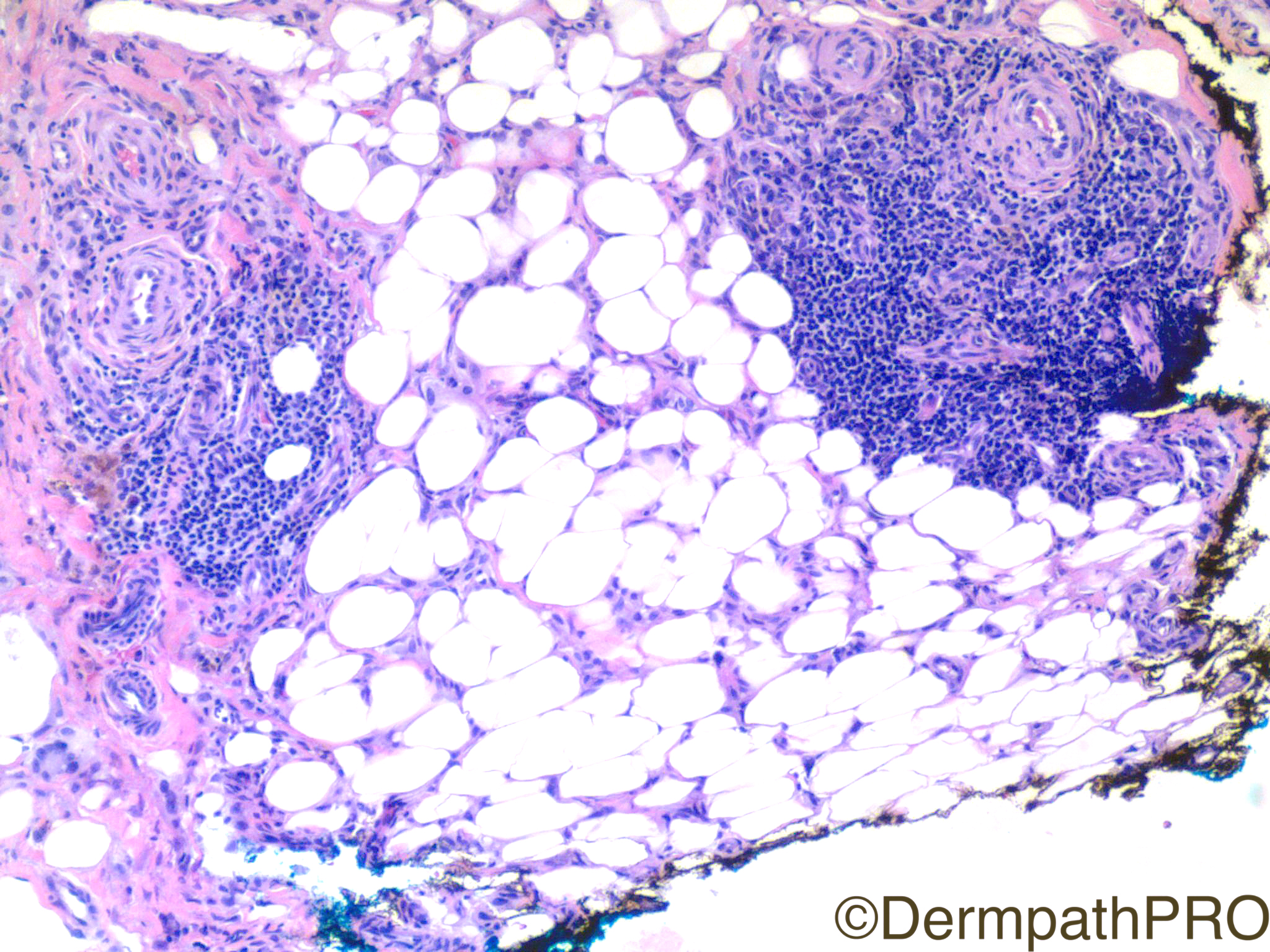
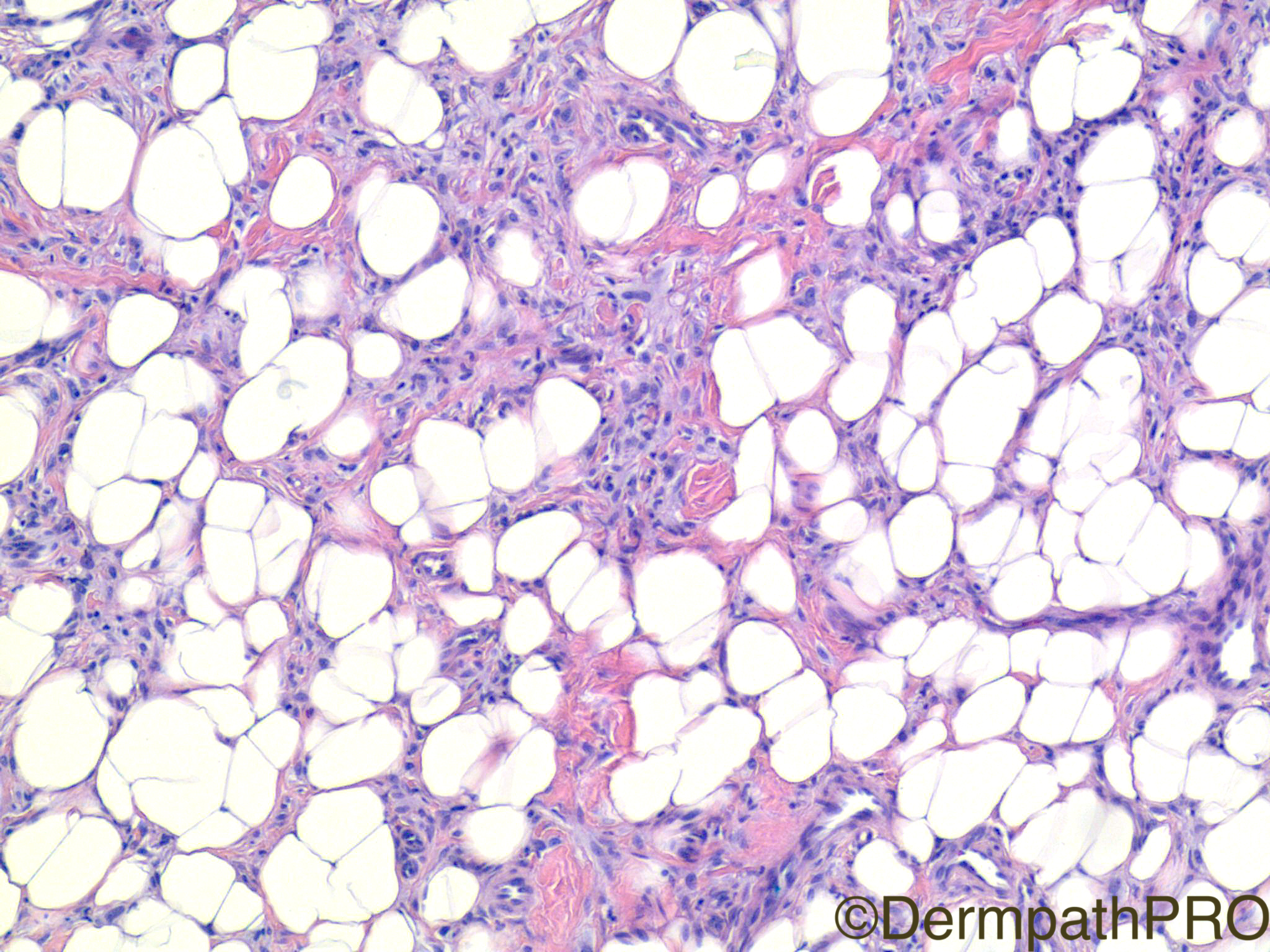
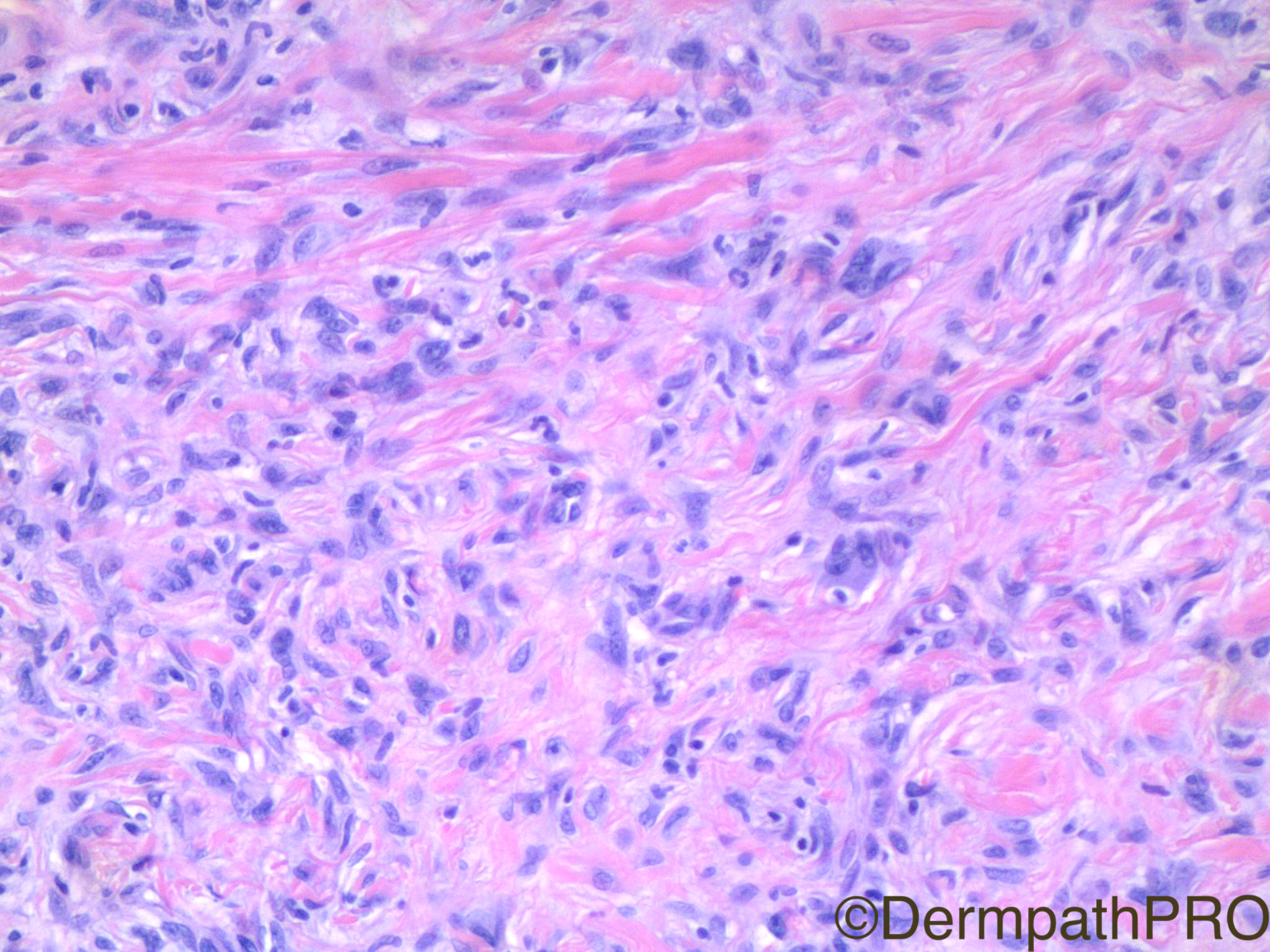
Clinical History: 49 year old woman with left anterior tibial lesion.


  Report Record

User Feedback


Operations Pathhub

Posted

Arash Daryakar  - Hard case. Infiltrative epithelioid histiocytic like lesion with extension to subcutaneous fat and lymphophagocytosis(?). I have no idea about cell proliferative activity . but i would like to include histiocytic sarcoma and deep fibrous hisiocytoma in differentials. I wish  to know expression of  histocytic and also melanocytic markers to determine line of differetiation.
   
Raul Perret - Due to the epidermal hyperplasia, presence of hemosiderine deposits and xanthomized cells in pic 5? I would consider dermatofibroma as the first diagnosis. In addition the cellularity and deep extension are seen in the cellular variant, cd34 should be performed. The presence of lymphoid nodules can be seen in df also but should always inspect them well to avoid a misdiagnosis of a lymphoma/leukemia (mainly LLC). 

Dimitris Chatzianastasiou - Fascinating little thing... Indeed on low power it recapitulates the pattern of a dermatofibroma with some layering (over fascicular intersecting pattern). However, in high power and deep lesional images it shows some vascular qualities with epitheloid/histiocytoid cells, possible intracytoplasmic vacuolation, scattered multinucleated cell forms. I m puzzled by the fact it spreads within the fat lobules, not only along the septa. The lymphoid hyperplasia appears vasculocentric and "out of the blue". No eosinophils. I would have asked for immunos (eg. CD34, FactorXIIIa, CD68, CD163, CD31, ERG) to see if I could end up favouring an epitheloid/histiocytoid hemangioma or some dermatofibroma variant. Lets see...

Dr. Mona Abdel-Halim - I am sharing with Raul the same line of thinking, I believe this is cellular DF with giant cells and lymphoid infiltrate. But it is good to verify the lymphoid cells with caution, maybe do some IHC... 

Vincenzo Polizzi - Dermatofibroma, with subcutaneous extension and lymphoid nodular reaction at the bottom of the lesion ( well known host abnormal reaction in this tumor        ( Inflammatory lymphadenoid reactions with dermatofibroma/histiocytoma https://www.ncbi.nlm.nih.gov/pubmed/3016046.). Agree with Raul: there are histiocytoid cells rather than slender hypercromatic and fibroblastic of DFSP. It needs IHC off course, but I consider unlikely CLL/SCL in this setting, because the infiltration of CLL is usually interstitial between adipocytes and radiating from nodules rather than well circumscribed micro nodular as here,...but "unlikely" doesn't mean impossible...

IgorSC - I believe this is a case of Deep DFB, but I would perform immunostains to exclude DFSP. I think that this infiltrate deep to the tumor is not concerning. CD34 and CD10 are a good choice for the differential, in my opinion.

Drdeepajacob - Deep cellular DF or a DFSP. But would always prefer to exclude a melanoma ( just a trainees thought)

Alice Roberts - Cellular DF. Would do F13A, CD34 and S100 to confirm.

Dr. Richard Carr - Nice discussion but I was thinking I'd be happy with dermatofibroma (subcutaneous extension).

Nitin Khirwadkar - I think this is a cellular DF with deep extension. Typical epidermal changes. Would definitely do a CD34.

Uma Sundram - Thanks everyone, great job. I fully concur with the ddx of cellular DF, DFSP, and possibly unusual desmoplastic melanoma (thank you trainee!). There are no secret traps here, factor 13a was positive and CD34 and S100 were negative. The lymphoid aggregates were non neoplastic. However, I think it's easy to get side tracked by the extensive infiltration into fat and the presence of lymphoid aggregates, so wanted to show a case that can have some confusing features which often leads to a send out to an expert.

Share this comment


Link to comment
share_externally


×
×
  • Create New...