Jump to content

Building Blocks of Dermatopathology

BAD DermpathPRO Learning Hub: Diagnostic Clues

Diagnostic Challenge
Interesting Case
Pitfalls

Case Number : CT0010 Admin_Dermpath

Please read the clinical history and view the images by clicking on them before you proffer your diagnosis.
Submitted Date :
   (0 reviews)

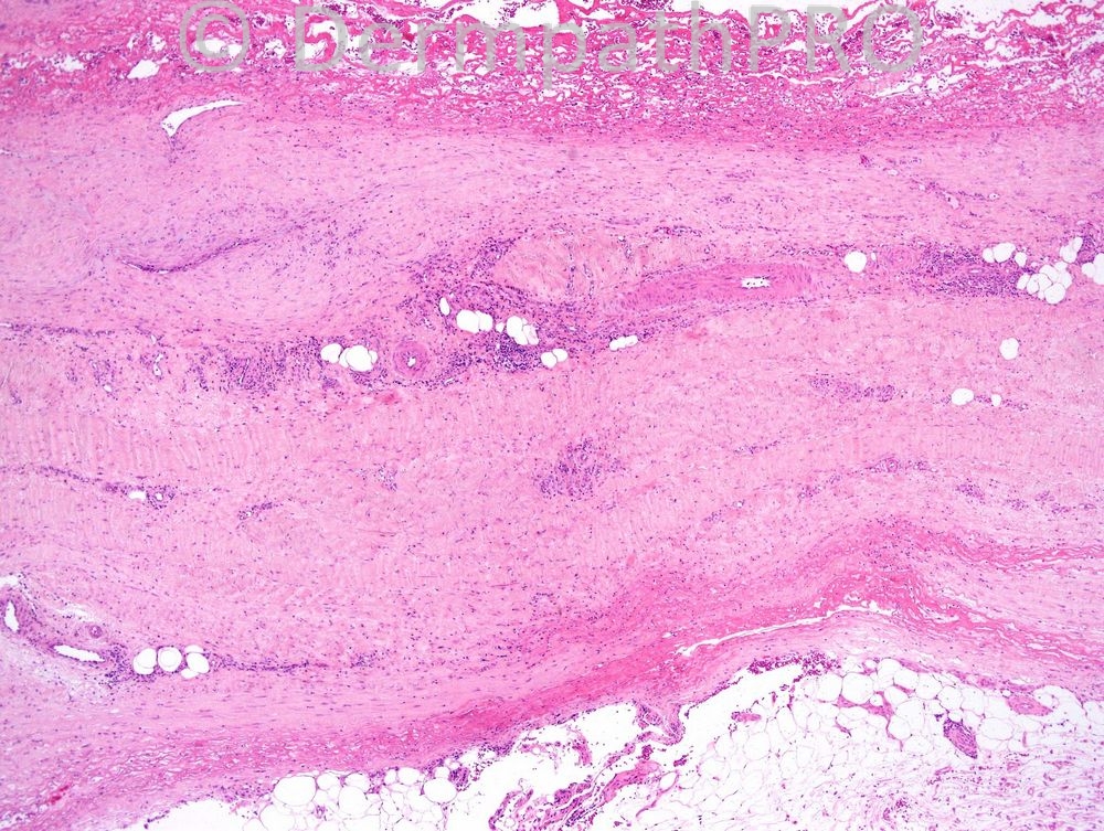
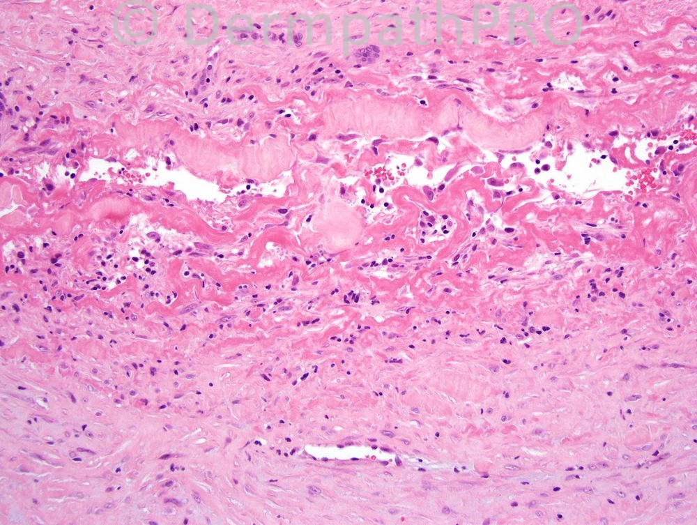
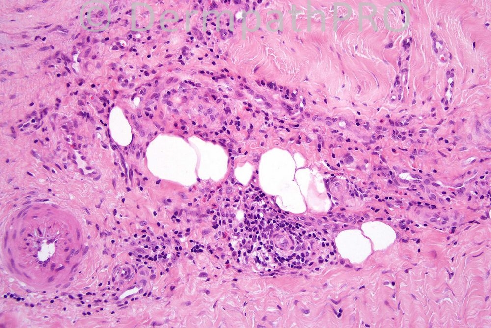
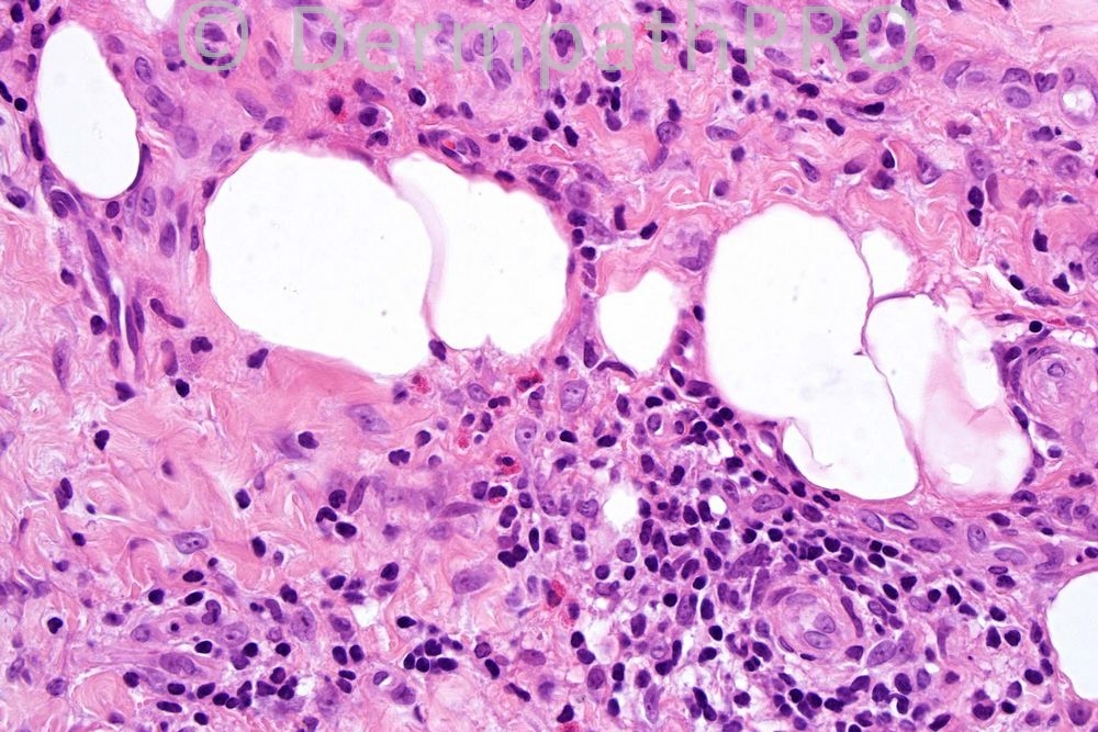
Female 38 years, pain and swelling of forearm.


  Report Record

User Feedback


Operations Pathhub

Posted

Atopica -Eosinophilic fasciitis.

Phillip H McKee - In the clinical history I left out the fact that the condition developed after strenuous activity. I think that the histology fits very well with eosinophilic fasciitis. Remember that in that condition, the histology may sometimes show few or even no eosinophils. Clinicopathological correlation is critical.

Sasi Attili - Been thinking about this all morning. There is a vessel with fibrinoid necrosis but not much inflammation. This is slightly atypical for an inflammatory process/ true vasculitis. Necrosis of surrounding subcutis also evident. There infiltrate is composed of eosinophils and lymphocytes. However I am not really able to come to a definitive diagnosis and would need further CPC. An injected toxin or an insect bite would be in my differentials. Looking forward to the answer

Mona Abdel Halim - I will diagnose this as esinophilic fasciitis, the 2nd image show fibrinoid necrosis in relation to a blood vessel seen in the acute phases of this condition.

I. Abdul-kadir - ?Nodular vasculitis.

Yüksel Okumuş - Lupus Panniculitis

Share this comment


Link to comment
share_externally


×
×
  • Create New...