Jump to content

Building Blocks of Dermatopathology

BAD DermpathPRO Learning Hub: Diagnostic Clues

Diagnostic Challenge
Interesting Case
Pitfalls

Case Number : CT0020 Dr. Richard Carr

Please read the clinical history and view the images by clicking on them before you proffer your diagnosis.
Submitted Date :
   (0 reviews)

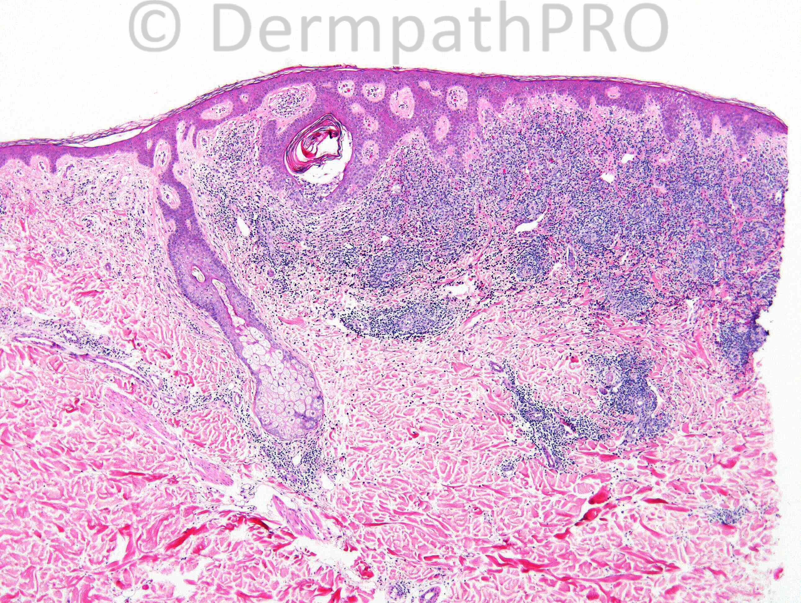
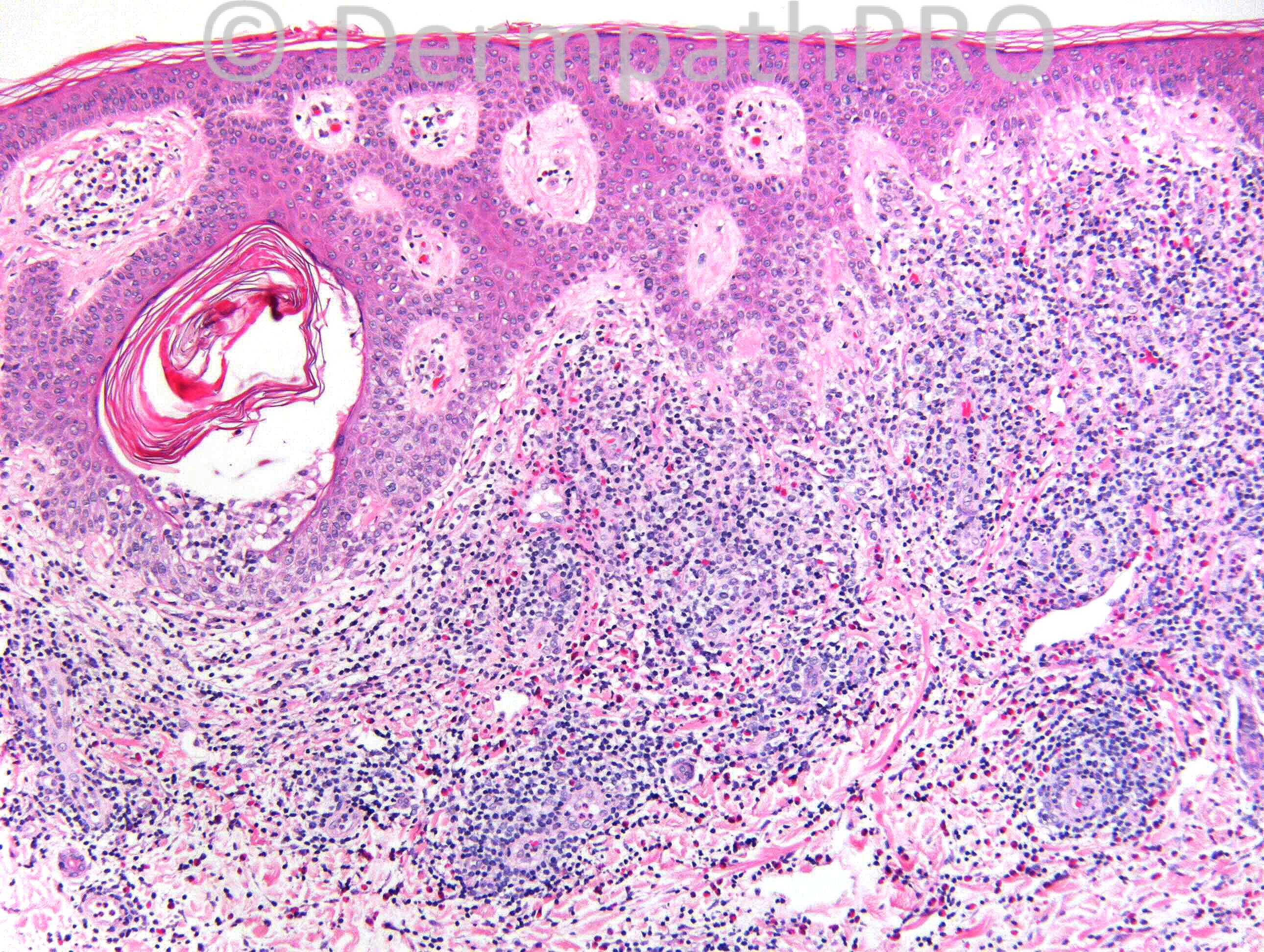
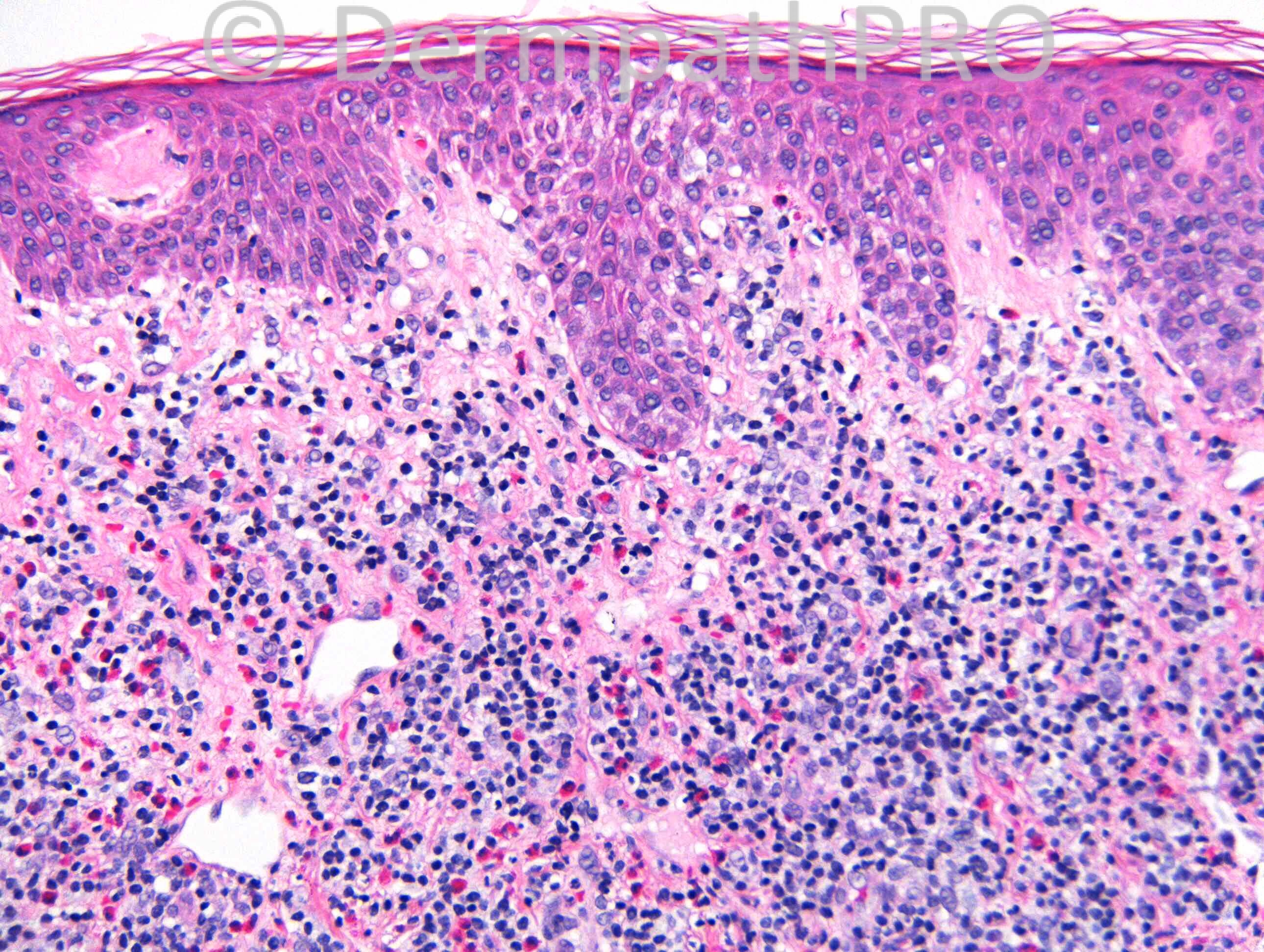
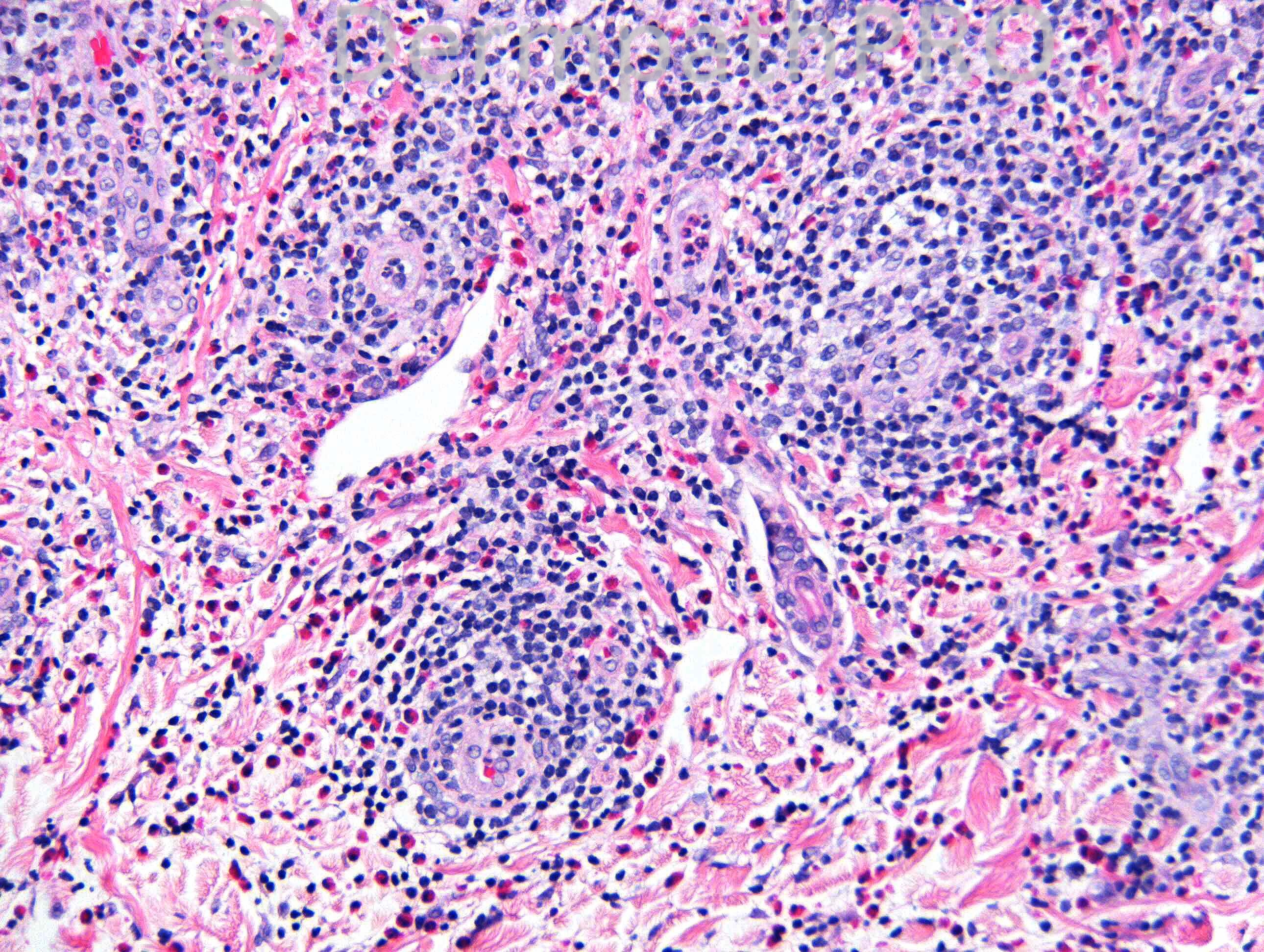
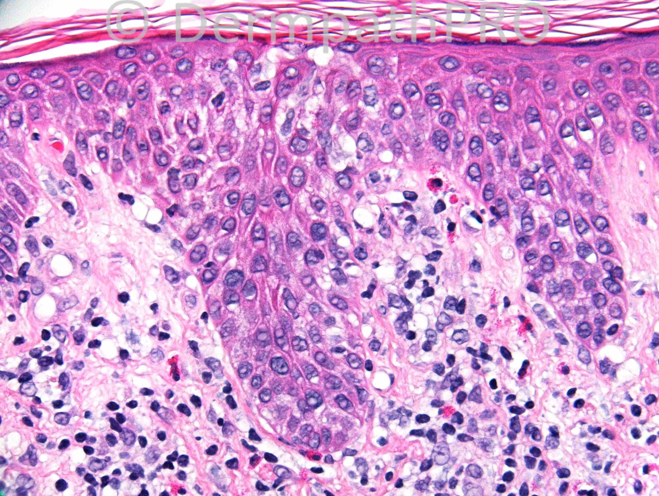
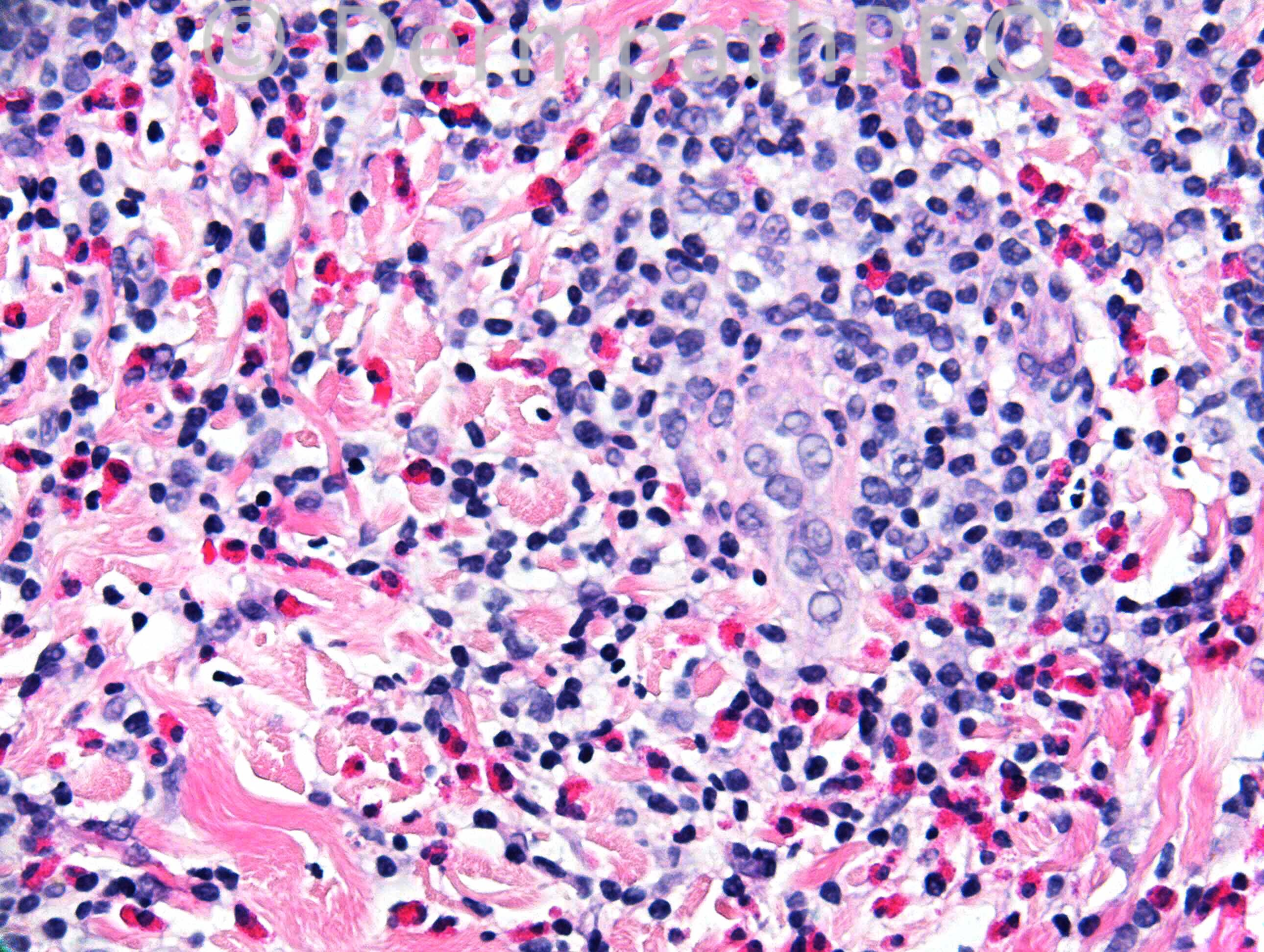
75 years old female. Persistent itchy plaques on back, almost blistering.


  Report Record

User Feedback


Admin_Dermpath

Posted

Dr. Mona Abdel-Halim - Esinophil rich infiltrate, spongiotic epidermis, I can see an esinophil along the basal cell layer, together with the clinical presentation, my first possibility is prodromal stage of pemphigoid.
 
Guest Saleem Taibjee - I agree with Mona. Would also need to exclude scabies.

Guest Mazen charaf iddin - I see the lesion is in the dermis and the epidermal changes are reactive may be I am far a little bit , and the sit is not typical, but in low power look like angiolymphoid hyperplasia with eosionphilia , or as dr saleem has said reaction to insect bits such as bed bugs

Guest Maria George - Low power points to insect bite ,lupus or drug erption.In HP there ire cell with cleaved nuclei .I want to add eosinophilic granuloma .Localzed LCH.
   
Guest Ali Y. Hashim - It could be due to reaction to insect bite or drug. CPC is essential to settle the Dx.
 
Guest Dr.Yüksel Okumuş - Early (prebullous) urticarial lesions of Pemfigoid.

Sasi Attili - Agree. DD includes insect bites and pre-bullous pemphigoid.
 
Guest - Pre-bullous pemphigoid.
 
Guest Romualdo - Agree with pre-bullous pemphigoid and insect bite reaction. Against the former is the near absence of intraepidermic eosinophils and against the latter the location of the lesions on sun protected part of the body. I prefer pre-bullous pemphigoid.
 
Eman El-Nabarawy - DD: Pre-bulbous pemphigoid and insect bite reaction.
 
Robledo F. Rocha - I’d add late inflammatory variant of epidermolysis bullosa acquisita. There are signs of epidermal re-epithelization from adnexal epithelial structures, fibrosis of the superficial dermis, and a miliary infundibular cyst. Against this hypothesis, site is not trauma-related.
 
Guest Ahmed F Lazim - Bullos pemphigoid , urticarial stage , there are both spongiosis and focus subepidermal early separation at hair follicle
 
Mark A. Hurt MD - Differential diagnosis includes bite, drug, contact (including Scabies), dermatophyte, Wells's syndrome, urticarial BP, some phases of LyP, mastocytosis, Langerhans cell disease. I suspect some type of hypersensitivity dermatitis.
 
Dr. Richard Carr - The diagnosis is post treatment hypersentitivity reaction to Scabies (often referred to as nodular scabies). I reported a pre-treatment biopsy as favouring an arthropod reaction. Another resident in the elderly care home had been diagnosed with Scabies and all the residents including this one had been treated. Have to say this case morphologically looks identical to other cases of the same I have seen and would be my first thought on H&E. Too dense lymphoid for bullous pemphigoid (in my experience). I did want you to mention angiolymphoid hyperplasia with eosinophils (but the vessels here just reactive and lack the distinctive amophilic abundant slightly glassy cytoplasm) and I have had trouble with granuloma faciale v's insect bites on the head. Well done to those who specifically mentioned Scabies - i.e. Mark!
Enjoy your weekends and have a wonderful 2014. Edited January 3, 2014 by Guest

Guest Jim Davie MD - Drug hypersensitivity reaction is my impression. Otherwise agree with the above comprehensive differentials.
  
IgorSC - Eosinophil-rich V-shaped inflammatory infiltrate with spongiosis. I think it is persistent insect bite reaction.

Share this comment


Link to comment
share_externally


×
×
  • Create New...