Jump to content

Building Blocks of Dermatopathology

BAD DermpathPRO Learning Hub: Diagnostic Clues

Diagnostic Challenge
Interesting Case
Pitfalls

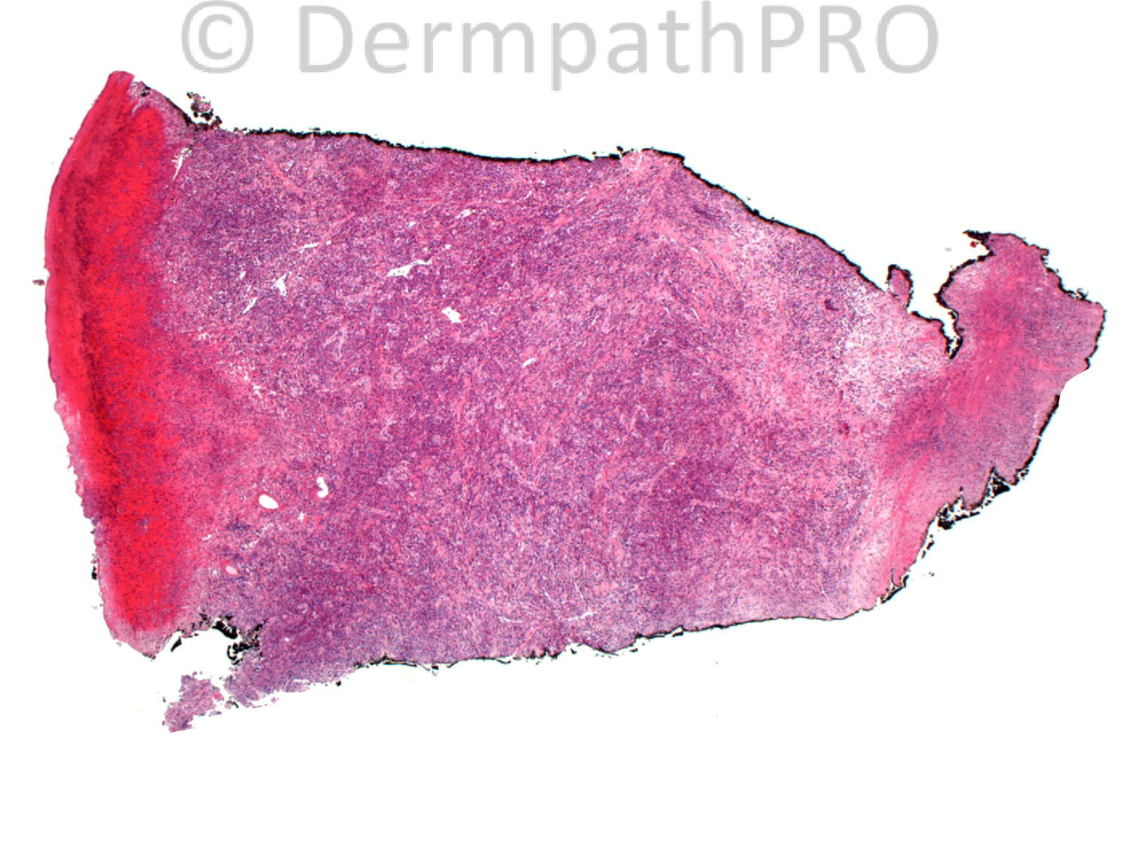
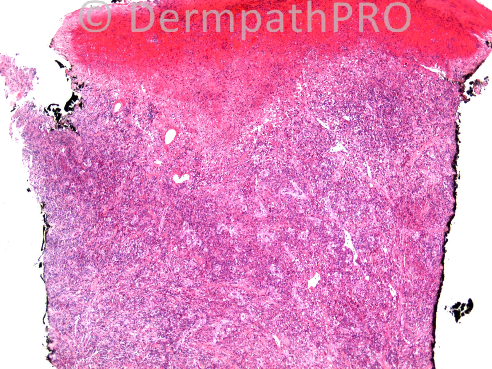
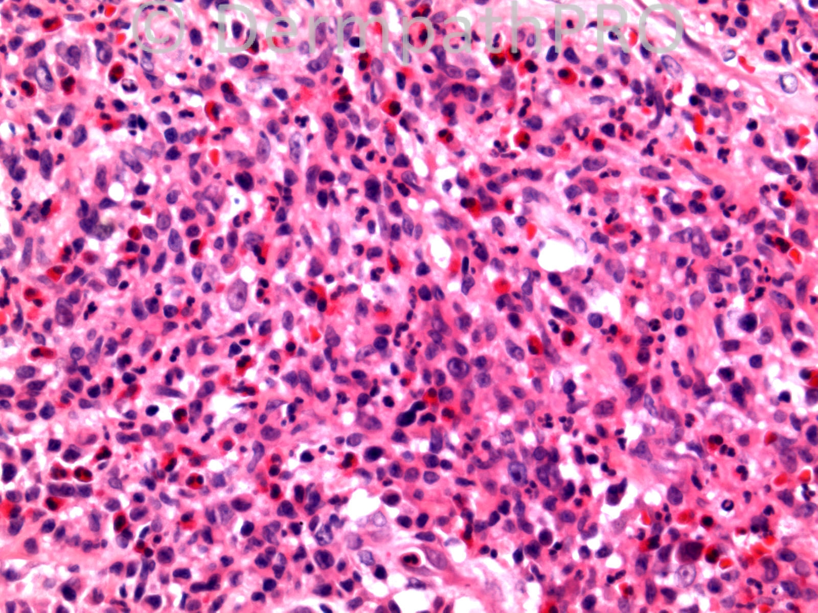
Case Number : CT0022 Dr. Richard Carr

Please read the clinical history and view the images by clicking on them before you proffer your diagnosis.
Submitted Date :
   (0 reviews)

M66. Dorsum 1st MCP joint. 7/52 indurated purple - red large nodule / plaque. ?atypical mycobacterium ?Orf


  Report Record

User Feedback


Admin_Dermpath

Posted

Eman El-Nabarawy  - After exclusion of infective etiology by Gram and PAS stains, I suggest solitary erythema elevatum diutinum.
 
Guest - I'm under the impression that EED is a bilateral, symmetrical process. I breifly thought about acral myxoinflammatory fibroblastic sarcoma, but most of the features of this are absent. My guess is that this is a suppurative process, caused by an infection of one sort or another. Edited June 13, 2014 by Guest
 
Mark A. Hurt MD - What about a cutaneous manifestation of Hodgkin's disease?
   
Dr. Richard Carr - One half-like so far but needs more comments please. In Edinburgh today enjoying the lectures!!

Dr. Mona Abdel-Halim - I favor infectious process, lots of eosinophils may point to a deep mycosis, ? Sporotrichosis... Further work with stains and culture is needed....

Robledo F. Rocha - Agree with Dr. Hurt. Cutaneous Hodgkin's lymphoma. A reactive background of lymphocytes, eosinophils and neutrophils, and at least in the center of the Image 6 there is a classic Reed-Sternberg cell. Edited June 13, 2014 by Guest
  
Guest Romualdo - I would like to add eosinophil-rich or neutrophil/ eosinophil-rich type of primary cutaneous anaplastic large cell lymphoma. This variant simulates infectious/ inflammatory processes and may have atypical munonucleated and binucleated cells resembling Hodgkin's lymphoma.
   
Dr. Richard Carr - I was hoping to have IHC posted by the dermpathpro team at 4pm (BST), hopefully it will appear soon. Edited June 13, 2014 by Guest
 
Guest Jim Davie MD - I like Romualdo's suggestion that this might be a primary cutaneous T-cell lymphoma. I would add to the differential: primary cutaneous CD4-positive small/medium T-cell lymphoma (which may have a proportion of larger pleomorphic cells). Also, a primary cutaneous [or metastatic] angioimmunoblastic T-cell lymphoma. Both can have a mixed inflammatory component with eosinophils, neutrophils, or plasma cells, resembling infectious process, as seen here. T-cell clonality studies might be helpful if the immunostains disappoint. That said, an infectious process would be a more common diagnosis than these 'zebras', and needs to be excluded by stains and clinical history. The infiltrate shows mixed eosinophils, neutrophils, rare plasma cells, and scattered small and large Reed-Sternberg like pleomorphic cells (some seem vacuolated). There is little mitotic activity. No reactive follicles. There may be some multinucleated giant cells and suspicious clear droplets. The solid extension into the deep dermis is impressive, but the biopsy doesn't give us epidermis to examine.[/font][/size] Edited June 13, 2014 by Jim Davie MD
 
Dr. Mona Abdel-Halim - I like the suggestion of Romualdo, waiting for immunos....
 
Guest - Cytotoxic, CD30-positive T-cell lymphoma. ALCL, as Romualdo suggested.
 
Dr. Mona Abdel-Halim - WAAAAAO,,, Chapeau Romualdo :-)). Anaplastic large TCL, the variant which is rich in esinophils and neutrophils mimicking an inflammatory/infectious process.... Amazing case,,, Thank u Dr Carr,,, this is a wounderful educational case.... Edited June 14, 2014 by Guest
 
Dr. Richard Carr - Well done Romualdo. This is a primary cutaneous, eosinophil-rich anaplastic large cell lymphoma. I shared the case with Werner Kempf who kindly excluded Orf. Clearly the sheet-like growth of large CD30 positive cells highlighted on CD30 is against an inflammatory mimic. Learning point being eosinophils can be a clue to CD30 lymphoproliferative disorders. The lesion responded to local steroids and radiotherapy and there has been no recurrence.
 Quote Edit Options  

Share this comment


Link to comment
share_externally


×
×
  • Create New...