Jump to content

Building Blocks of Dermatopathology

BAD DermpathPRO Learning Hub: Diagnostic Clues

Diagnostic Challenge
Interesting Case
Pitfalls

Case Number : CT0042 Adam_Bates

Please read the clinical history and view the images by clicking on them before you proffer your diagnosis.
Submitted Date :
   (0 reviews)

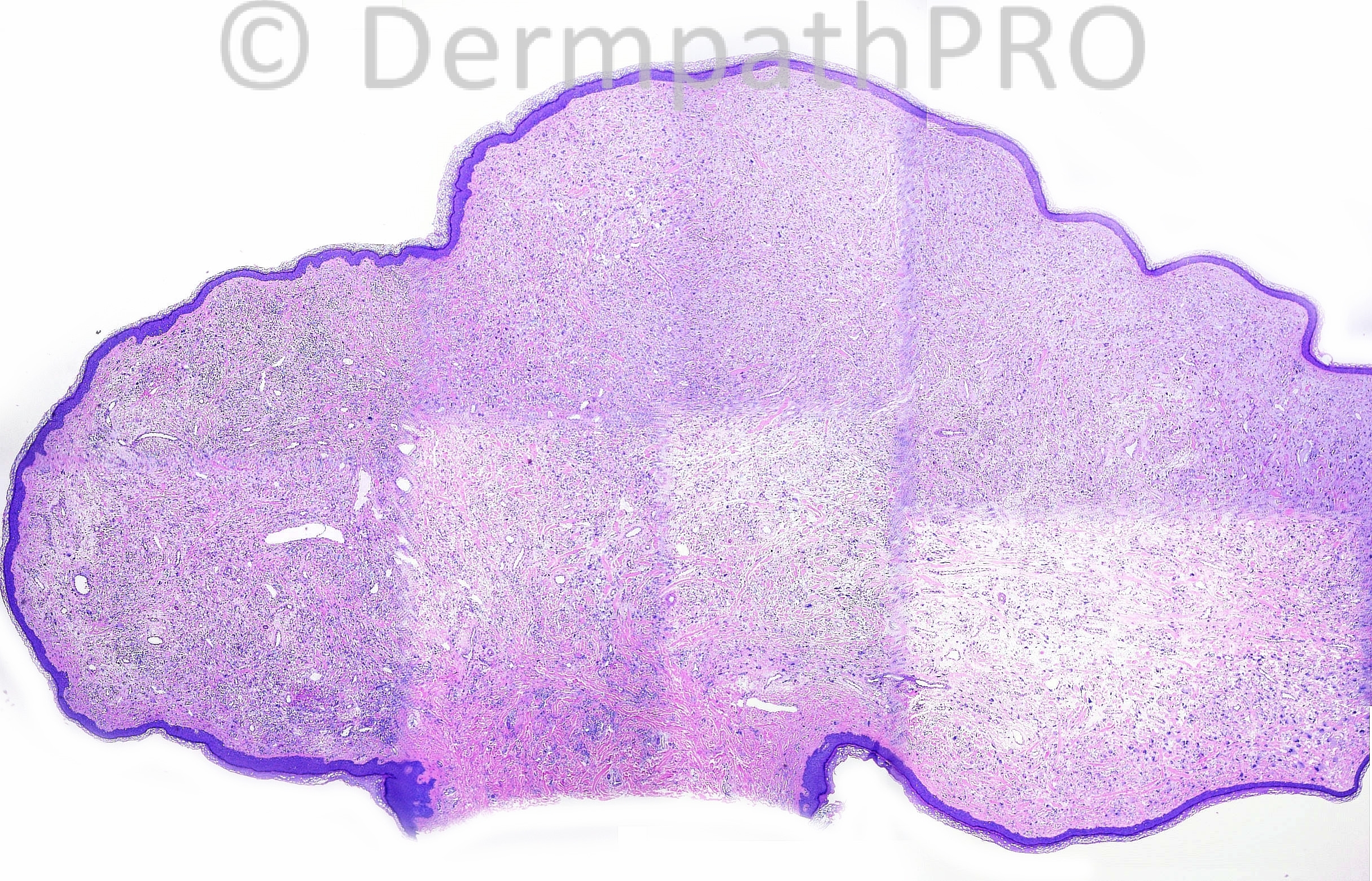
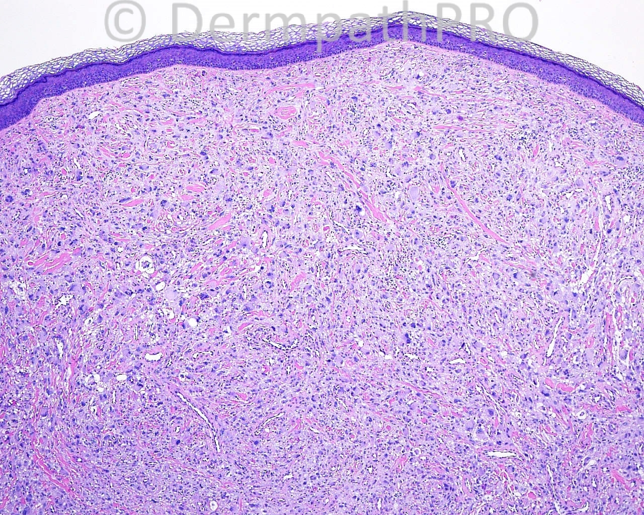
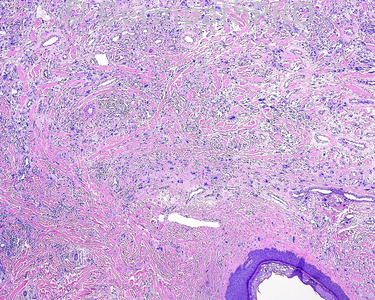
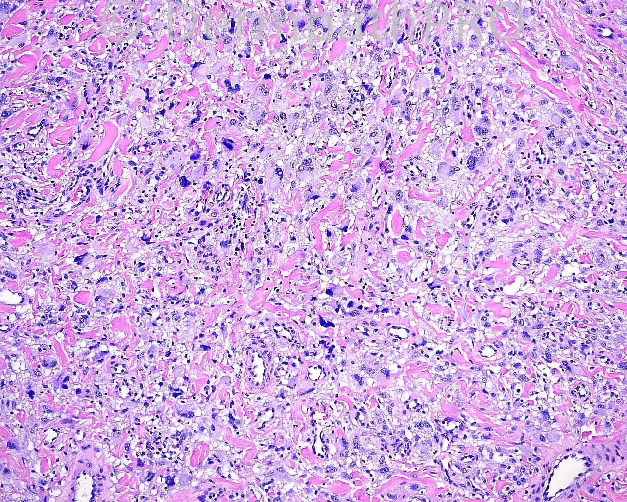
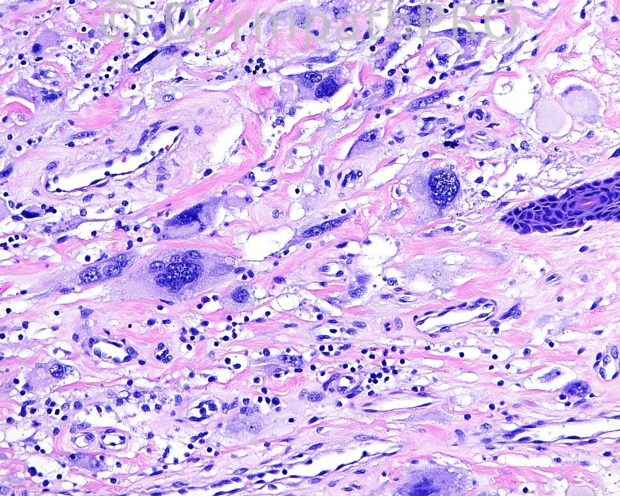
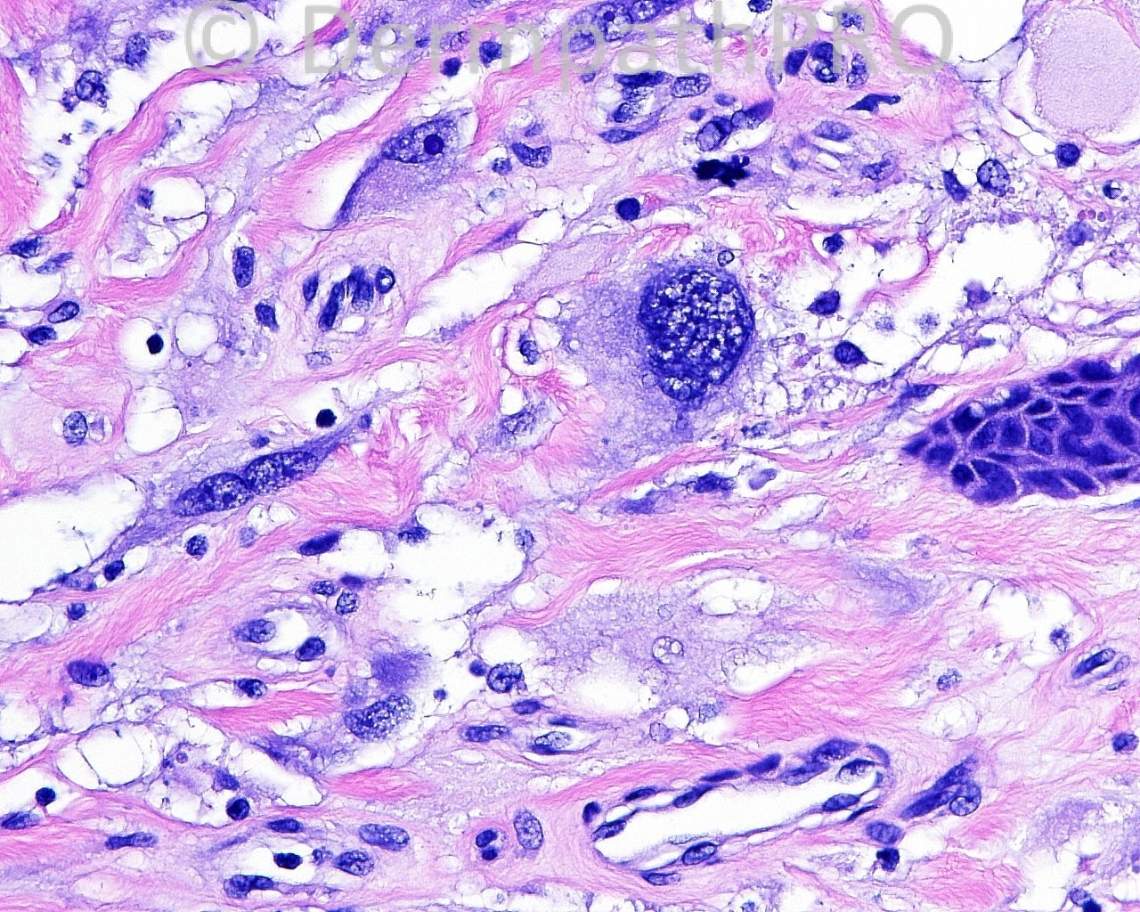
50 year old female with a shave biopsy from right upper thigh.


  Report Record

User Feedback


Adam_Bates

Posted

AFX with osteoclast-like giant cells
 

Share this comment


Link to comment
share_externally

Adam_Bates

Posted

Scanning magnification points to a small exophytic tumor without neoplastic involvement of the stalk. Those findings, in addition to the marked nuclear pleomorphism, make me think of atypical fibroxanthoma. Since this is a diagnosis of exclusion and sun-protected skin of the limb is a rare location, others anaplastic sarcomatoid tumors must be first ruled out, including nodular melanoma, a tumor much more commonly found at this site in a woman in her fifties.
 

Share this comment


Link to comment
share_externally

Adam_Bates

Posted

Pleomorphic sarcoma, NOS
 

Share this comment


Link to comment
share_externally

Adam_Bates

Posted

The lesion is polypoidal, well circumscribed, dermal lesion. I do not think this fits with pleomorphic sarcoma. The lesion not in a sun exposed site, I think this R/O AFX. I thought of malignant giant cell tumor of soft tissue. It is usually subcutaneous but I read that dermal and polypoidal lesions have been reported.
 

Share this comment


Link to comment
share_externally

Adam_Bates

Posted

Atypical fibrous histiocytoma of the skin. IHQ is necessary. Clear margins and follow-up are in order, since there is a small risk of recurrence and metastasis.
 

Share this comment


Link to comment
share_externally

Adam_Bates

Posted

I would add to the differential: Dermatofibroma, Monster-cell variant (DF with Monster Cells).

I agree that this is a fibrohistiocytic neoplasm with very unpleasant nuclei and foamy cytoplasm, resembling AFX. There is overall pattern of multinucleate giant cells, some with osteoclast-like or Touton-type foamy cytoplasmic features, and thickened keloidal collagen bundles. No tumor necrosis, ulceration, or solid growth pattern. Location on non-actinic site, overall symmetry, and low mitotic activity would support DFMC, if the rare mitoses in the above photos are representative of the true mitotic density of the neoplasm.

Immunostains would be worthwhile to help rule out Spitzoid or rare cutaneous leiomyosarcomas that show convincing AFX-like cytologic features. Edited September 30, 2013 by Jim Davie MD

Share this comment


Link to comment
share_externally

Adam_Bates

Posted

AFX. If this were a melanoma, I would expect S-100 to be positive. I reported an AFX with positive HMB45 about 8 years ago (the patient is alive and well), which was S-100 negative. Atypical fibrous histiocytoma/dermatofibroma with monster cells would be in my differential.
 

Share this comment


Link to comment
share_externally

Adam_Bates

Posted

Favour Dermatofibroma with monster cells.
 

Share this comment


Link to comment
share_externally

Adam_Bates

Posted

Nice case :-)
 

Share this comment


Link to comment
share_externally


×
×
  • Create New...