Jump to content

Building Blocks of Dermatopathology

BAD DermpathPRO Learning Hub: Diagnostic Clues

Diagnostic Challenge
Interesting Case
Pitfalls

Case Number : CT0054 Adam_Bates

Please read the clinical history and view the images by clicking on them before you proffer your diagnosis.
Submitted Date :
   (0 reviews)

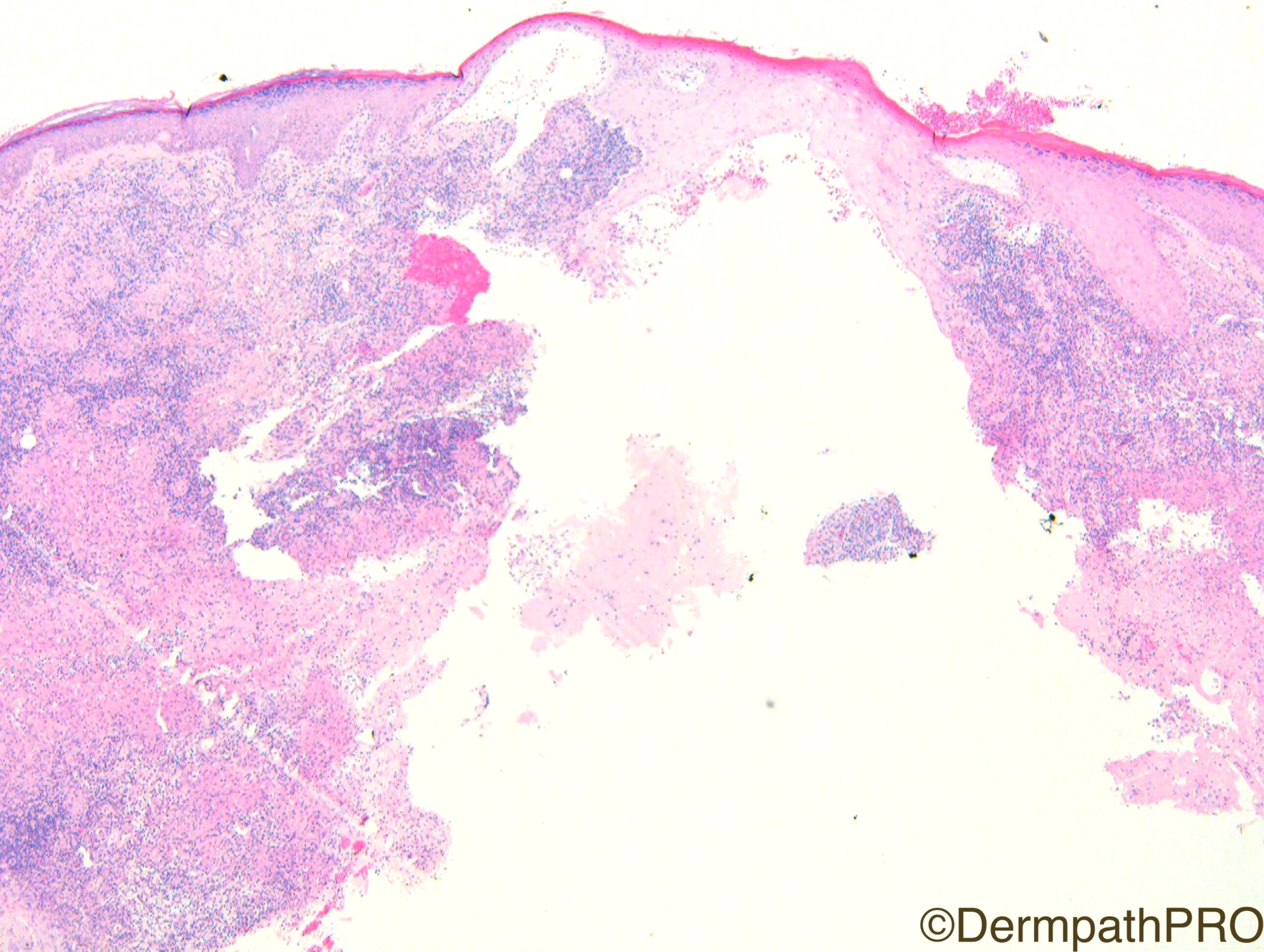
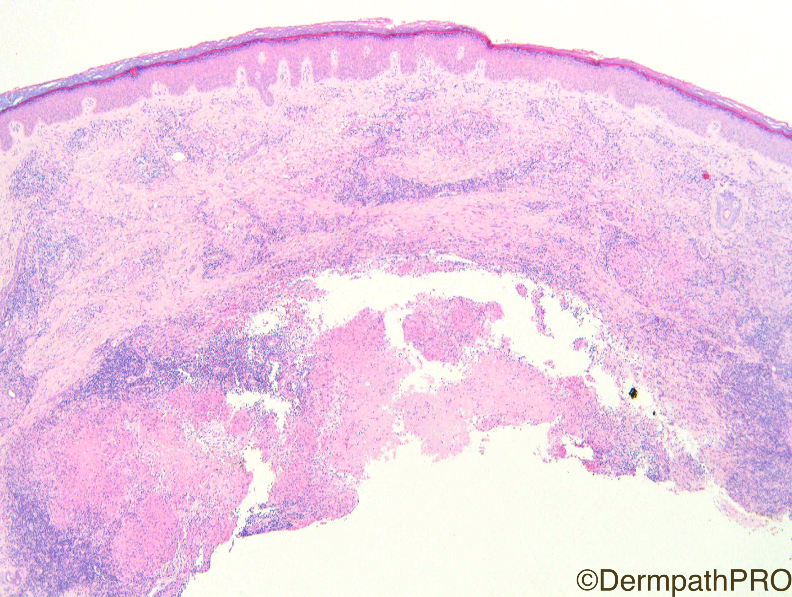
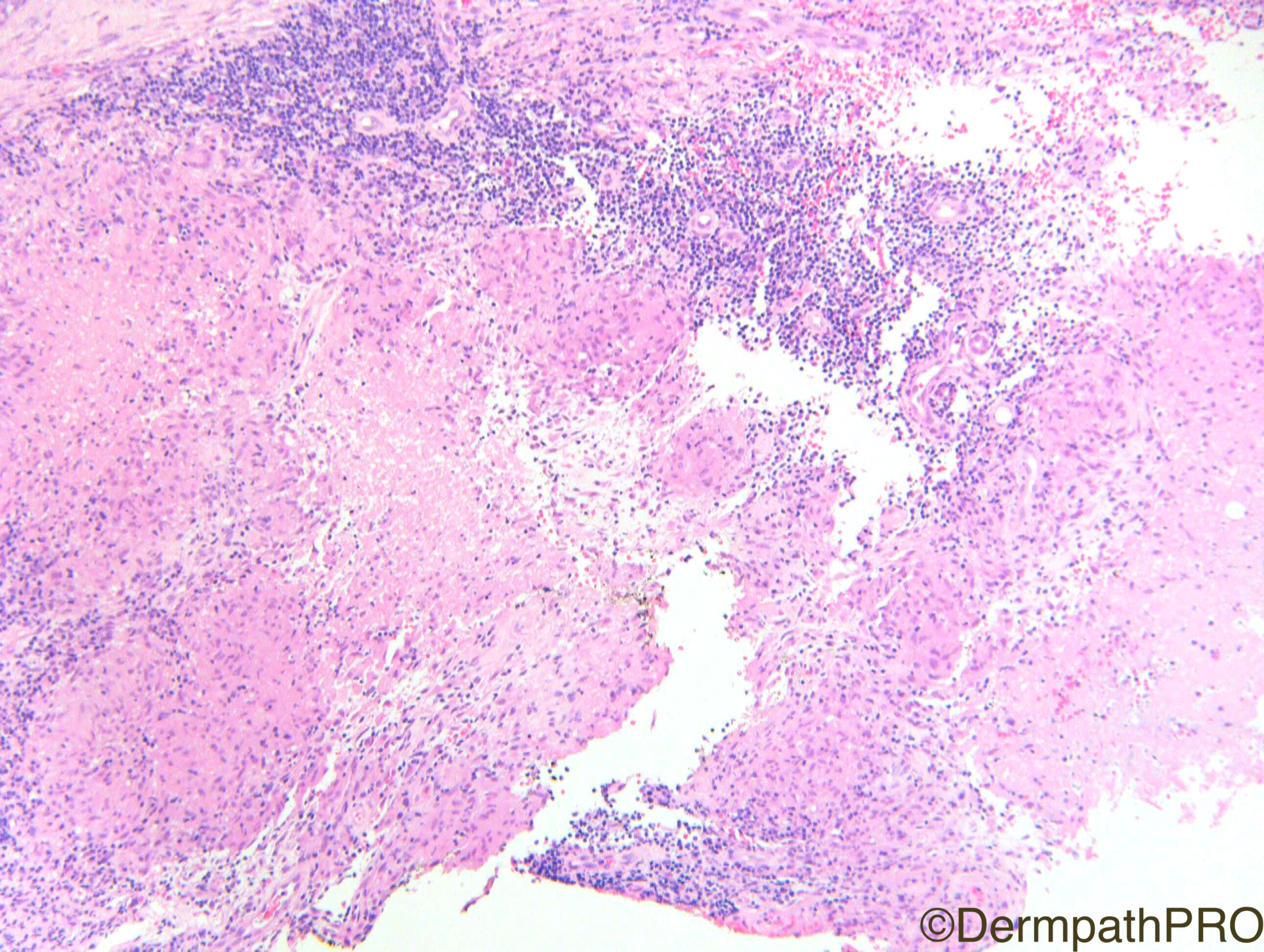
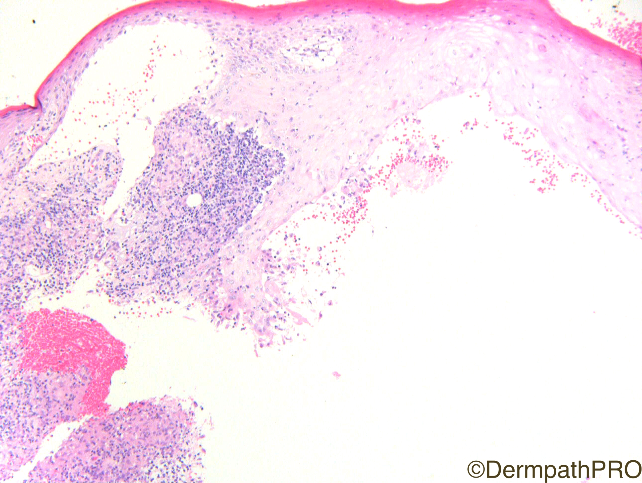
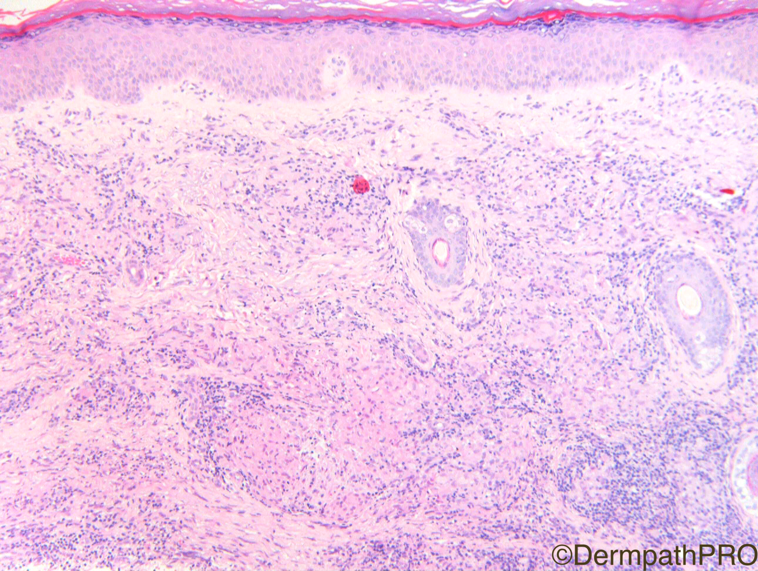
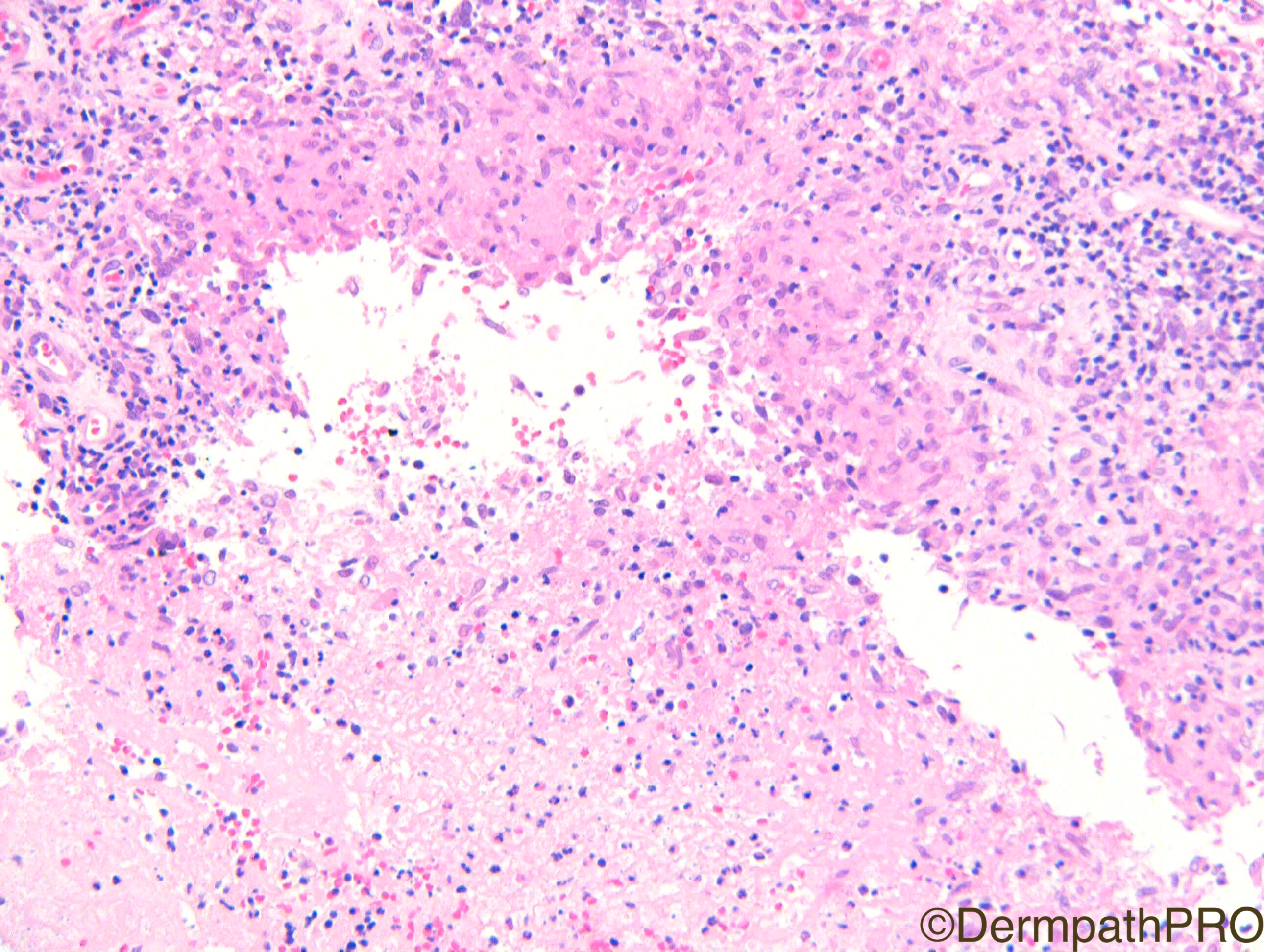
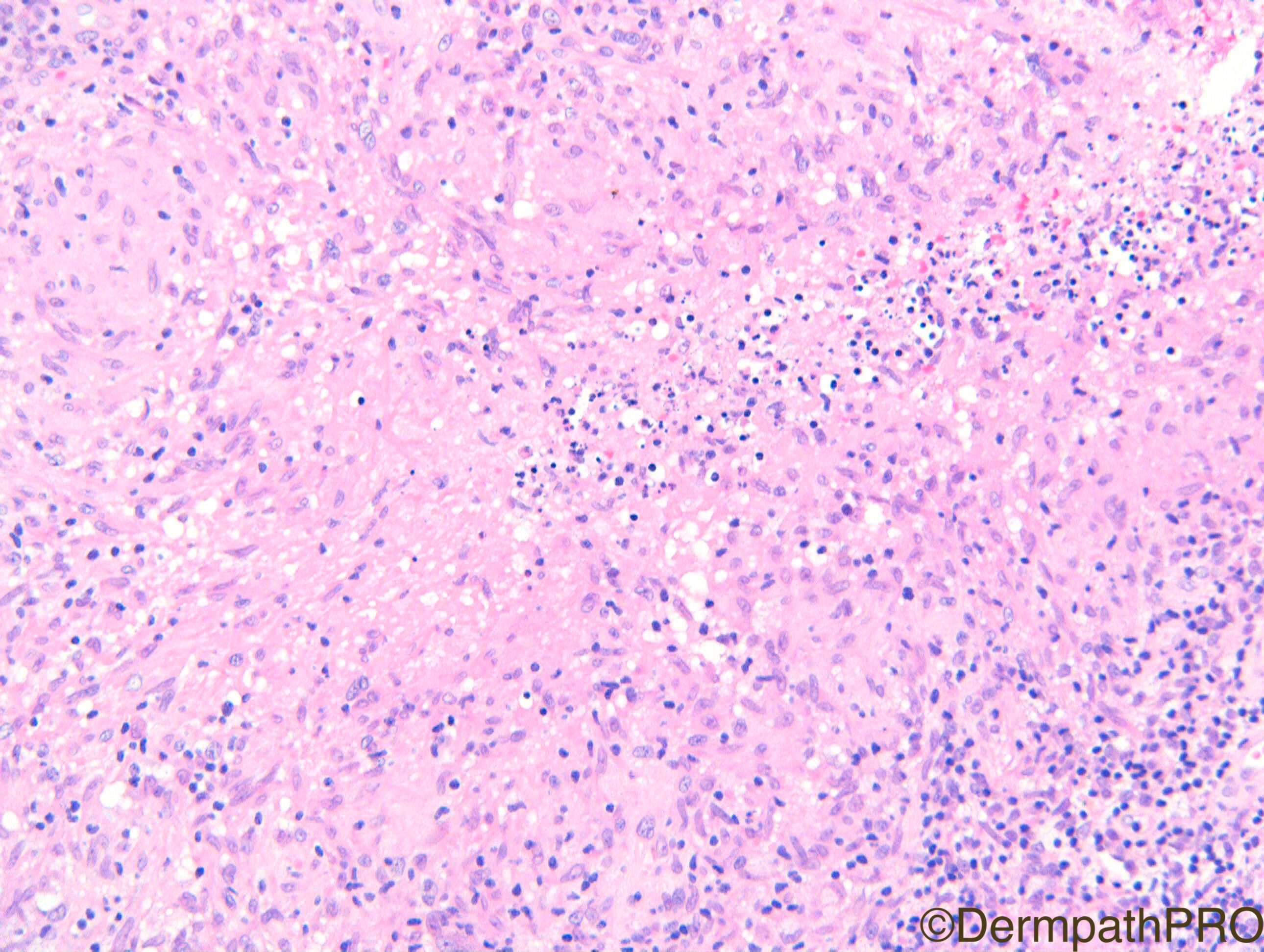
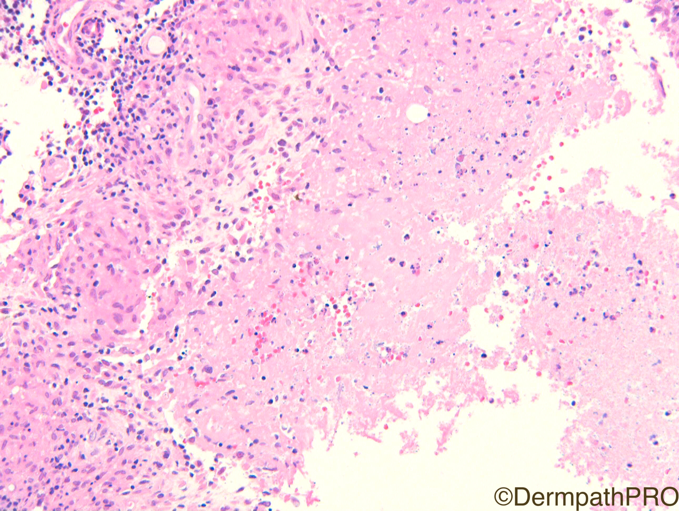
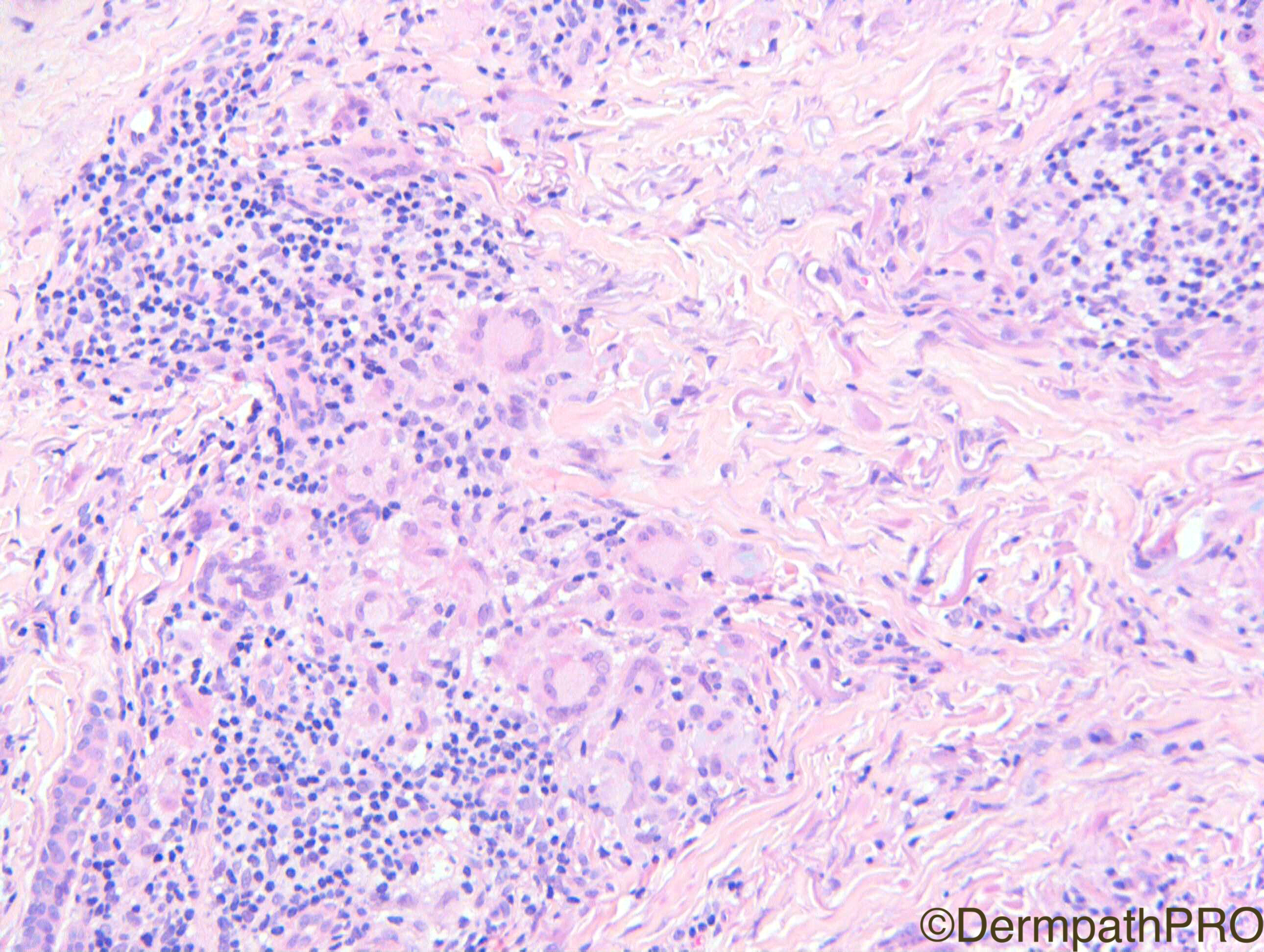
F50. Several crusted nodules along the radial border of the thumb and radial border of the left wrist extending proximally.


  Report Record

User Feedback


Adam_Bates

Posted

This is a very interesting case. There are several granulomas types including suppurative, tuberculoid and histiocytic with necrosis and giant cells. My bet is for Sporotrichosis. There is some blue-gray material...Granulomatous foreing body reaction and Rheumatoid Nodule are my differential diagnosis.

Share this comment


Link to comment
share_externally

Adam_Bates

Posted

Atypical mycobacteria vs. Sporotrichosis for stains and culture. 

Share this comment


Link to comment
share_externally

Adam_Bates

Posted

Sporotrichosis is also my first impression. It is very difficult to see the fungi. The suppurative granulomas and the clinical description resembling a lymphangitic spread of the disease favor this diagnosis.

Share this comment


Link to comment
share_externally

Adam_Bates

Posted

Given the clinical and H&E favor infectious, especially sporotichosis. Would do PAS and AFB and recommend cultures, even if negative.

Share this comment


Link to comment
share_externally

Adam_Bates

Posted

Suppurative granulomatous inflammation. Agree with Sporotrichosis ( clinical picture fits). Would also consider an atypical mycobacterial infection/ ' Fish tank' granuloma.

Share this comment


Link to comment
share_externally

Adam_Bates

Posted

The clinical description of lesional location reminds me of some wound patch, so agree with Sporotrichosis, but also with Fish Tank Granuloma.  I don't know the exactly answer....

Share this comment


Link to comment
share_externally

Adam_Bates

Posted

Well done all on the correct differential.  This woman worked in a pet shop washing out fish tanks without protective equipment and this was clinically suspected to be Fish Tank granuloma. I reported it as suppurative granulomas in keeping with mycobacterium marinum.  I did not actually do Zn but I think I read in Weedon years ago that it is positive in up to 50% of cases.  I have not seen a case of sporotrichosis in the UK and will be very grateful to receive a case to photograph for Dermpathpro spot diagnosis.  I understand that it is one of the few conditions in which serial sections may be indicated to help find the organisms in the centre of the granulomas. It is one of my bug bears (pun intended) hearing people talk about serial sections when they mean step sections.  Serial sections produce a lot of slides! The blue grey material is elastophagocytosis - rather dramatic in this case but basically a secondary process - I'd hope it would lead to some confusion but you guys are too good!!

Share this comment


Link to comment
share_externally


×
×
  • Create New...