Jump to content

Building Blocks of Dermatopathology

BAD DermpathPRO Learning Hub: Diagnostic Clues

Diagnostic Challenge
Interesting Case
Pitfalls

Case Number : CT0073 Adam_Bates

Please read the clinical history and view the images by clicking on them before you proffer your diagnosis.
Submitted Date :
   (0 reviews)

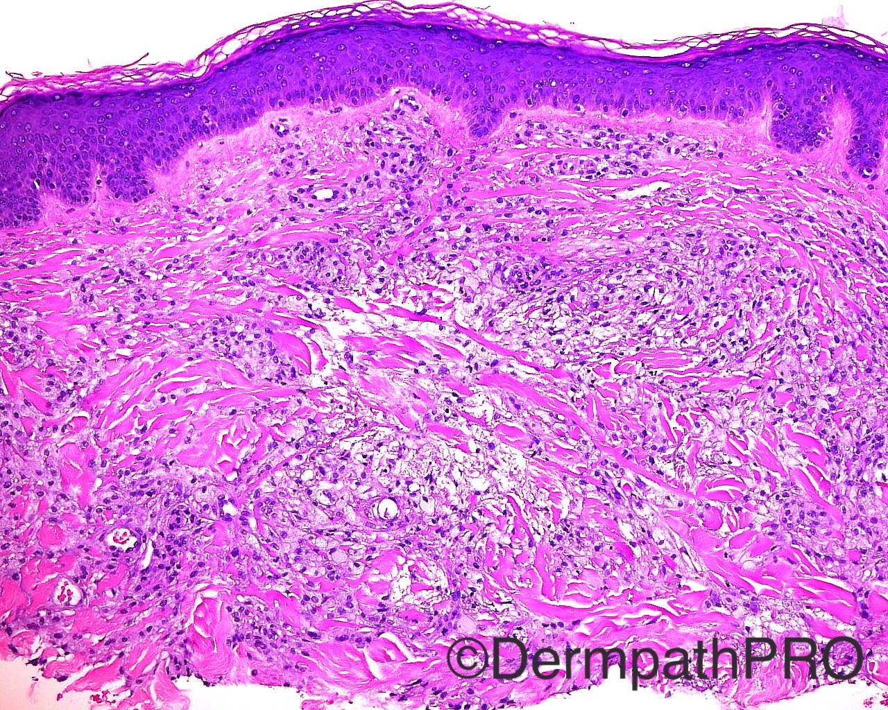
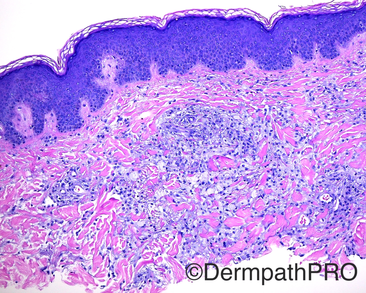
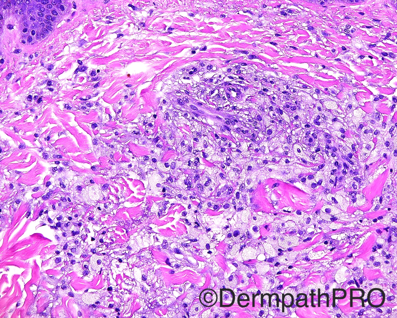
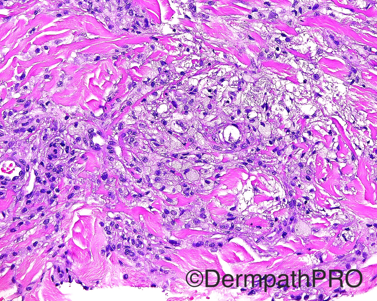
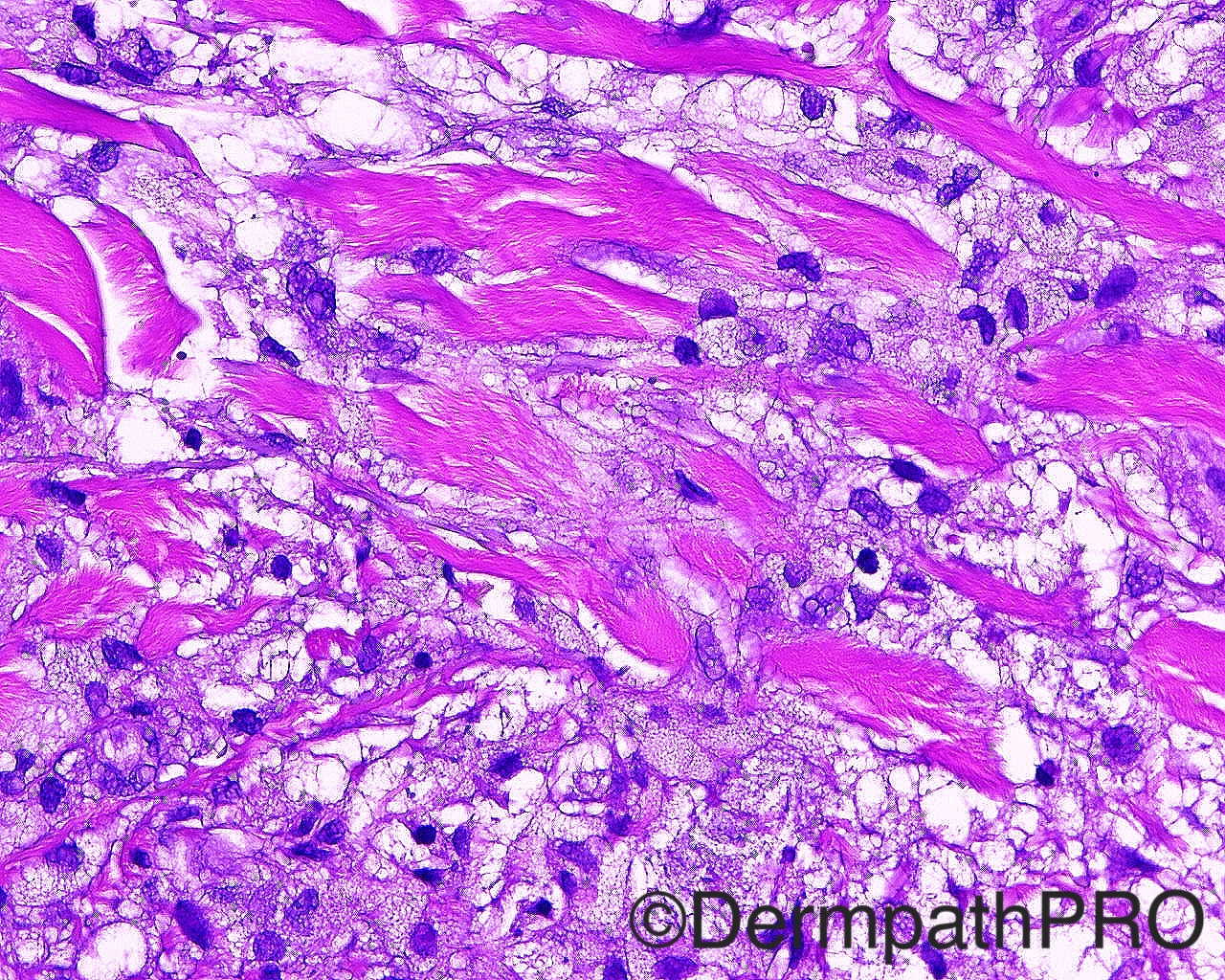
Case History: The patient is a 41-year-old man with an eruption of small yellow papules over the trunk, extremities and penis. A shave biopsy is taken from the right upper back.


  Report Record

User Feedback


Adam_Bates

Posted

Papular xanthoma
 

Share this comment


Link to comment
share_externally

Adam_Bates

Posted

I will call it eruptive xanthoma.
 

Share this comment


Link to comment
share_externally

Adam_Bates

Posted

Agree, eruptive xanthoma.
Image 3 has a slight resemblance to granuloma annulare, which can occur with these lesions, the foamy nature of the histiocytes and lack of true necobiosis, being the differentiating point.
Also, important to correlate with clinical findings as eruptive xanthoma is usually associated with hyperlipedemia. If normolipaemic, then xanthoma disseminatum would also come in the d/d.

Share this comment


Link to comment
share_externally

Adam_Bates

Posted

Agree with Arti´s comments
 

Share this comment


Link to comment
share_externally

Adam_Bates

Posted

Dear Friends, just so I can learn, what histological difference would you expect between papular and eruptive xanthoma?. I thought in papular xanthoma we see only foamy histyocytes whether in eruptive xanthoma there is usually lymphocytes and/or leukocytes intermingled with the histiocytes. By the way, great webpage and community!
 

Share this comment


Link to comment
share_externally


×
×
  • Create New...