Jump to content

Building Blocks of Dermatopathology

BAD DermpathPRO Learning Hub: Diagnostic Clues

Diagnostic Challenge
Interesting Case
Pitfalls

Case Number : CT0087 Adam_Bates

Please read the clinical history and view the images by clicking on them before you proffer your diagnosis.
Submitted Date :
   (0 reviews)

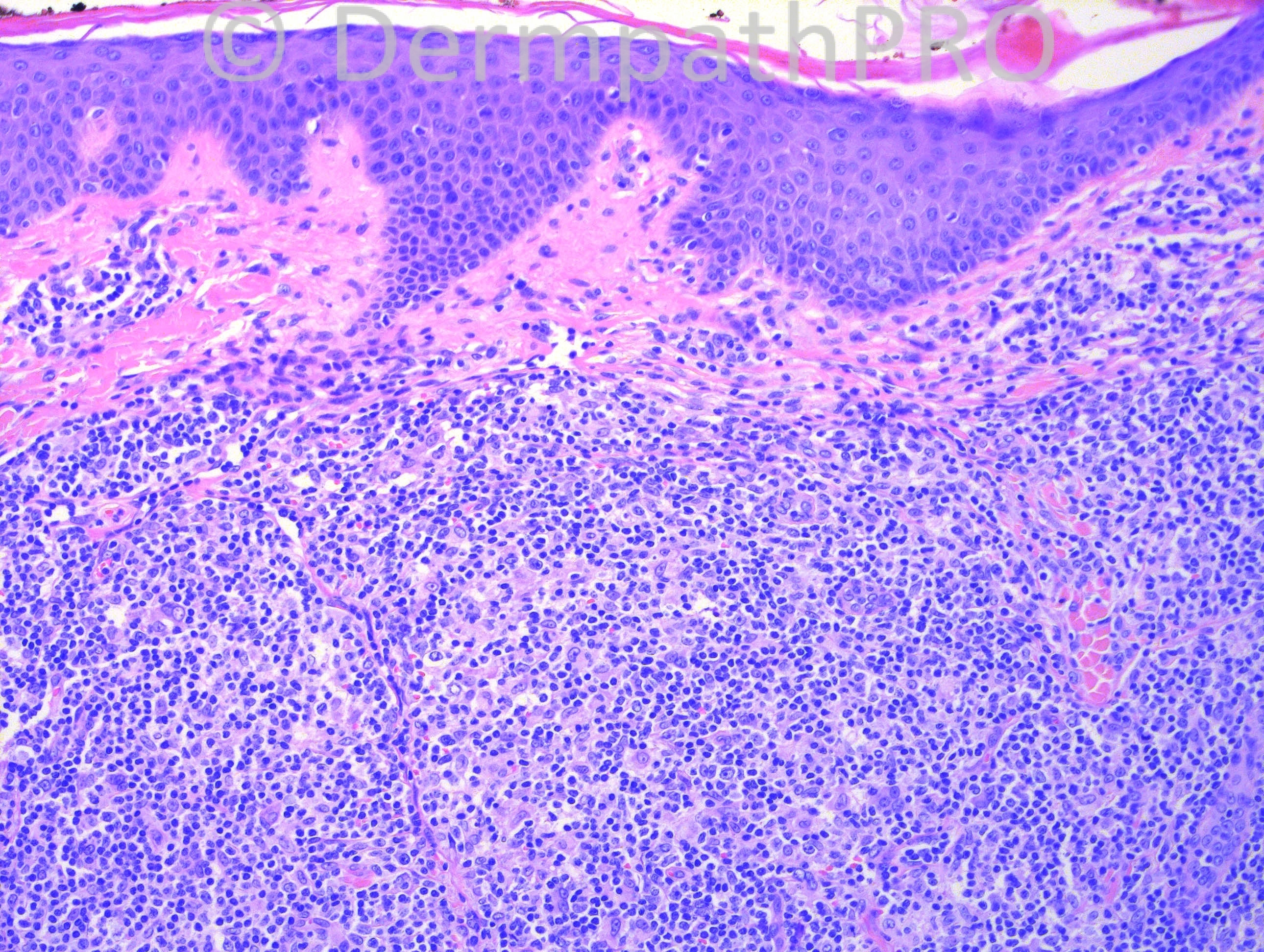
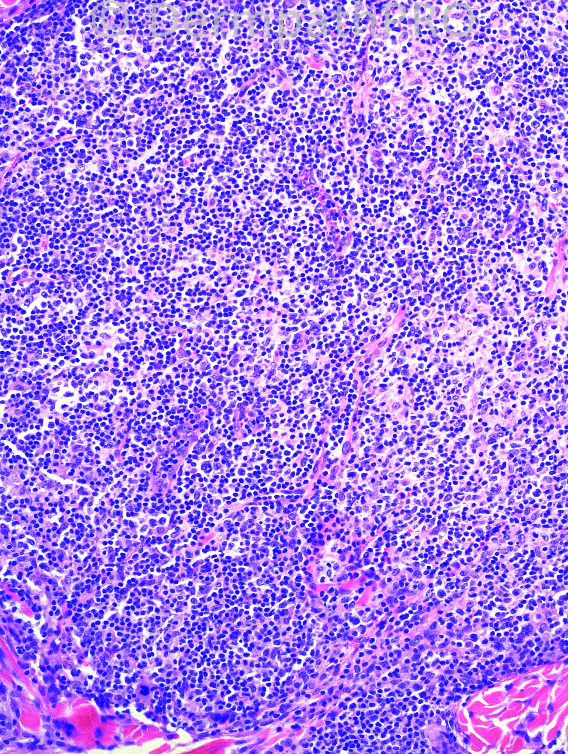
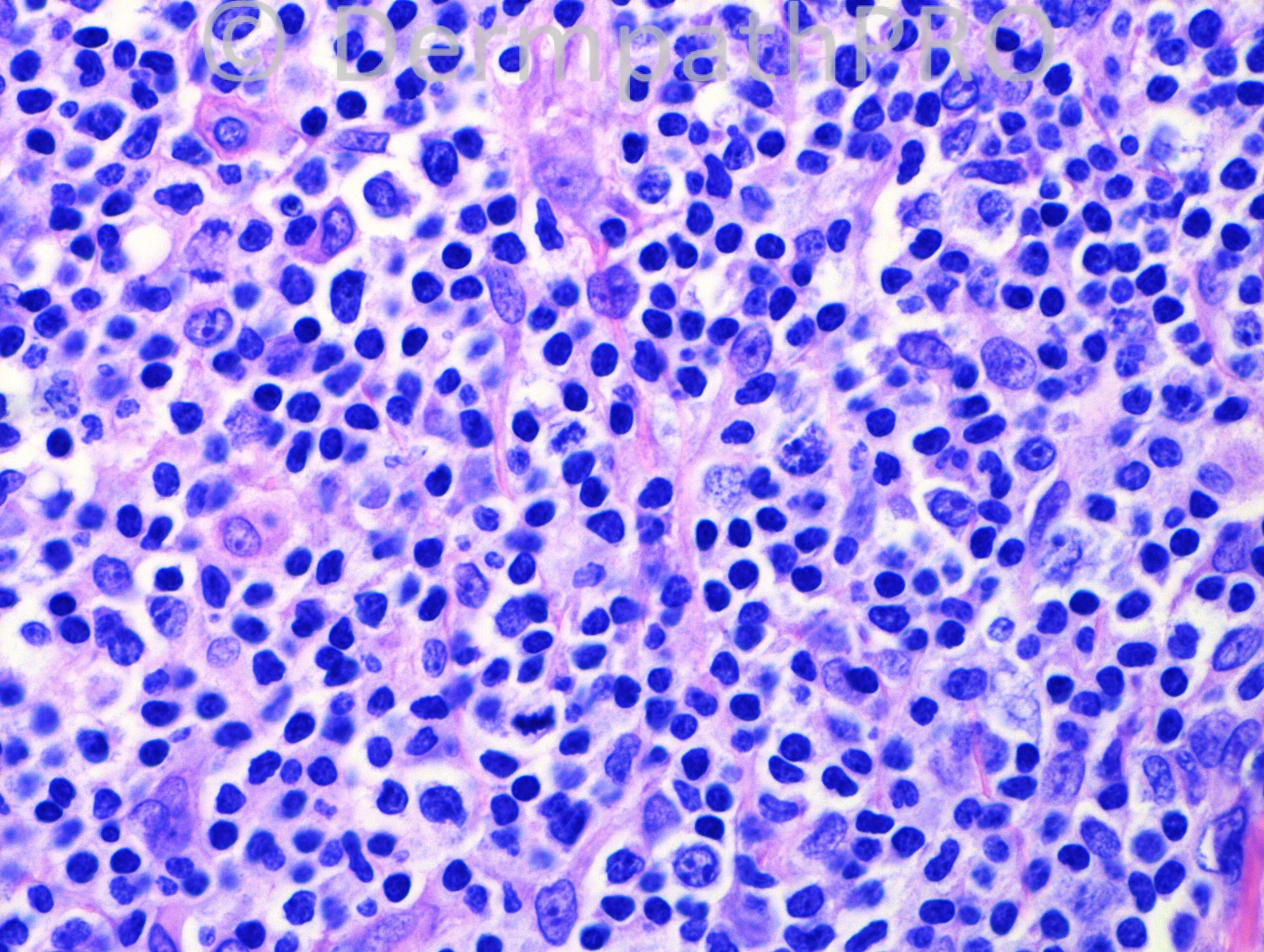
46-year-old male with a lesion on his right upper back.


  Report Record

User Feedback


Adam_Bates

Posted

Crosti lymphoma.
 

Share this comment


Link to comment
share_externally

Adam_Bates

Posted

Yes, primary cutaneous follicle-center lymphoma.
 

Share this comment


Link to comment
share_externally

Adam_Bates

Posted

Primary cutaneous follicle-center lymphoma x PCMZL x Cutaneous lymphoid hyperplasia (less probably). I think it is a primary cutaneous follicle center lymphoma and the reasons are the deep dermal involvement, extensive nodular architecture and upper back localization (Crosti lymphoma). I can not forget systemic follicular lymphoma with secondary cutaneous involvement. I had a case like this a couple of months ago. Waiting for Immunohistochemistry study.
 

Share this comment


Link to comment
share_externally

Adam_Bates

Posted

Crosti lymphoma... PCFCL
 

Share this comment


Link to comment
share_externally

Adam_Bates

Posted

Unless immunohistochemistry study prove differently, it's a malignant lymphoid proliferation with nodular growth pattern since the fullthickness of the dermis is involved, mantle zone is nothing but a discontinuous narrow strip, and cell population do not include tingible body macrophages.

I did not find, surrouding the nodules, a pale area composed of cleaved cells, plasma cells, and lymphoplasmacytoid cells, a feature that, if present, would made me favor marginal zone B-cell lymphoma.

So, I will agree with the diagnosis of follicle center cell lymphoma, variant so-called Crosti's reticulohistiocytoma of the dorsum.

Share this comment


Link to comment
share_externally

Adam_Bates

Posted

Agree with the above excellent differentials.
I favor PCMZL (primary cutaneous marginal zone lymphoma). Differential would include a diffuse-pattern high-grade FCL.

I believe there are rare scattered plasma cells (bottom left photo), and mixed monocytoid and larger centroblastic-type cells with frequent, focally aberrant mitotic figures, and absence of follicular architecture. There are nodular and focally infiltrative aggregates, that preferentially encase and infiltrate eccrine coils and follicular adnexa.

Share this comment


Link to comment
share_externally

Adam_Bates

Posted

The differential diagnosis is excellent. This turned out to be cutaneous lymphoid hyperplasia. The plasma cells were polyclonal, there were abundant CD3 positive cells with scattered clusters of CD20 positive and Bcl-6 positive cells.
 

Share this comment


Link to comment
share_externally

Adam_Bates

Posted

Hafeez,
Fascinating case (I was favouring follicular lymphoma). This reminds me of a recent case I saw on biopsy (sizeable scalp nodules) strongly favouring follicular lymphoma (large expanded germinal centres on CD21) but on excision (with flow cytometry) favouring cutaneous follicular lymphoid hyperplasia (based on relatively high proliferation on Ki67 in the follicles). Plasma cells were mixed and flow cytometry favoured reactive. However it is well to remember this distinction can at times be impossible (possibly largely academic based on prognosis) but some cases may go on in time to more obvious lymphoma. What was the CD21 and CD10 in your case?
Regards

Share this comment


Link to comment
share_externally

Adam_Bates

Posted

Out of curiosity, what was the kappa/lambda ratio for the PCs?
 

Share this comment


Link to comment
share_externally

Adam_Bates

Posted

Yes, I was worried about this case as well. CD10 was negative. I didn't do a CD21. The kappa to lambda ratio was about 1:1. Since I was concerned about this case, I showed to our hemepath folks who went with CLH as well.
 

Share this comment


Link to comment
share_externally


×
×
  • Create New...