Jump to content

Building Blocks of Dermatopathology

BAD DermpathPRO Learning Hub: Diagnostic Clues

Diagnostic Challenge
Interesting Case
Pitfalls

Case Number : CT0114 Adam_Bates

Please read the clinical history and view the images by clicking on them before you proffer your diagnosis.
Submitted Date :
   (0 reviews)

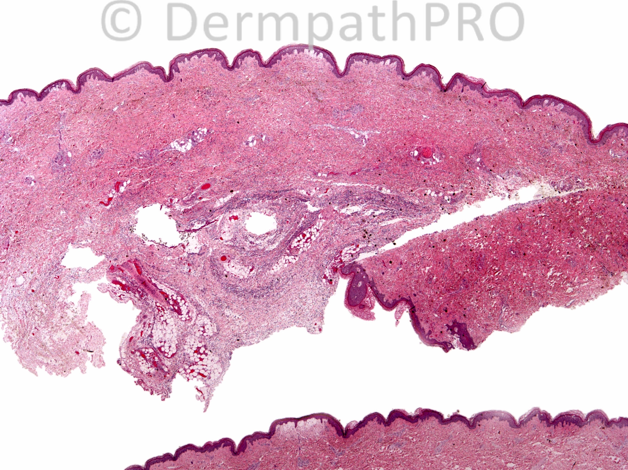
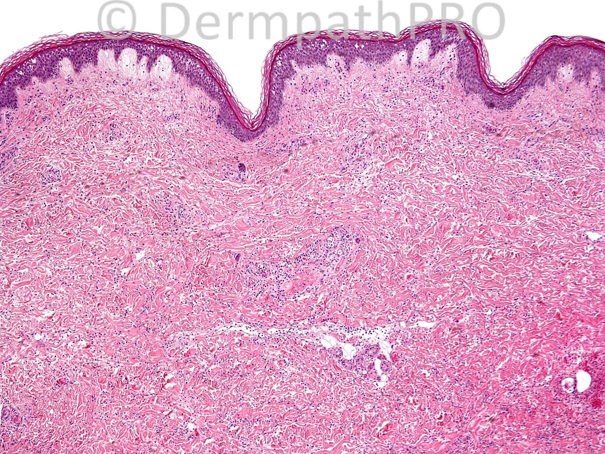
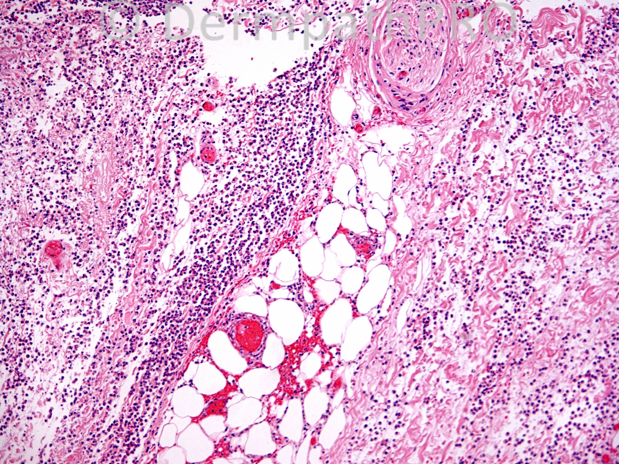
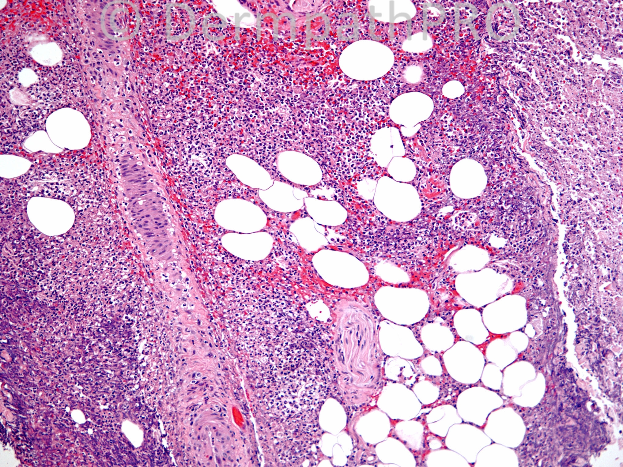
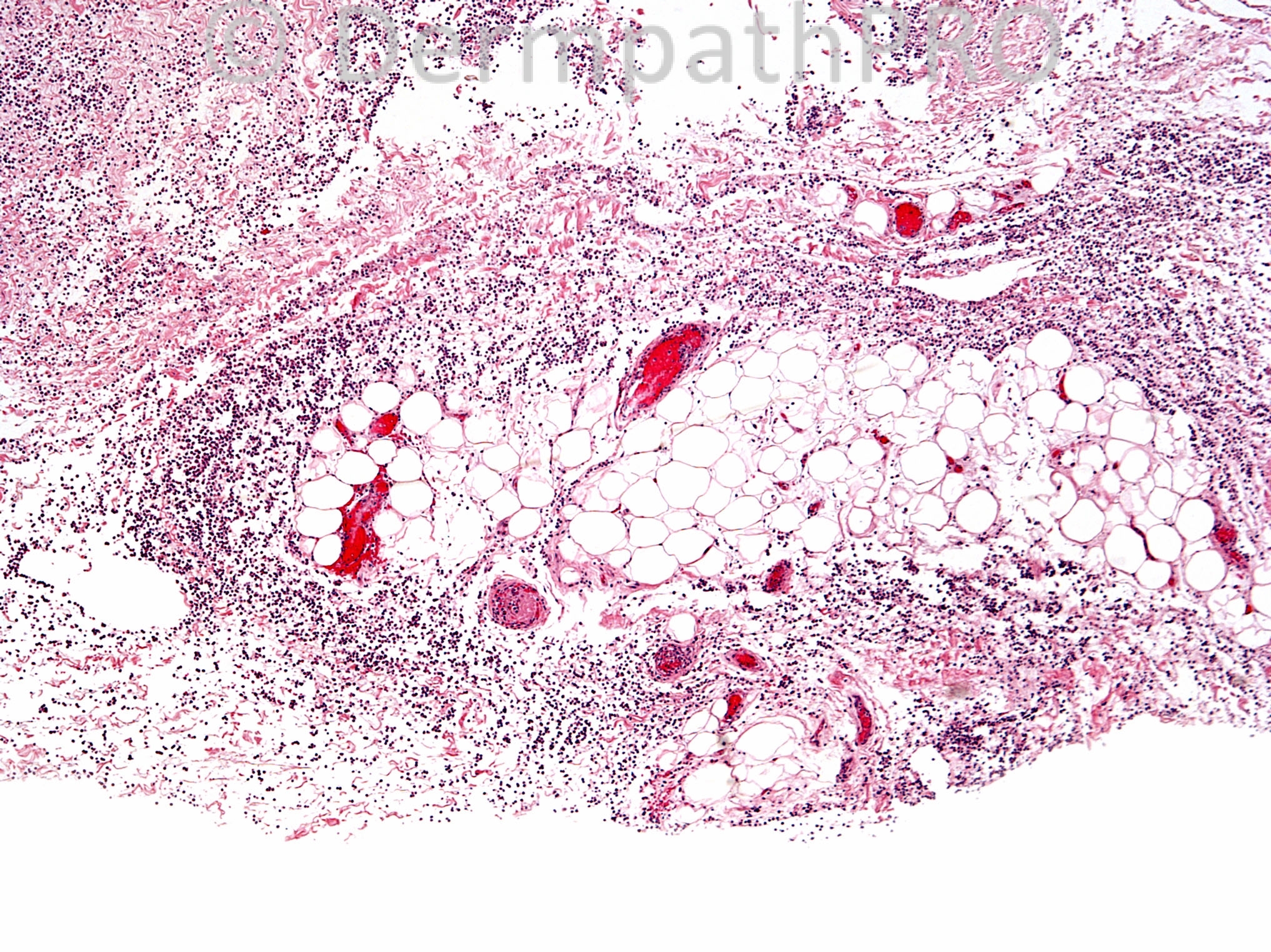
30 year old female with right ankle, foot and heel ulcer, s/p recent traumatic injury. This is an excisional biopsy of the right ankle lesion.


  Report Record

User Feedback


Adam_Bates

Posted

Deep neutrophilic infiltrate associated with thrombosis. This could be echthyma gangrenosum. Work up with stains and culture is needed.
 

Share this comment


Link to comment
share_externally

Adam_Bates

Posted

Neutrophilic panniculitis, infective panniculitis being likeliest, but I'll put A1AD panniculitis out here

Share this comment


Link to comment
share_externally

Adam_Bates

Posted

Ecthyma gangrenosum is a nice idea. Gram and culture!.
 

Share this comment


Link to comment
share_externally

Adam_Bates

Posted

Hi everyone, I wanted to say this is such a great forum and I'm glad to meet all of you. Sorry for the mix up yesterday but you all arrived at the correct answer just fine!
 

Share this comment


Link to comment
share_externally

Adam_Bates

Posted

The diffuse neutrophilic infiltrate throughout the dermis accompanied by subcutaneous fat suppuration and thrombi are features that make me think of infectious cellulitis, presumably caused by bacterial organisms traumatically inoculated. Special stains to highlight infective agents are required, but those stains are likely to fail in demonstrating pathogenic microorganisms.
 

Share this comment


Link to comment
share_externally

Adam_Bates

Posted

Diagnosis: Necrotizing fasciitis. This is a dermatological emergency which may call for a frozen section diagnosis from the dermatopathologist. It is a rare disorder that is related to cellulitis but rapidly involves fascial planes. Necrosis of the skin and underlying soft tissues, such as fat and muscle, is present. It commences as a poorly defined area of erythema and usually involves the lower extremity or perineum. It is often preceded by a penetrating injury, such as in this case. Constitutional symptoms may be present and there is significant associated morbidity and mortality. Usually the culprit is group A streptococcus, but it is often polymicrobial (as was the case here). Clinically, the patient is treated with surgical debridement and broad spectrum antibiotics so the stakes are high. On histopathology, necrosis is seen involving the skin at all levels and extending down to the fascial plane and muscle. Numerous neutrophils extend throughout the biopsy and surround viable tissue. A key feature is the presence of necrosis and neutrophils down in the muscle, hence the need for a deep incisional biopsy. Vascular changes are seen, as in this case, but are thought to be secondary to inflammation. Cellulitis is a related entity, in which Staphylococcus aureus] is often implicated. However, cellulitis does not extend as deeply as necrotizing fasciitis and it is important to realize that they are part of a spectrum. In this case the patient was an otherwise healthy athlete with a penetrating lower extremity injury, a clinical set up for cellulitis/necrotizing fasciitis. The excisional biopsies had positive cultures, which argues against a neutrophilic panniculitis secondary to alpha 1 antitrypsin deficiency. Gram stains identified Gram positive cocci within areas of acute inflammation.
 

Share this comment


Link to comment
share_externally


×
×
  • Create New...