Spot Diagnosis
Adam_Bates 47 Posted March 24, 2024 A tidy case to wrap up the week by Dr Richard A Carr. Share this comment Link to comment share_externally More sharing options...
Adam_Bates 47 Posted March 24, 2024 Tubular apocrine adenoma Share this comment Link to comment share_externally More sharing options...
Adam_Bates 47 Posted March 24, 2024 Looks like something with oncocytic differentiation, similar to, for example, a lymphocytic stroma poor Warthin tumor or a oncocytoma. May be I'm wrong...we don't know the topography of the lesion, but...I was thinking of an ocular adnexal oncocytoma. Share this comment Link to comment share_externally More sharing options...
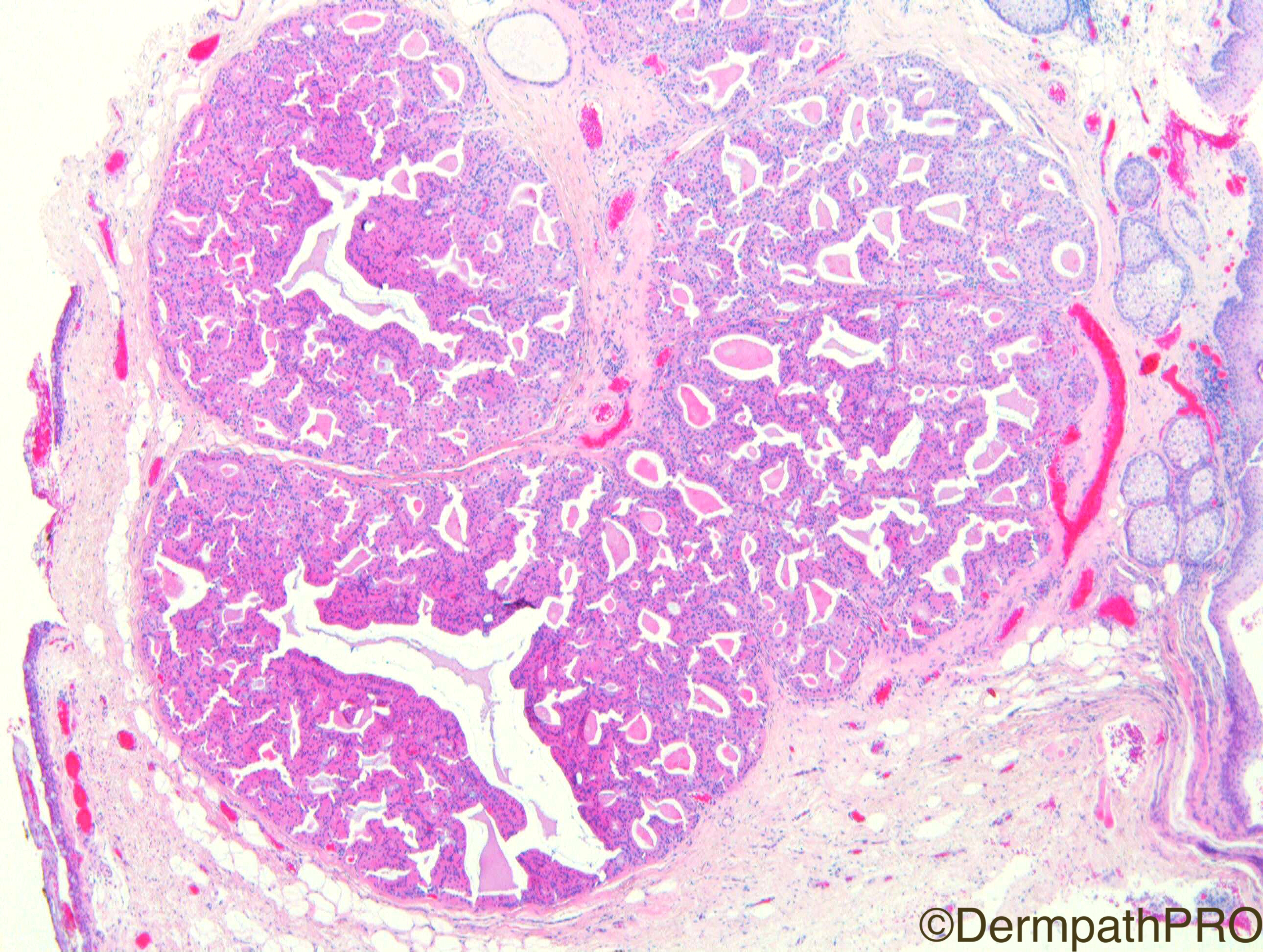
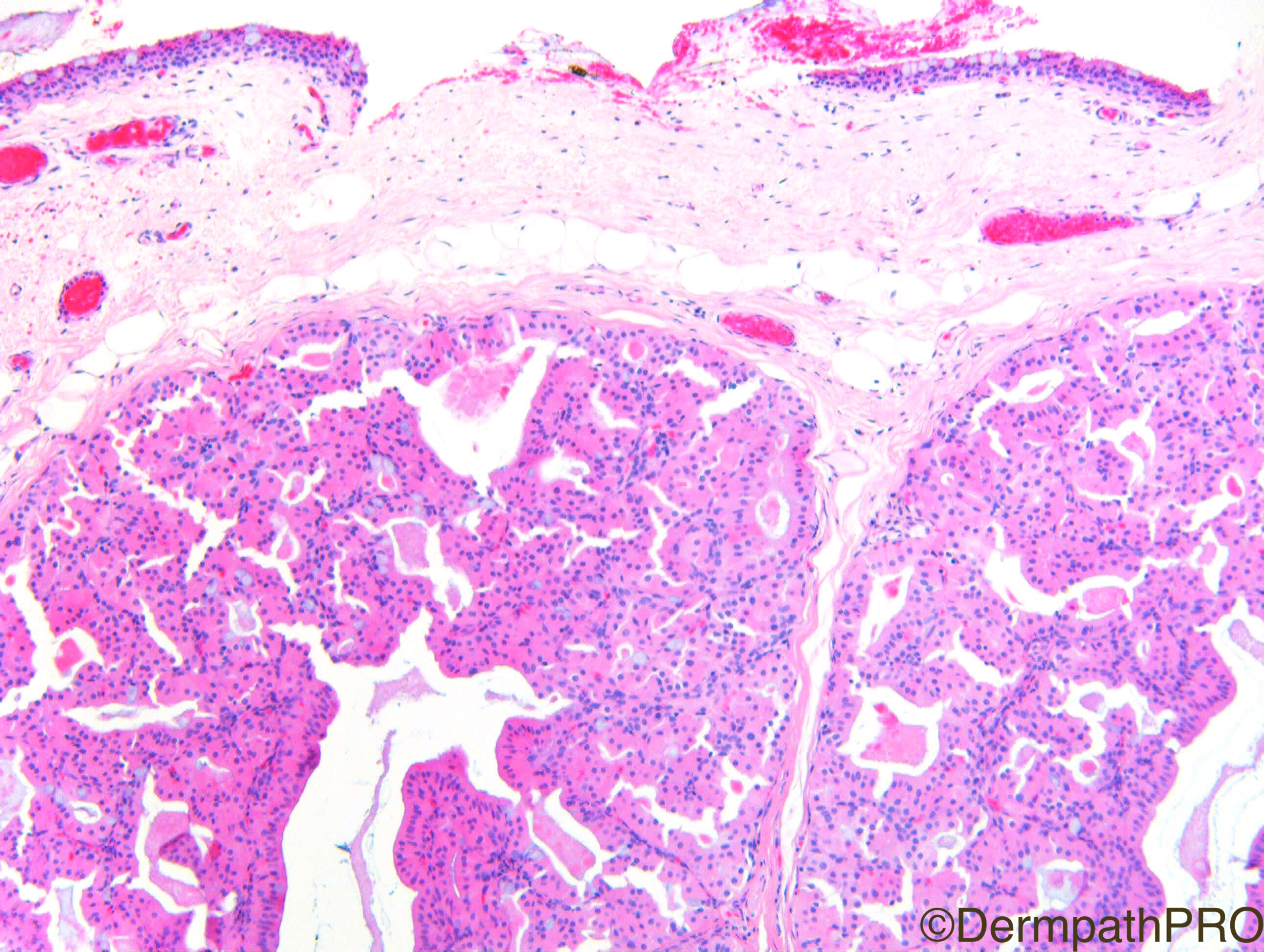
Adam_Bates 47 Posted March 24, 2024 Agree completely with Vincenzo! This is probably an eyelid lesion, as can see conjunctival epithelium on one surface. Interestingly, I was at the Royal Society of Medicine dermatology meeting in London yesterday and a rather similar case of primary cutaneous oncocytoma was presented in the CPC section. Share this comment Link to comment share_externally More sharing options...
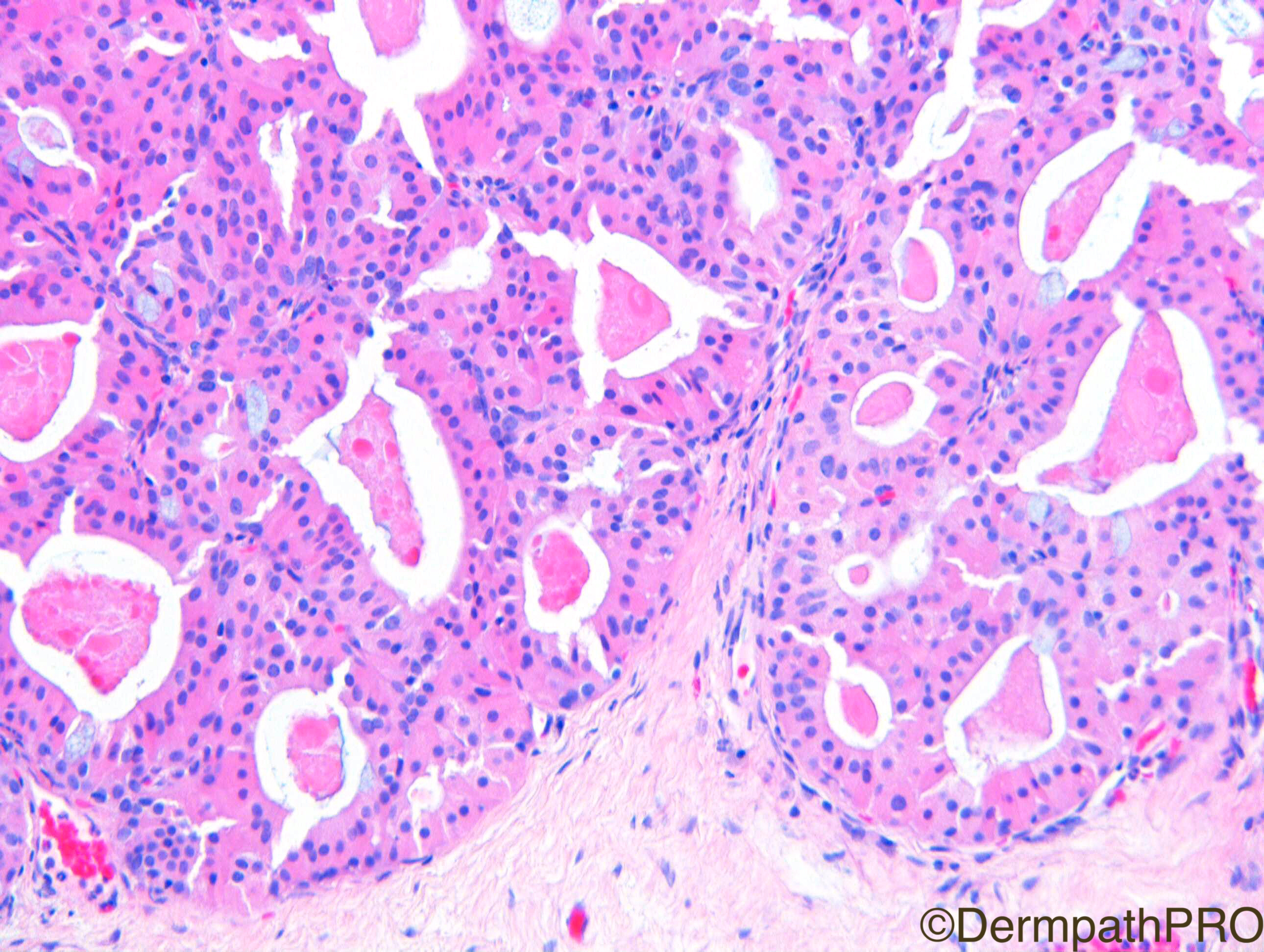
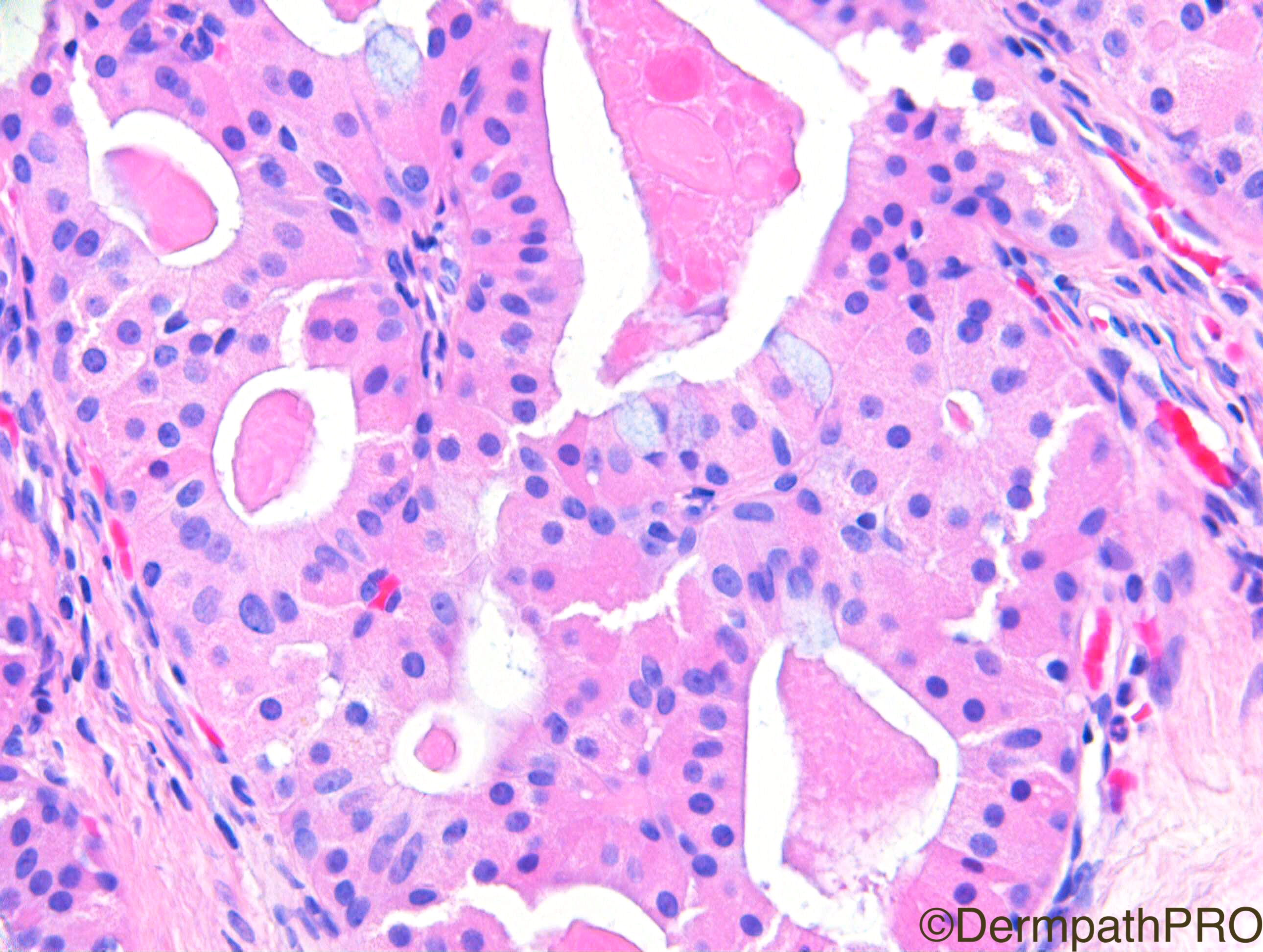
Adam_Bates 47 Posted March 24, 2024 Agree with Vicenzo. Solid-cystic lobulated oncocytic adenoma with some mucinous cells lining cystic spaces. Hidradenoma with predominant oxyphilic cellular population may be a point, but I cannot imagine a hidradenoma exclusively composed of this type of cells. Also, site favors oncocytic adenoma as at the periphery of the images one can find structures of the eyelid, including Meibomian sebaceous glands of the tarsal plate, and squamous stratified conjunctival epithelium with mucinous goblet cells. Share this comment Link to comment share_externally More sharing options...
Adam_Bates 47 Posted March 24, 2024 Agree, Oncocytic adenoma. Share this comment Link to comment share_externally More sharing options...
Adam_Bates 47 Posted March 24, 2024 Well done this is an oncocytic adenoma of the conjunctival caruncle. A nice spot diagnosis. Share this comment Link to comment share_externally More sharing options...


User Feedback