Jump to content

Building Blocks of Dermatopathology

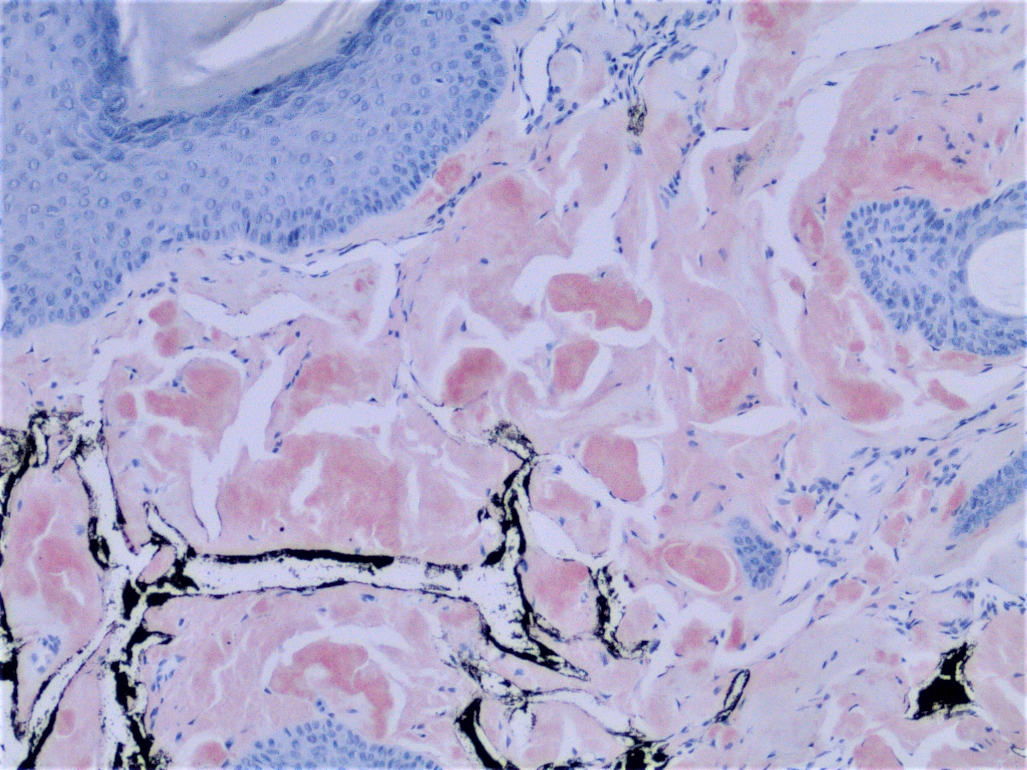
BAD DermpathPRO Learning Hub: Special Stains

Dermatopathology
Course Overview
Quizze

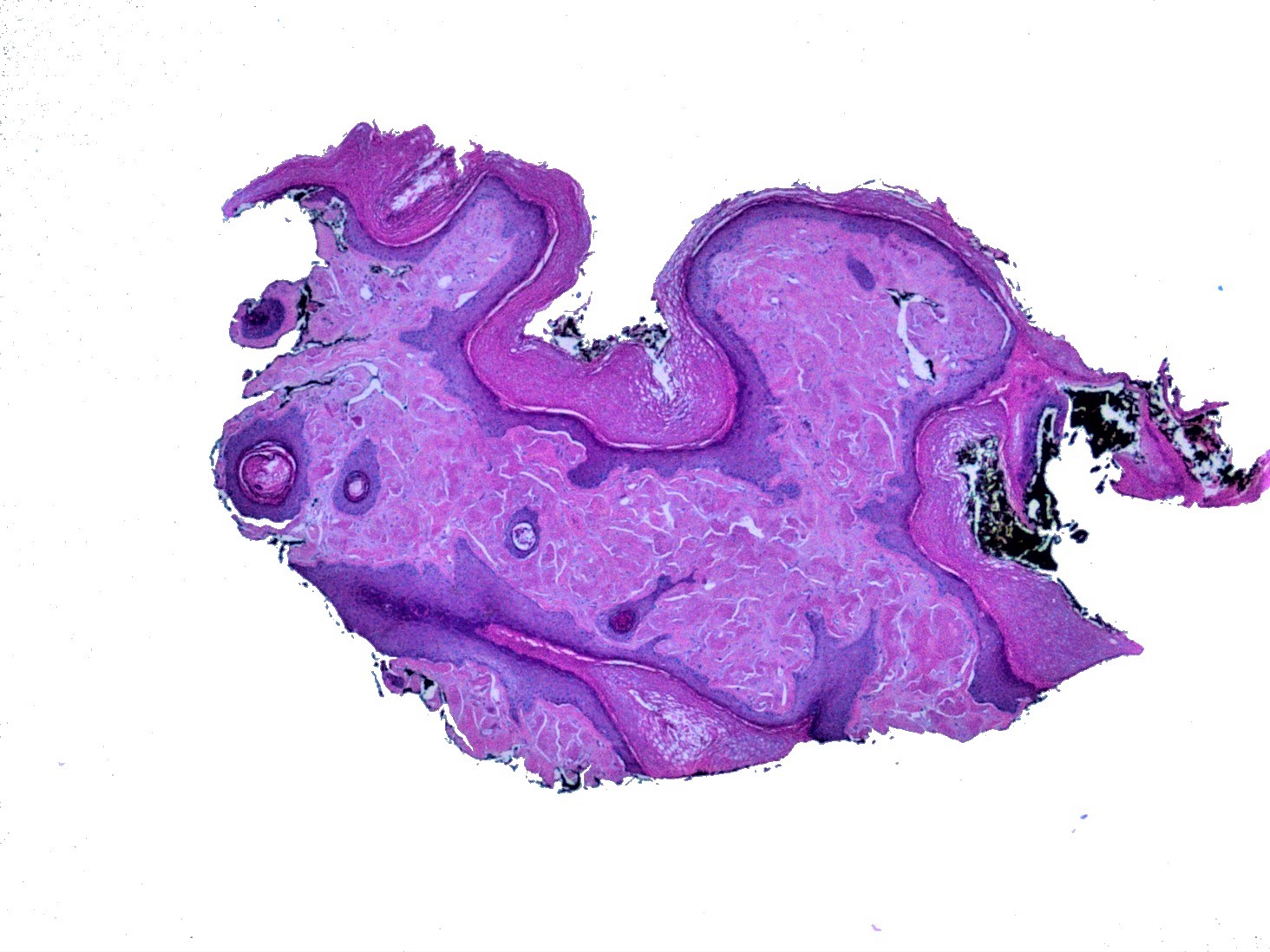
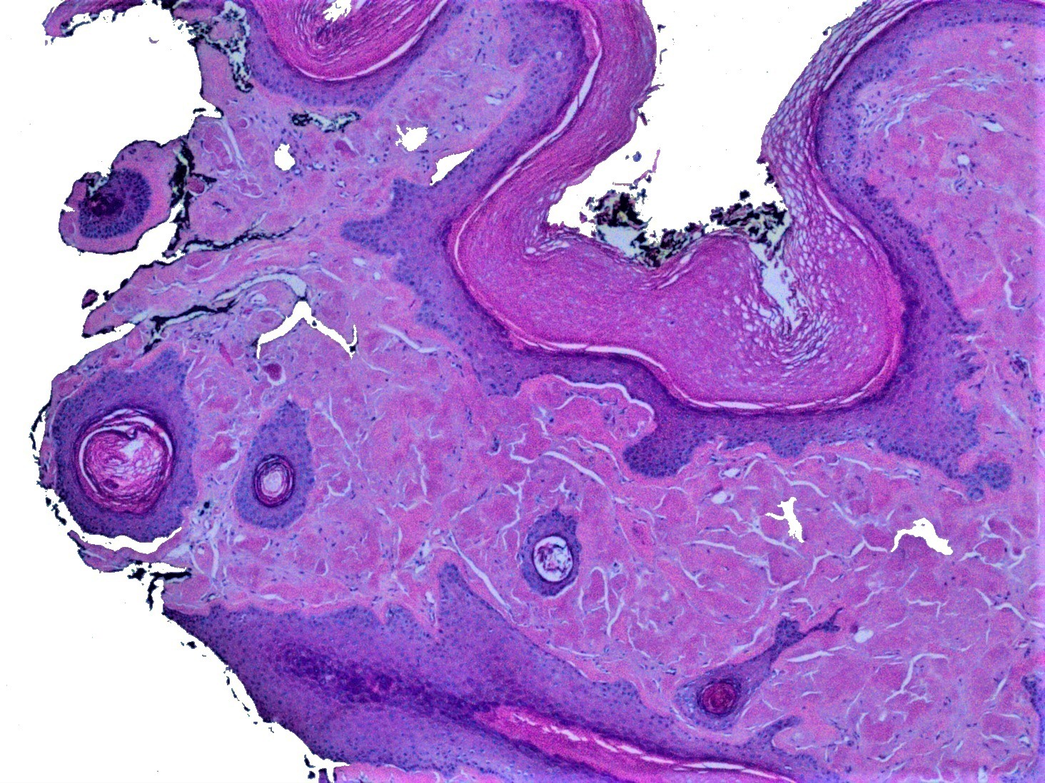
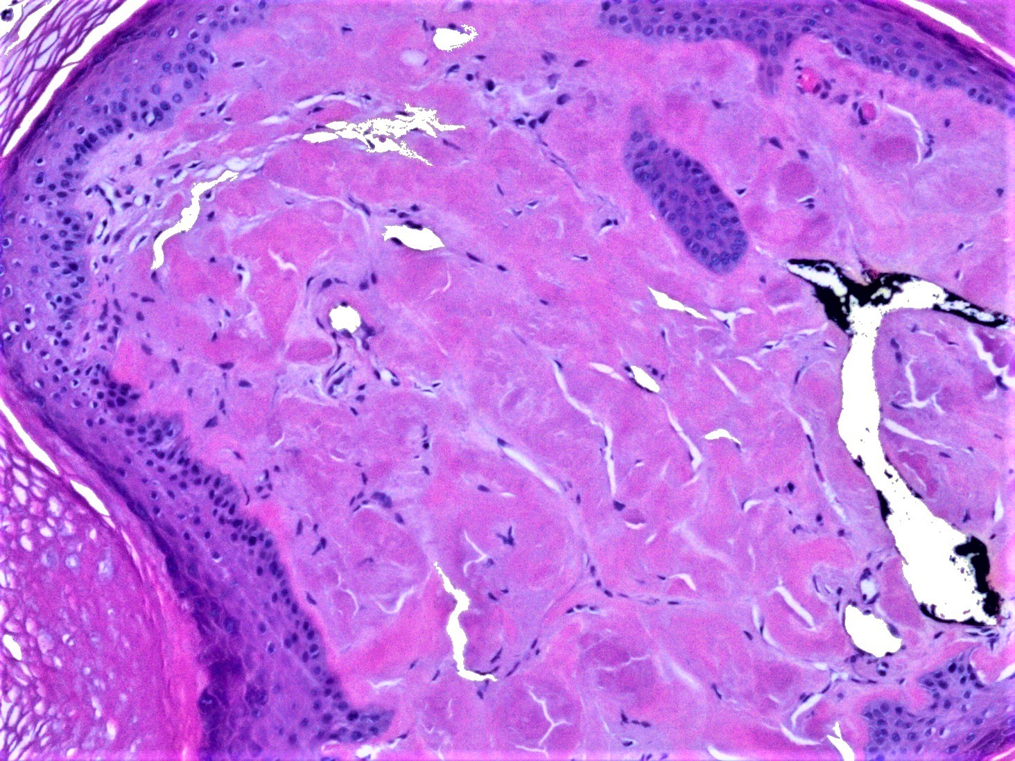
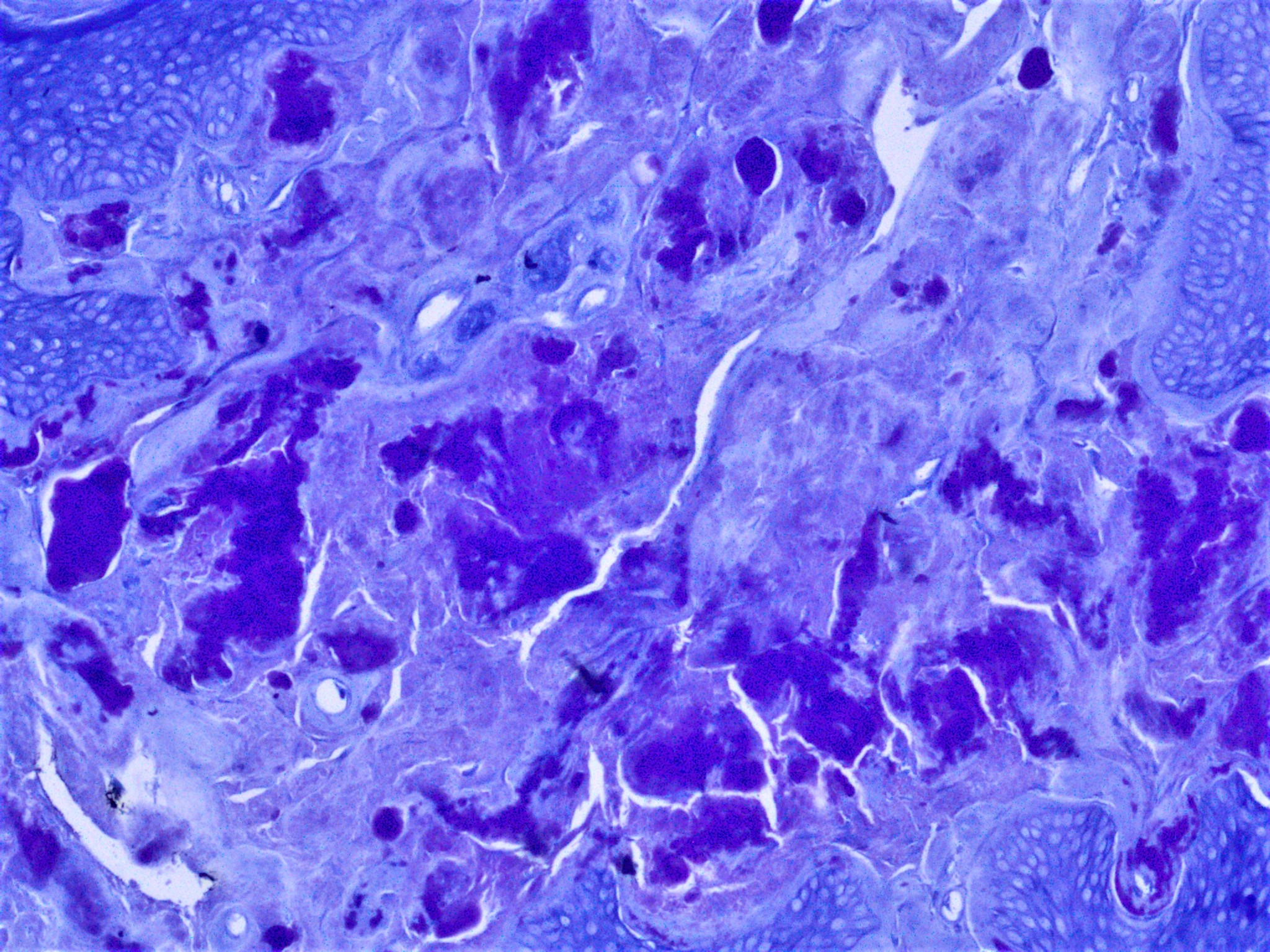
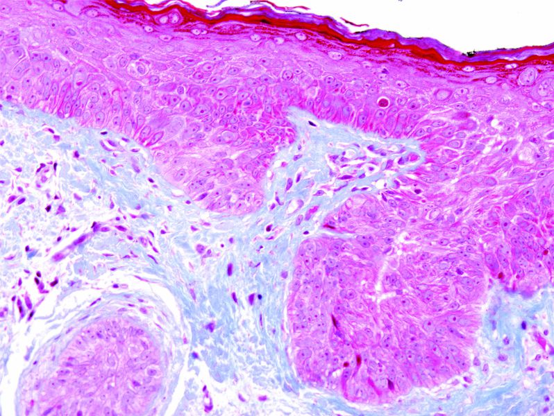
Case Number : ST0002 Limin Yu

Please read the clinical history and view the images by clicking on them before you proffer your diagnosis.
Submitted Date :
   (0 reviews)

78F, Biopsy of left ear external canal.


  Report Record

User Feedback


Operations Pathhub

Posted

Urmilapandey - Amyloid

Robledo F. Rocha - Agree, amyloidosis.

Dr. Mona Abdel-Halim - Amyloidosis of the ear, lovely case :-)

Vincenzo Polizzi - Agree with Colleagues. Nice case. 

Nitin Khirwadkar - Agree, amyloid.

Admin_Dermpath - Hi all Image 5 is Congo Red and Image 6 Crystal violet stain. Provided by Dr Limin Yu. 

Urmilapandey - ?Colloid Milium

Raul Perret - Type IV collagen, laminin or electron microscopy would help for this differential, but do we really need to do it? I would just mention it in the report.

Limin Yu - Thank everyone for the profound discussion. Actually this patient has a history of smoldering multiple myeloma. The case was signed-out as amyloidosis. Colloid milium is a great differential diagnosis. Sometimes, it's indistinguishable from amyloidosis histologically. In this clinical setting (patient's history and lesion location), amyloidosis is more appropriate. I thought about using the term "auricular amyloidosis" to be more specific in terms of the biopsy location. But I didn't use it due to the potential confusion to my clinician colleagues. 

Share this comment


Link to comment
share_externally


×
×
  • Create New...