Jump to content

Building Blocks of Dermatopathology

BAD DermpathPRO Learning Hub: Special Stains

Dermatopathology
Course Overview
Quizze

Case Number : ST0008

Please read the clinical history and view the images by clicking on them before you proffer your diagnosis.
Submitted Date :
   (0 reviews)

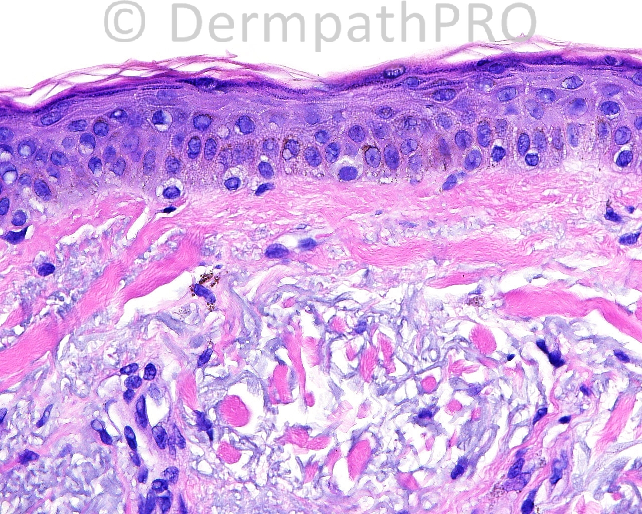
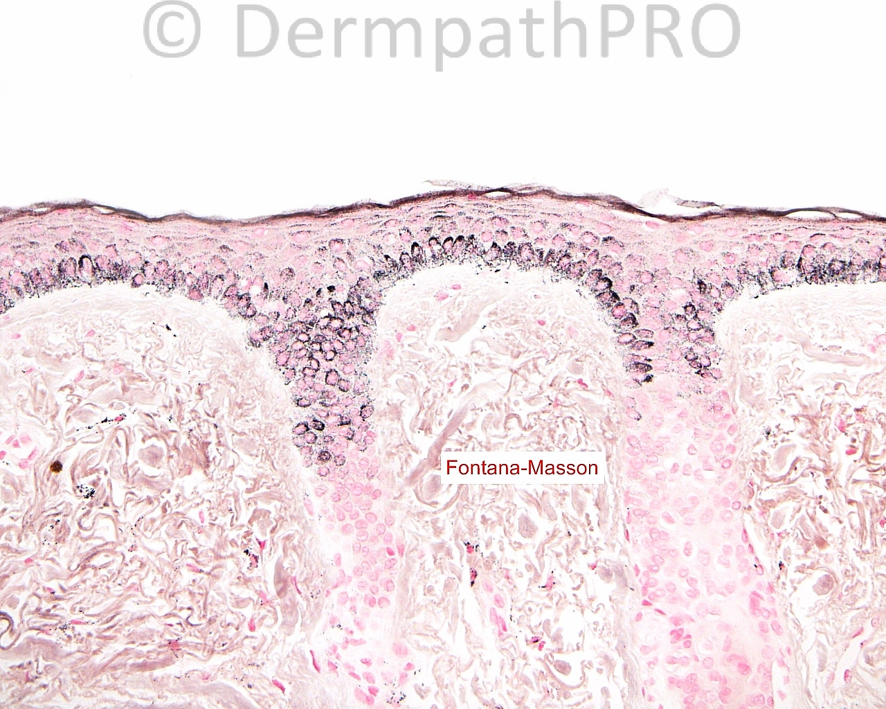
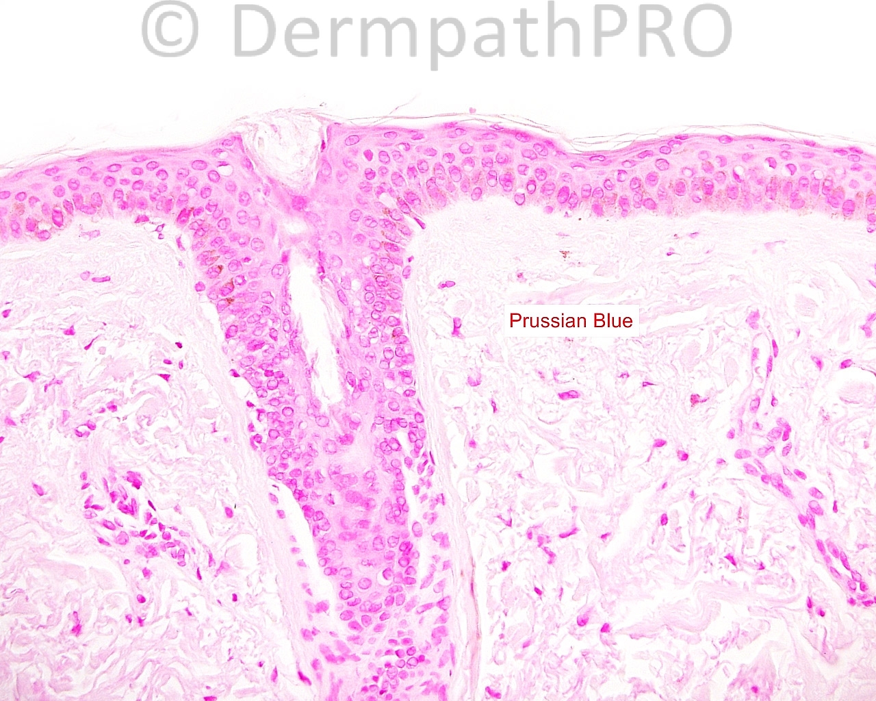
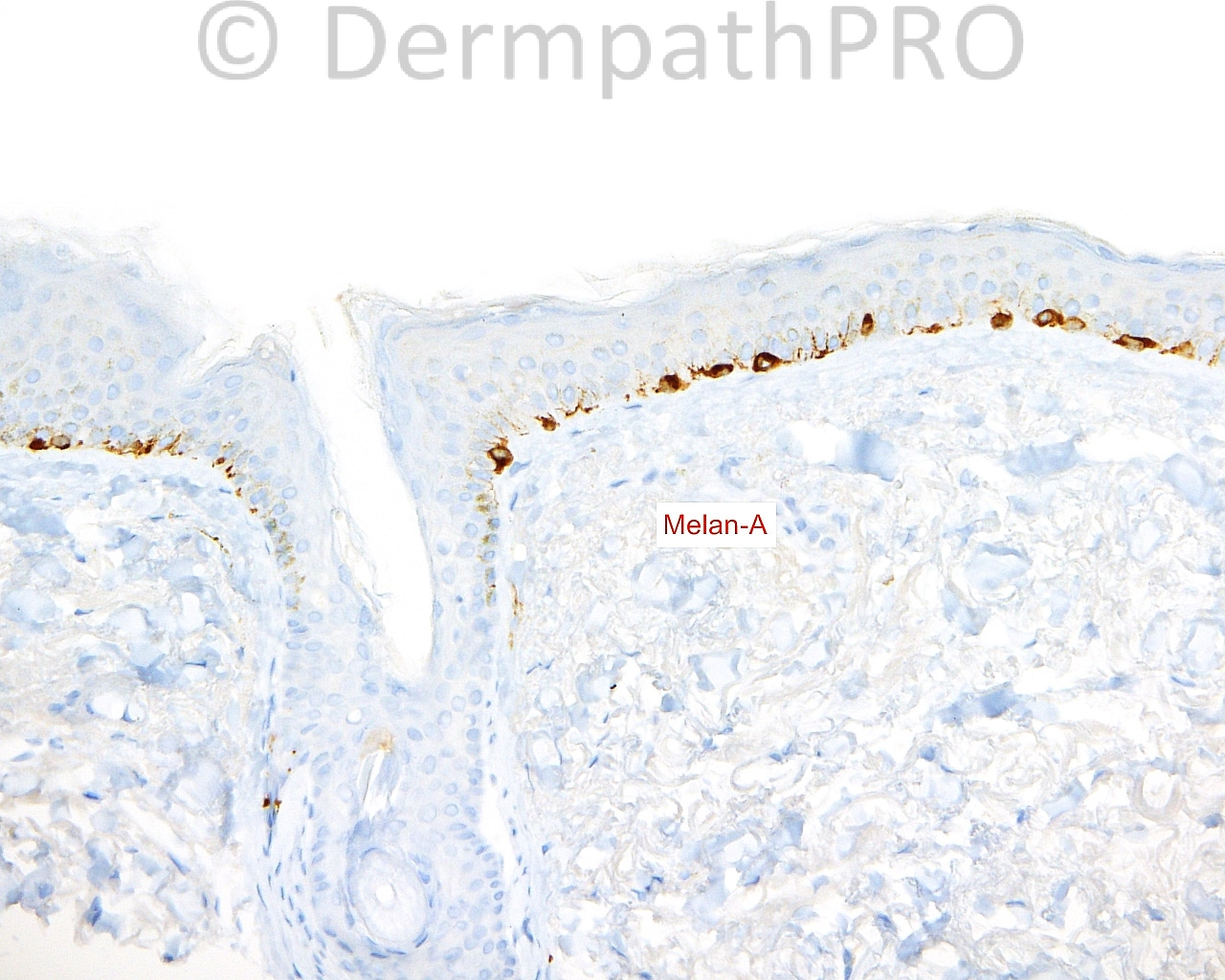
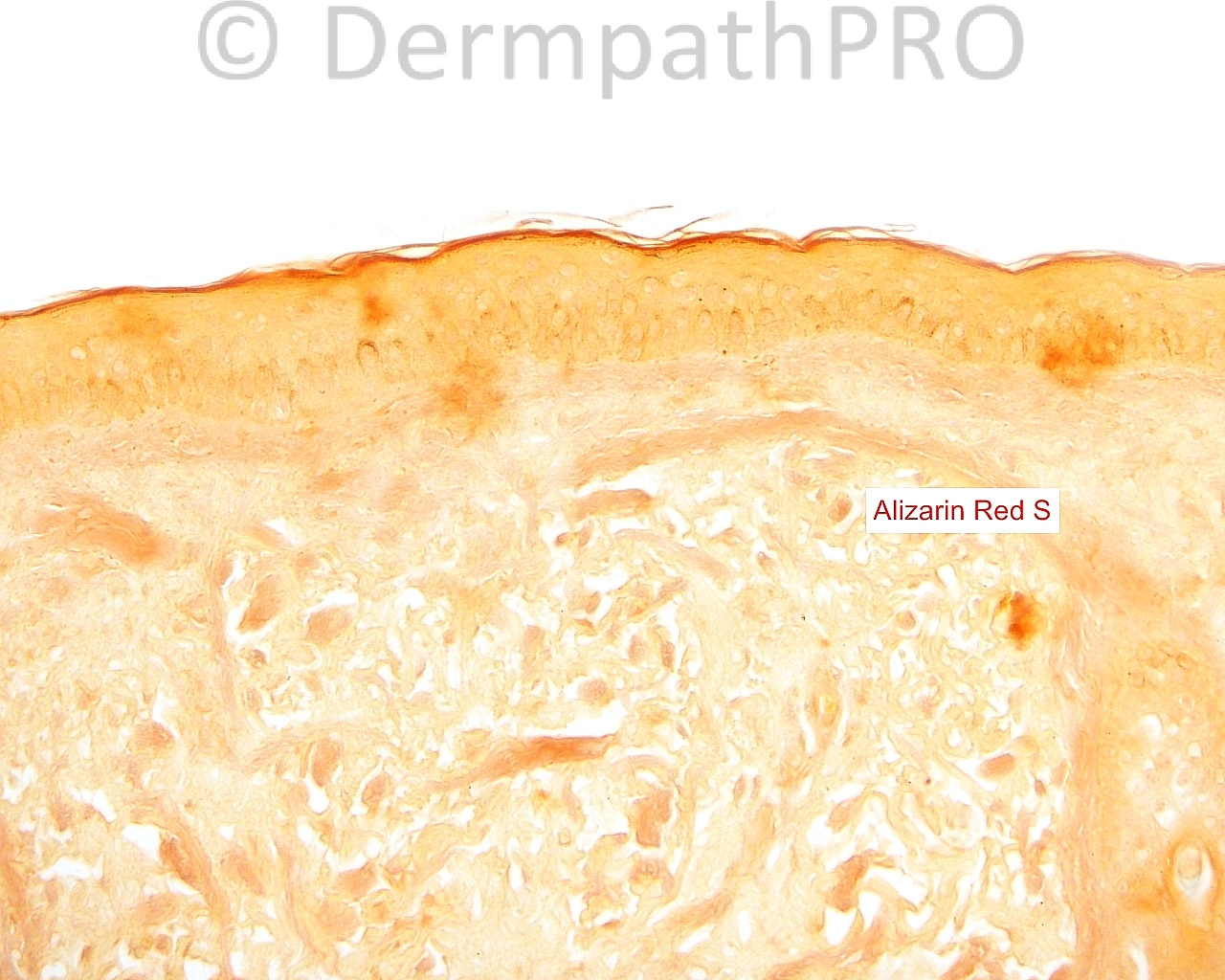
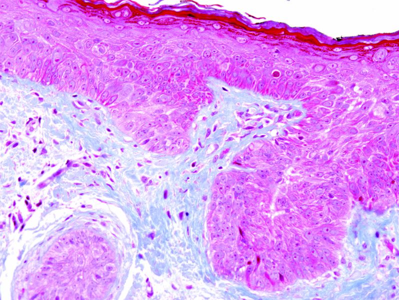
The patient is a 68-year-old woman who takes medication for ocular rosacea. A shave biopsy is taken of asymptomatic, blue-gray, macular pigment on the left cheek.


  Report Record

User Feedback


Operations Pathhub

Posted

Dr. Mona Abdel-Halim - Possibly she is taking minocycline, minocycline induced pigmentation.
 
Guest Romualdo - Agree: minocycline induced pigmentation.
  
Robledo F. Rocha - I don't think this is minocycline-induced pigmentation since it is negative for Prussian blue, but I agree this is postinflammatory pigmentation due to photosensitizing agent.
 
Dr. Mona Abdel-Halim - There is a report about a type IV minocycline induced pigmentation that was confined to acne scars and was negative for iron stain but positive with calcium stain. Could it be this case.

 
Mark A. Hurt MD - I think this is a form of Minocycline hyperpigmentation. By the way, I did not think there was calcium deposition on Alizarin Red S or von Kossa.

from my report:

-- HYPERPIGMENTATION
COMMENT: The presence of hyperpigmentation with Fontana-Masson indicates melanin hyperpigmentation in the basal layer, presumably from minocycline, which has been described as "type III". Other types of pigmentation with minocycline use include a hemosiderin and iron chelate of minocycline (type I) and a metabolite protein complex of chelated iron and calcium (type II). I see neither hemosiderin nor calcium deposition in this skin, and thus presume, by default, that the pigmentation is due to melanin in the basal layer of the epidermis.


And from Weedon (2010):

Type I: bluish-black pigmentation of scars and old inflammatory foci, including sites of immunobullous diseases, related to hemosiderin or an iron chelate of minocycline, a variant of this (proposed type IV), with blue-gray pigmentation of acne scars on the back was characterized by calcium-containing melanin deposits within dendritic cells and in an extracellular location.

Type II: blue-gray circumscribed pigmentation of the lower legs and arms due to a pigment which is probably a drug metabolite–protein complex chelated with iron and calcium. The recently reported cases with deposits of pigment localized to the subcutaneous fat of the lower extremity appear to be a different type (proposed type V).

Type III: a generalized muddy brown pigmentation due to increased melanin in the basal layer, and accentuated in sun-exposed areas.

Share this comment


Link to comment
share_externally


×
×
  • Create New...