Jump to content
In this section we have spot diagnoses posted on a daily basis since June 2010, now over 4000! You can review the archived cases and read the suggested diagnoses by users and the final comment by the contributors.
Case are uploaded each week day by 10 am UK time with the correct diagnosis will generally be posted at 8 pm UK time. Why not view the most recent spot diagnosis and proffer a diagnosis?

Case Number : 114 Posted By: Guest

Please read the clinical history and view the images by clicking on them before you proffer your diagnosis.
Submitted Date :
   (0 reviews)

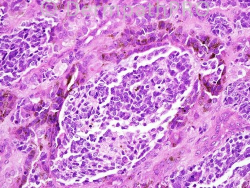
retinal anlage tumor (pigmented neuroectodermal tumor of infancy)


  Report Record

User Feedback


Admin_Dermpath

Posted

Phillip McKee - Overseas consultations (Sedona, Arizona, USA) Wrote:

Dear Mona, the pleasure is mine. In the near future, teaching videos will come on line which you all may enjoy. I look forward to feed back when you view them. Best wishes Phillip

Submitted on 16/11/2010 21:07
________________________________________
Juan Carlos Garcés - Hospital Oncológico / Hospital Luis Vernaza (Guayaquil Ecuador) Wrote:

Great case!!! Thanks a lot!

Submitted on 16/11/2010 21:06
________________________________________
Mona Abdel-Halim - Dermatology Department, Faculty of Medicine, Cairo University (Egypt) Wrote:

This is such an amazing case. I just can't believe it. Once in a life time!! Thank u very much Sir!! You are really doing a great and wounderful job by sharing your very valuable collection with us. I am so happy today!!

Submitted on 16/11/2010 20:16
________________________________________
Phillip McKee - Overseas consultations (Sedona, Arizona, USA) Wrote:

This is a very difficult spot diagnosis which was very kindly shared with me by Sook-Bin Woo from Boston. It is a high power view of a retinal anlage tumor (pigmented neuroectodermal tumor of infancy) which presented in the oral cavity from an underlying maxillary lesion. The clue to the diagnosis is the presence of small blue cells (neuroblastoma-like) surrounded by melanin containing epithelioid cells. With the eye of faith you can see some eosinophilic fibrillary material adjacent to some of the blue cells. It is very much a case of "once seen never forgotten" You just have to add it to the differential diagnosis list of small blue cell tumors.
Submitted on 16/11/2010 19:56
________________________________________
ANDREW DANCKWERTS - WITS UNIVERSITY (JOHANNESBURG, RSA) Wrote:

MALIGNANT MELANOMA WITH LYMPHOVASCULAR SPACE INVASION / PERMEATION.

Submitted on 16/11/2010 16:08
________________________________________
Eman El-Nabarawy - Cairo University (Egypt) Wrote:

Brilliant idea that of Dr Carlos ( a collision tumor). So I'd like to add squamo-melanocytic tumor to the DD?

Submitted on 16/11/2010 14:41
________________________________________
Juan Carlos Garcés - Hospital Oncológico / Hospital Luis Vernaza (Guayaquil Ecuador) Wrote:

Good morning.. I am not sure of anything in this case.. It looks like melanoma.. but there is a group of different kind of cells in the middle..a colision tumor?

Submitted on 16/11/2010 13:59
________________________________________
Marcela Saeb Lima - Instituto Nacional de Ciencias Médicas y Nutrición Salvador Zubirán (Mexico City) Wrote:

I was thinking of intravascular invasion of melanoma.

Submitted on 16/11/2010 12:51
________________________________________
Mona Abdel-Halim - Dermatology Department, Faculty of Medicine, Cairo University (Egypt) Wrote:

Melanocytic nests as well as solitary displaced melanocytes within the epidermis. Melanocytes look atypical. The nuclei are pleomorphic, large, hyperchromatic and very irregular in outlines, assuming geometrical patterns. I will consider this malignant melanoma.

Submitted on 16/11/2010 04:56
________________________________________
Eman El-Nabarawy - Cairo University (Egypt) Wrote:

Malignant melanoma.

Submitted on 16/11/2010 04:26
________________________________________

Share this comment


Link to comment
share_externally


×
×
  • Create New...