Jump to content
In this section we have spot diagnoses posted on a daily basis since June 2010, now over 4000! You can review the archived cases and read the suggested diagnoses by users and the final comment by the contributors.
Case are uploaded each week day by 10 am UK time with the correct diagnosis will generally be posted at 8 pm UK time. Why not view the most recent spot diagnosis and proffer a diagnosis?

Case Number : Case 318 Posted By: Guest

Please read the clinical history and view the images by clicking on them before you proffer your diagnosis.
Submitted Date :
   (0 reviews)

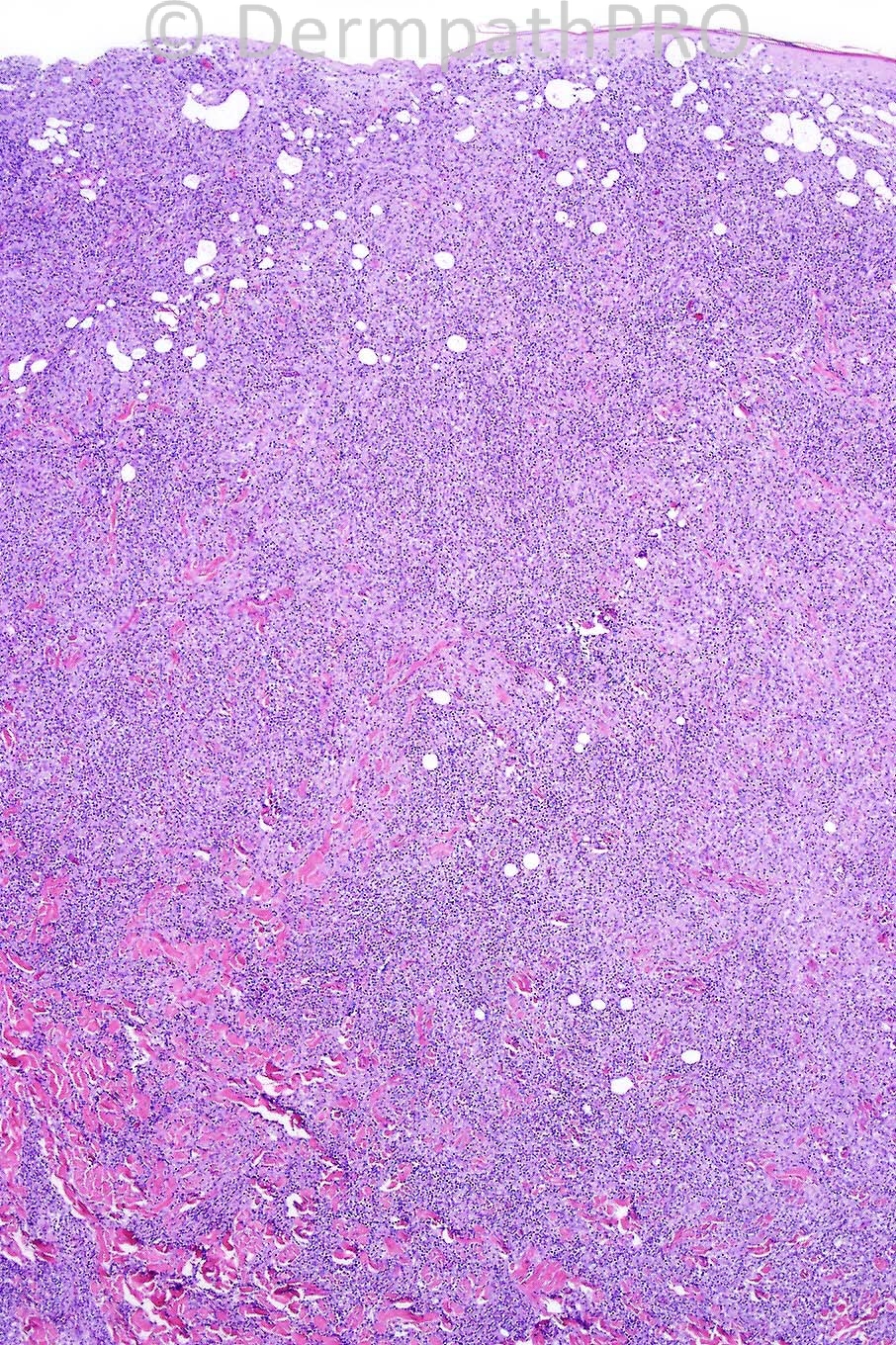
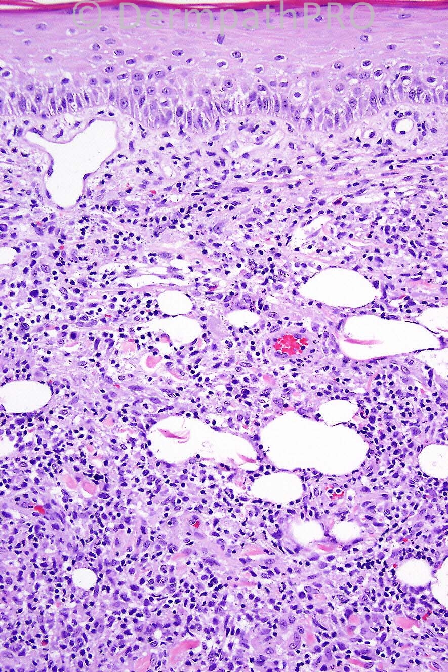
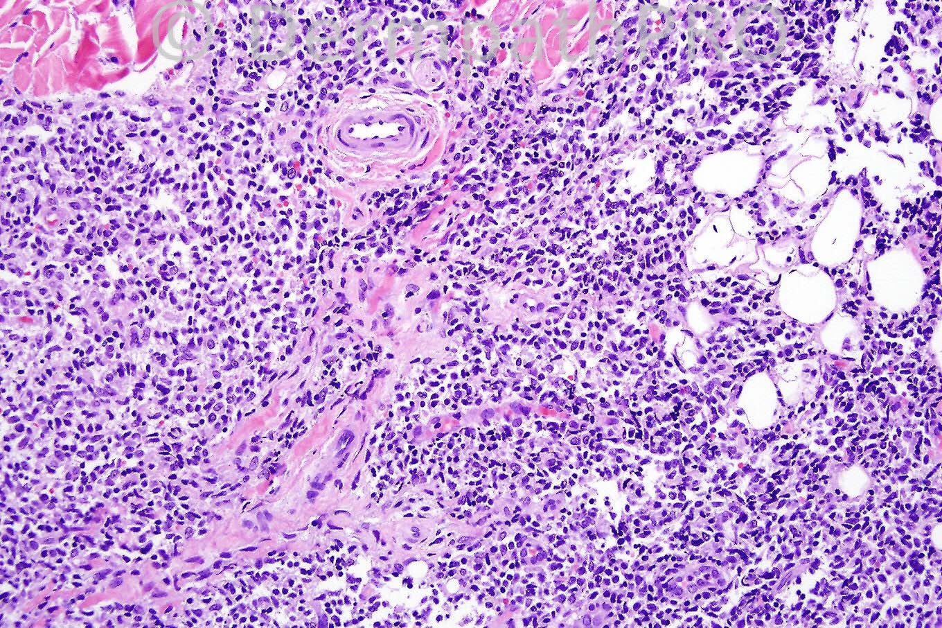
Male 62 years, widespread scaly plaques and nodule on forearm.


  Report Record

User Feedback


Admin_Dermpath

Posted

Phillip McKee - Overseas consultations (Arizona, USA) Wrote:

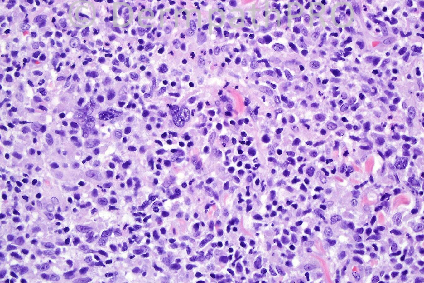
Well done, not an easy case but with this history and histology as you say the correct diagnosis is Mycosis fungoides in transformation.

Submitted on 30/08/2011 06:03
________________________________________
Andrew Danckwerts - NHLS / WITS University (Johannesburg, RSA) Wrote:

Tumour stage MF is favoured, with possible blastic transformation (>25% large cells in dermis). The differential diagnosis must also include cutaneous ALCL. I would like to do CD 3 / CD 4 / CD 8 / CD 30 and ALK1 to differentiate.

Submitted on 30/08/2011 02:50
________________________________________
Rodrigo Restrepo - UPB/CES (Mdlln/Col) Wrote:

Lymphomatoid papulosis

Submitted on 29/08/2011 22:28
________________________________________
Mohamad briak - King fahed medical center (Usa) Wrote:

lymphomatoid papulosis Thanks for this nice web site.

Submitted on 29/08/2011 19:12
________________________________________
Mona Abdel Halim - Dermatology Dept. Faculty of Medicine, Cairo University (Egypt) Wrote:

Liked the idea of tumor stage MF suggested by Eman, will add the possibility of large cell transformation in this context.

Submitted on 29/08/2011 18:34
________________________________________
Marwa Fawzi - Dermatology-Cairo University (Egypt) Wrote:

Thought of lymphomatoid papulosis,mixed cellularity

Submitted on 29/08/2011 18:26
________________________________________
Eman El-Nabarawy - Cairo University (Egypt) Wrote:

Tumor stage MF vs large anaplastic T cell lymphoma.

Submitted on 29/08/2011 18:20
________________________________________
Mona Abdel Halim - Dermatology Dept., Faculty of Medicine, Cairo University (Egypt) Wrote:

I guess this biopsy is taken from the nodule on the forearm. I can see a dense lymphocytic infiltrate encroaching on the epidermis (1st image), admixed with histiocytes, few esinophils and few neutrophils. 8 can also percieve many cells with large vesicular nuclei and prominent nuclei, thought of anaplastic large cell lymphoma maybe developing against a background of MF (widespread scaly plaques). Will do CD3,4,8 and CD30 and ALK-1.

Submitted on 29/08/2011 17:59
________________________________________

Share this comment


Link to comment
share_externally


×
×
  • Create New...