Jump to content
In this section we have spot diagnoses posted on a daily basis since June 2010, now over 4000! You can review the archived cases and read the suggested diagnoses by users and the final comment by the contributors.
Case are uploaded each week day by 10 am UK time with the correct diagnosis will generally be posted at 8 pm UK time. Why not view the most recent spot diagnosis and proffer a diagnosis?

Case Number : Case 393 Posted By: Guest

Please read the clinical history and view the images by clicking on them before you proffer your diagnosis.
Submitted Date :
   (0 reviews)

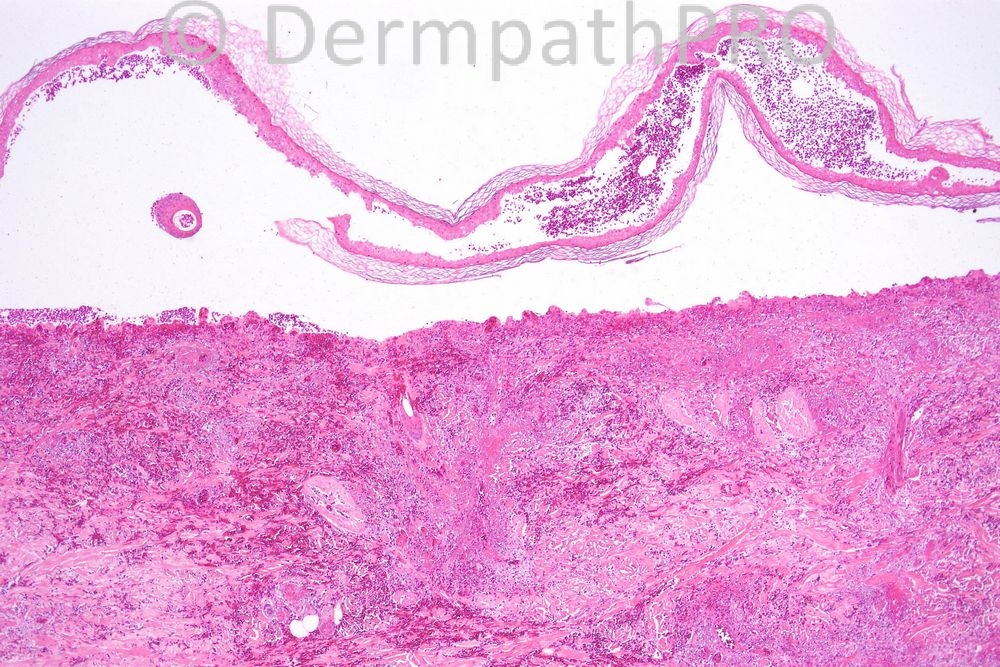
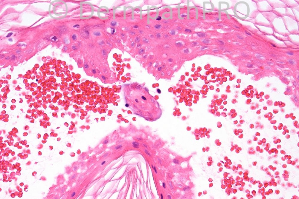
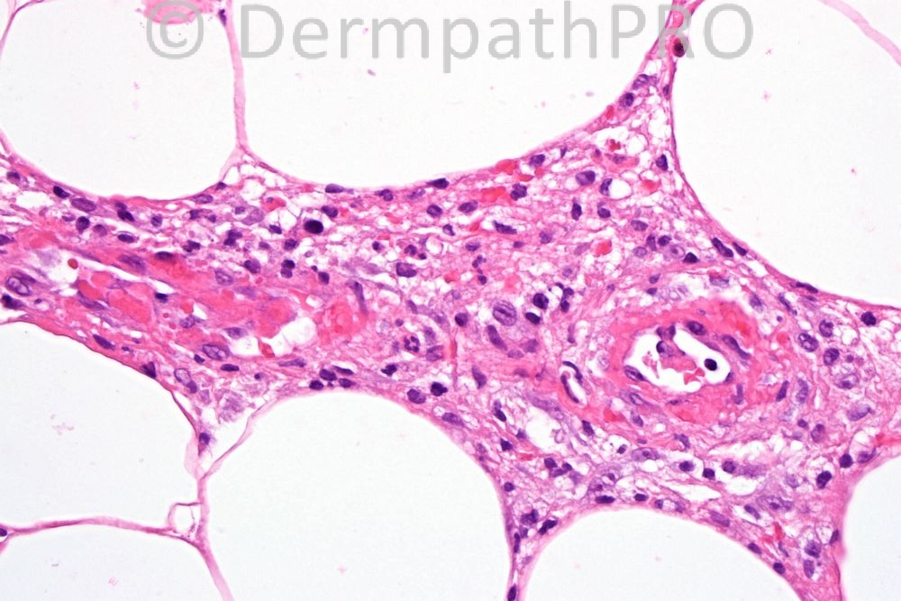
44 year old, purpuric lesion lower leg with blistering.


  Report Record

User Feedback


Admin_Dermpath

Posted

Phillip McKee - Overseas Consultations (Cave Creek, Arizona, USA) Wrote:

Well done Marcela. This is just an extreeme example of LCV with epidermal necrosis giving rise to a blister. PCT is invariably cell poor/free and does not show vasculitis.

Submitted on 12/12/2011 22:45
________________________________________

Peter Karolyi - () Wrote:

Leucocytoclasic vasculitis, I think the best alternative that can be based on the available clinical data and pictures.

Submitted on 12/12/2011 22:12
________________________________________

Diva Uribe Cordova - Hospital de Clínicas (La Paz) Wrote:

Hypersensitivity Vasculitis (Leukocytoclastic Vasculitis)

Submitted on 12/12/2011 20:11
________________________________________

Rodrigo Restrepo - UPB/CES (Mdlln/Col) Wrote:

Small vessell vasculitis

Submitted on 12/12/2011 19:21
________________________________________

Mona Abdel Hali - Dermatology Dept. Cairo Univ. (Egypt) Wrote:

I thought of vasculitis, clinical information and laboratory investigations are needed to establish a definite cause, deep extension will make me think of microscopic polyangiitis, the associated thrombosis could be a clue also to septic vasculitis,,

Submitted on 12/12/2011 18:33
________________________________________

Sasi Attili - () Wrote:

This I think might be tricky. There is a sub-epidermal re-epithelialising blister with festooning of dermal papillae. A follicular unit lies free in the blister suggesting adnexal extension. There is definite small vessel vasculitis with vessel wall necrosis and extravasation of RBC. I can understand why colleagues were thinking of PCT and I did consider this. However I think the pink stuff around the vessels might just be fibrin as vasculitis is not usually a feature of PCT and also the clinical does not quite fit. I would like to see the character of the inflammatory cells. If the figure on the bottom left is deep i.e fat, then there does appear to be a panniculitis too, associated with the vasculitis. In the middle there is a large cell with a coffee bean shaped nucleus. However I am not convinced the histo is anything like LCH described in books. So in summary I think the biopsy shows a small vessel vasculitis. CPC needed to look for systemic infective, drug and neoplastic causes

Submitted on 12/12/2011 18:12
________________________________________

Rok Kokol - (Graz) Wrote:

PCT

Submitted on 12/12/2011 16:15
________________________________________

Marcela Saeb Lima - INCMNSZ (Mexico City) Wrote:

small vessell necrotizing vasculitis superficial and deep with epidermal necrosis the differential diagnosis includes a list of entities which needs clinicopath correlation

Submitted on 12/12/2011 15:51
________________________________________

Hazem Hamed - Department of Histopathology, Imperial College (London) Wrote:

Porphyria cutanea tarda

Submitted on 12/12/2011 15:44
________________________________________

I. Abdul-kadir - SJUH (Leeds, UK) Wrote:

Porphyria

Submitted on 12/12/2011 14:55
________________________________________

Share this comment


Link to comment
share_externally


×
×
  • Create New...