Jump to content
In this section we have spot diagnoses posted on a daily basis since June 2010, now over 4000! You can review the archived cases and read the suggested diagnoses by users and the final comment by the contributors.
Case are uploaded each week day by 10 am UK time with the correct diagnosis will generally be posted at 8 pm UK time. Why not view the most recent spot diagnosis and proffer a diagnosis?

Case Number : Case 396 Posted By: Guest

Please read the clinical history and view the images by clicking on them before you proffer your diagnosis.
Submitted Date :
   (0 reviews)

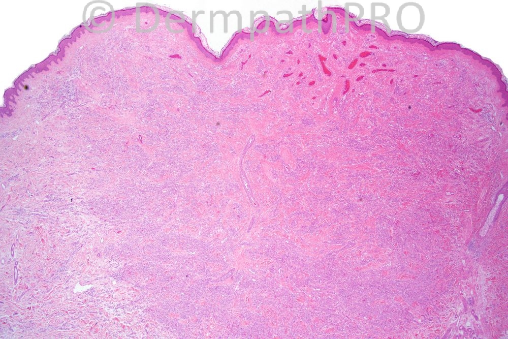
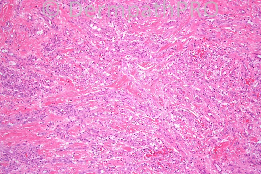
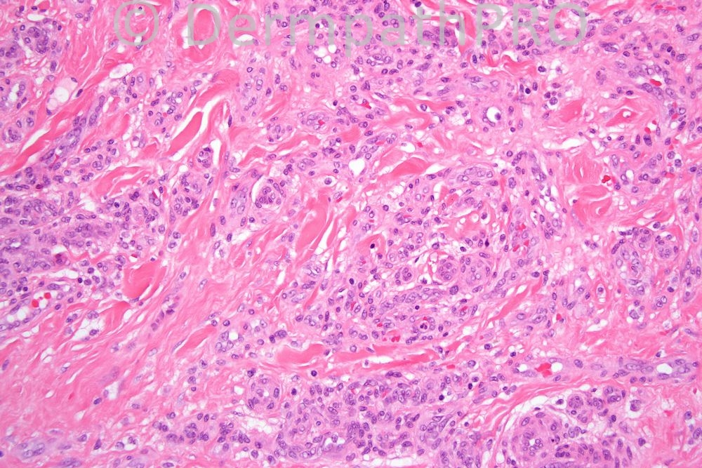
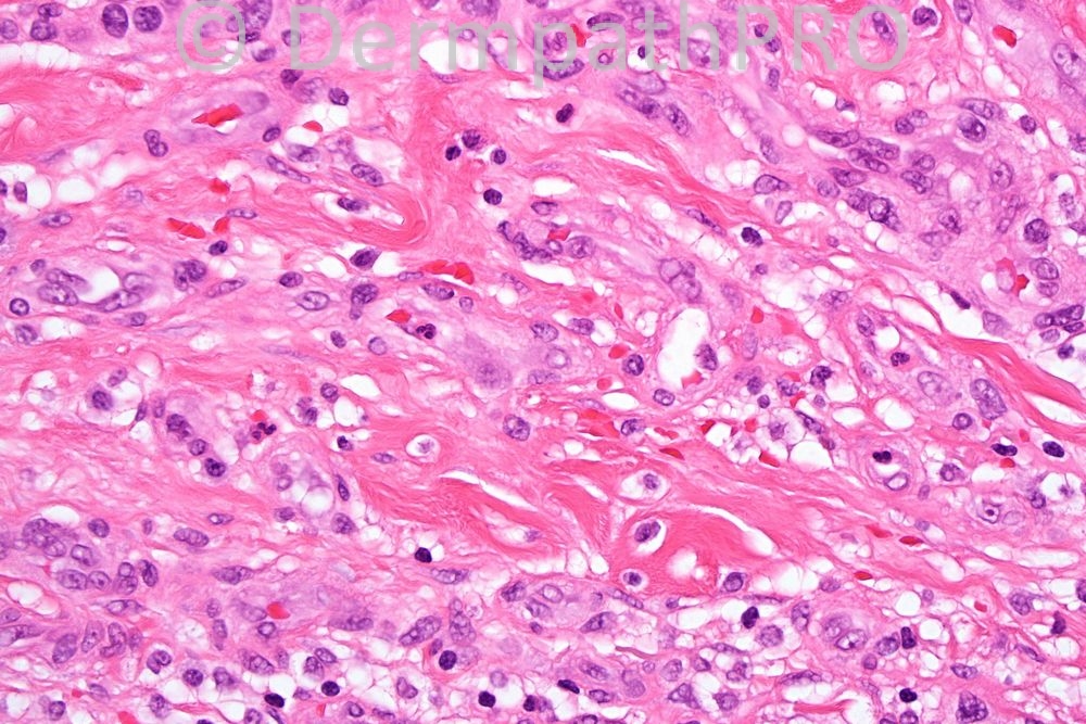
Female 30 years, lesion on thigh clinically hemangioma.


  Report Record

User Feedback


Admin_Dermpath

Posted

Phillip McKee - Overseas Consultations (Cave Creek, Arizona, USA) Wrote:

This is a difficult case and demonstrates how many "different" vascular tumors there currently are. It is a florrid microvenular hemangioma with most of the vessels cut at right angles. The bottom right figure shows the vessels infiltrating between somewhat hyalinized dermal collagen. Involvement of arrector pili muscles (not shown) is a typical feature. Epithelioid hemangioendothelioma does not show much in the way of full vascular differentiation. One tends to see clusters of cells some with intracytoplasmic vacuoles occasionally containing erythrocytes (if you are lucky). In addition there is an accompanying myxoid and hyalinized stroma.

Submitted on 15/12/2011 22:40
________________________________________

Marcela M. Escanamé - Medical Univesity of Graz (Austria) Wrote:

Epithelioid Hemangioendothelioma

Submitted on 15/12/2011 22:25
________________________________________

Sasi Attili - (UK) Wrote:

I would favour an EAN rather than an Epithelioid hemangioendothelioma

Submitted on 15/12/2011 22:00
________________________________________

Mona Abdel Halim - Dermatology Dept. Cairo Univ. (Egypt) Wrote:

I thought of cutaneous epithelioid angiomatous nodule due to the presence of focal vascular channels, and the back ground of fibrosis. I think in epithelioid fibrous histiocytoma, cells appear more spitzoid like and there is no fibrosis, however , it is a good DDx. Epithelioid hemangioendothelioma usually shows no well formed vascular channels and is more diffuse and infiltrative, the first image gives me a rather circumscribed architecture,,,

Submitted on 15/12/2011 18:38
________________________________________

Eman El-Nabarawy - Cairo University (Egypt) Wrote:

Epithelioid fibrous histiocytoma.

Submitted on 15/12/2011 18:11
________________________________________

W Chen - () Wrote:

Can this be an epithelioid cell histiocytoma or a histiocytic variant of DF?

Submitted on 15/12/2011 17:33
________________________________________

Francisco Vilchez - Department of Dermatology. Virgen de la Victoria Clinical Universitary Hospital. (Málaga (Spain)) Wrote:

Ufff, this time I´m not late...Epitheloid hemangioendothelioma

Submitted on 15/12/2011 17:14
________________________________________

Marcela Saeb Lima - INCMNSZ (Mexico City) Wrote:

epithelioid hemangioendothelioma

Submitted on 15/12/2011 16:51
________________________________________

M A Rahman - North Cumbria University Hopsitals NHS Trust (carlisle) Wrote:

epithelioid haemangioendothelioma

Submitted on 15/12/2011 15:34
________________________________________

Share this comment


Link to comment
share_externally


×
×
  • Create New...