Jump to content
In this section we have spot diagnoses posted on a daily basis since June 2010, now over 4000! You can review the archived cases and read the suggested diagnoses by users and the final comment by the contributors.
Case are uploaded each week day by 10 am UK time with the correct diagnosis will generally be posted at 8 pm UK time. Why not view the most recent spot diagnosis and proffer a diagnosis?

Case Number : Case 287 Posted By: Guest

Please read the clinical history and view the images by clicking on them before you proffer your diagnosis.
Submitted Date :
   (0 reviews)

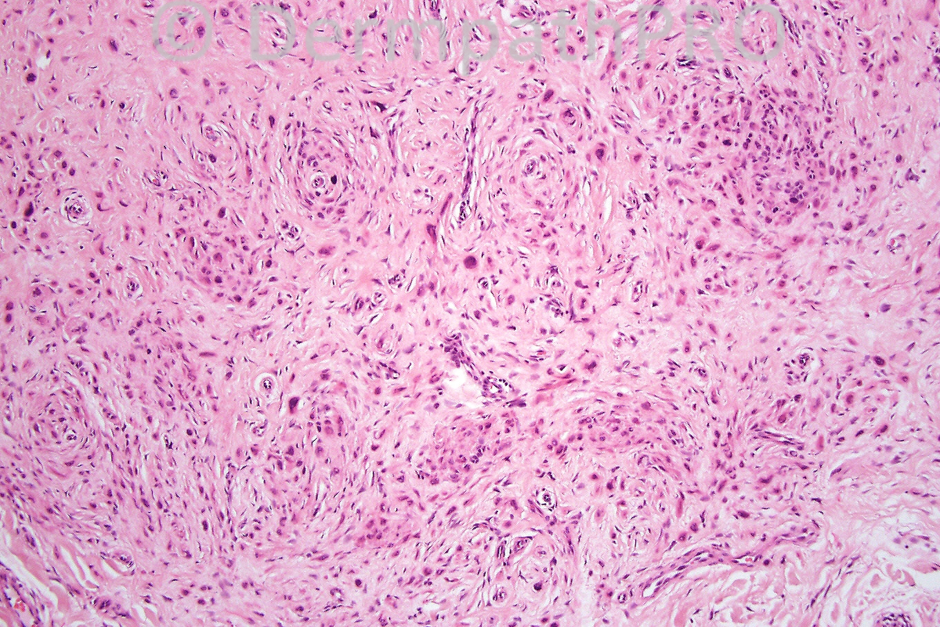
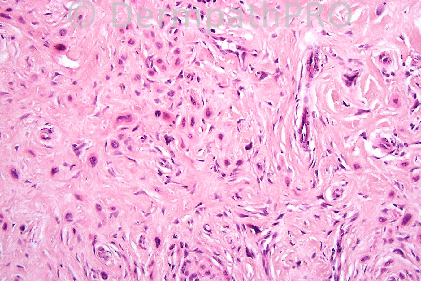
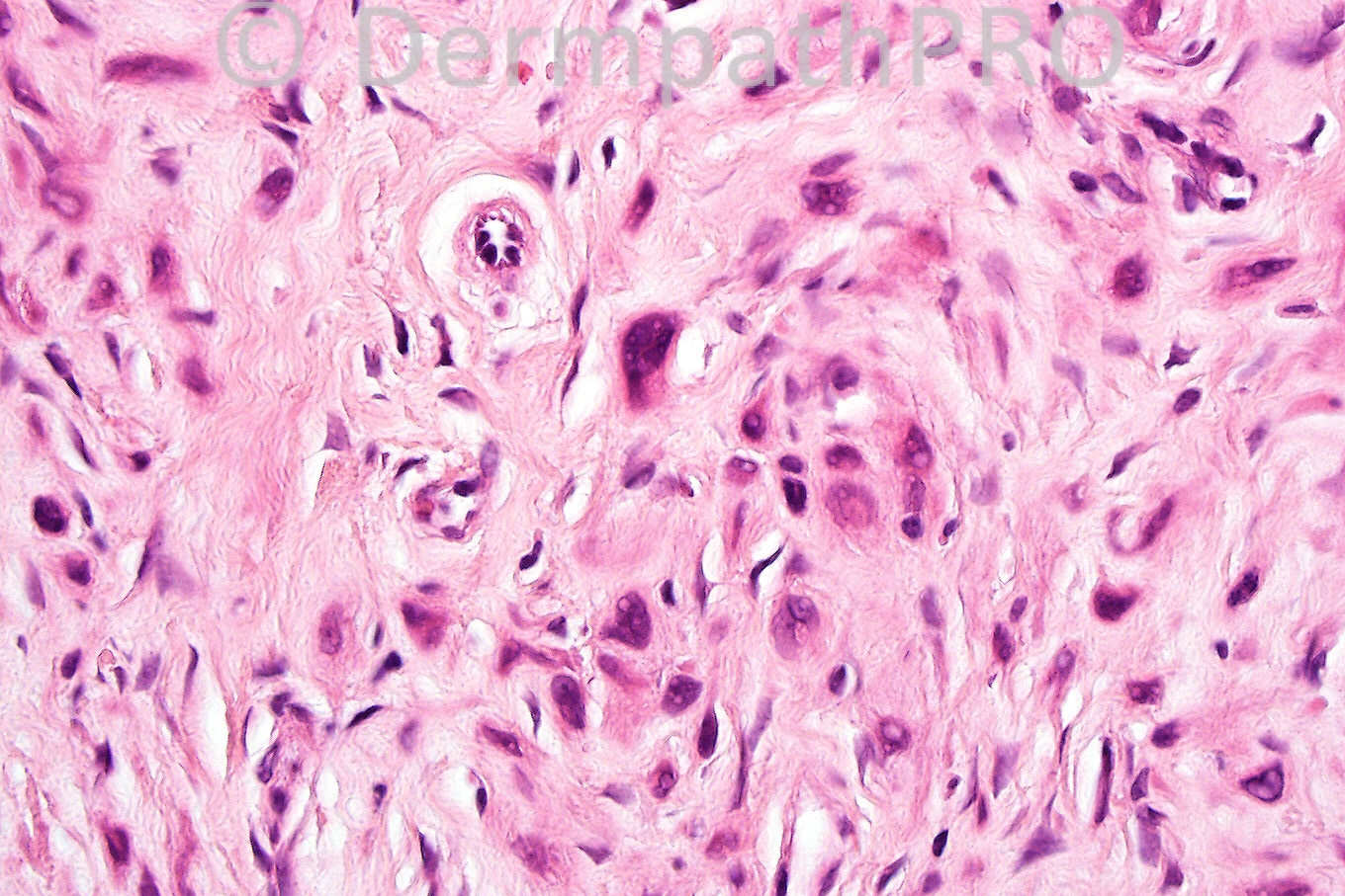
Female 27 years with flesh colored nodule on cheek.


  Report Record

User Feedback


Admin_Dermpath

Posted

Marie MD - () Wrote:

Desmoplastic Spitz'

Submitted on 18/07/2011 17:36
________________________________________
Phillip McKee - Overseas consultations (Cave Creek, USA) Wrote:

The correct diagnosis is desmoplastic Spitz nevus. The second figure emphasizes the nested superficial population whereas the 3rd figure shows diminution in cell size and a dispersed cellular population indicative of maturation. Nuclei are vesicular and intranuclear cytoplasmic psdeudo-inclusion are present. There is a background of desmoplasia. Have a great weekend.

Submitted on 15/07/2011 12:08
________________________________________
Rodrigo Restrepo - UPB/CES (Colombia) Wrote:

Vesicular nuclei and pseudoinclusions: Desmoplastic nevus

Submitted on 15/07/2011 11:29
________________________________________
Eman El-Nabarawy - Cairo University (Egypt) Wrote:

Difficult case.Thought of neural neoplasm: atypical cellular neurothekoma, dysplastic neurofibroma.

Submitted on 15/07/2011 10:29
________________________________________
Juan Carlos Garcés - Hospital Oncológico / Hospital Luis Vernaza (Guayaquil Ecuador) Wrote:

Desmoplastic nevus?

Submitted on 15/07/2011 09:33
________________________________________
Marwa Fawzi - Dermatology Department, Faculty of Medicine, Cairo University (Egypt) Wrote:

Fibrous papule of the face

Submitted on 15/07/2011 08:54
________________________________________
Mona Abdel Halim - Dermatology Dept. Faculty of Medicine, Cairo University (Egypt) Wrote:

Thinking of multinucleate cell angiohistiocytoma. Vascular proliferations, thin walled, some appear collapsed like MVH, together with fibrohistiocytic cells and many multinucleated cells with angulated cytoplasm.

Submitted on 15/07/2011 05:16
________________________________________

Share this comment


Link to comment
share_externally


×
×
  • Create New...