Jump to content
In this section we have spot diagnoses posted on a daily basis since June 2010, now over 4000! You can review the archived cases and read the suggested diagnoses by users and the final comment by the contributors.
Case are uploaded each week day by 10 am UK time with the correct diagnosis will generally be posted at 8 pm UK time. Why not view the most recent spot diagnosis and proffer a diagnosis?

Case Number : Case 272 Posted By: Guest

Please read the clinical history and view the images by clicking on them before you proffer your diagnosis.
Submitted Date :
   (0 reviews)

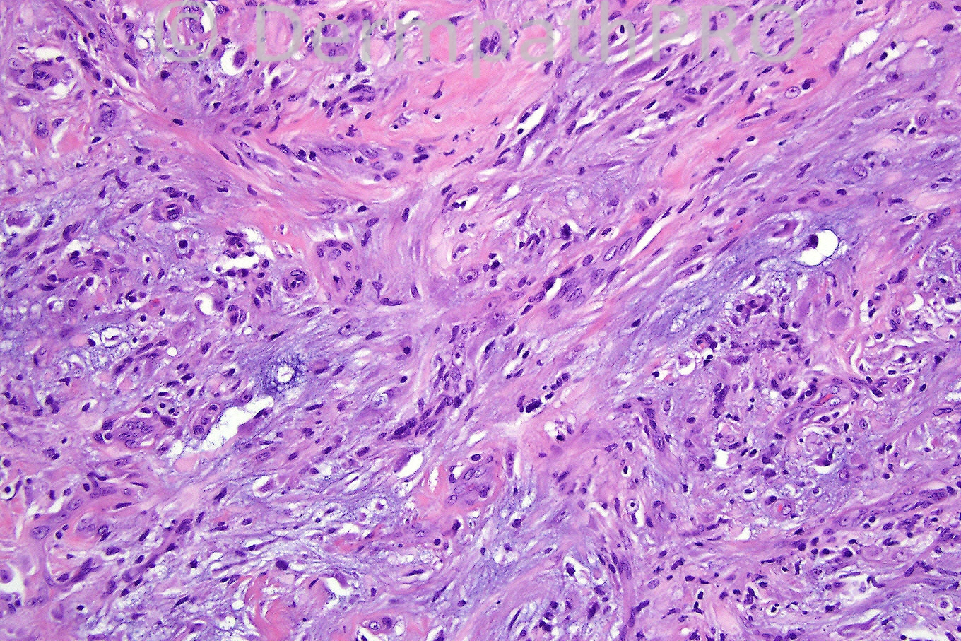
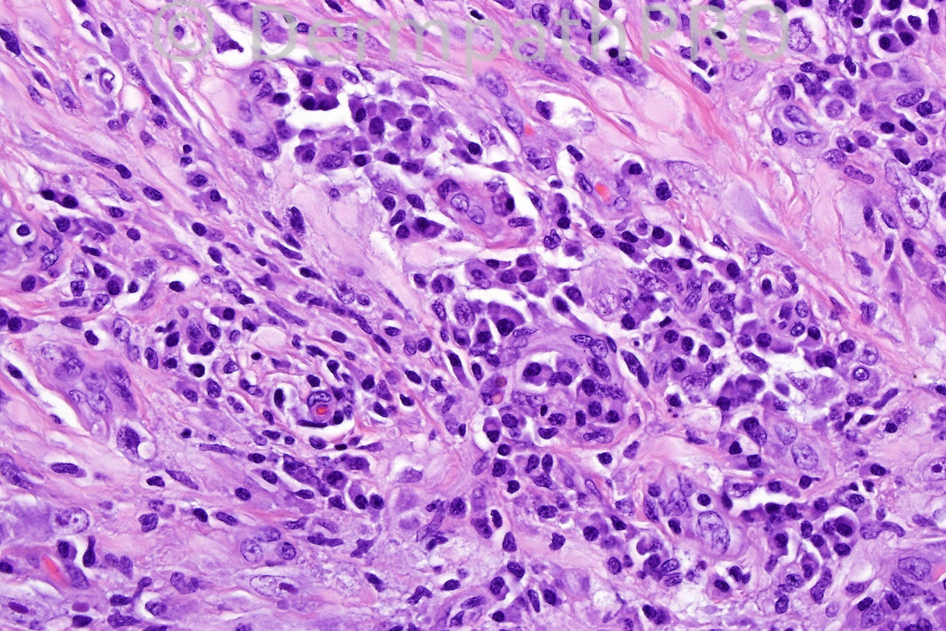
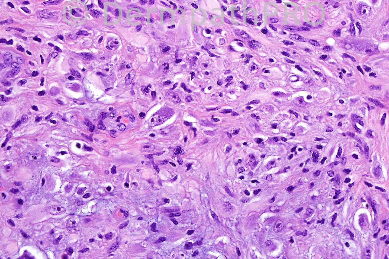
Male 42 years, subcutaneous nodule in forearm.


  Report Record

User Feedback


Admin_Dermpath

Posted

Phillip McKee - Overseas consultations (Cave Creek, Arizona, USA) Wrote:

Yes Richard, these connective tissue tumors do leave one battered and bruised. They are very difficult. The diagnosis in this case is acral myxoinflammatory fibroblastic sarcoma. Mona's comments are excellent. Unfortunately I do not have the results of ALK1 but I assume that it was positive. Have a wonderful weekend and see you on Monday

Submitted on 24/06/2011 13:31
________________________________________
Juan Carlos Garcés - Hospital Oncológico / Hospital Luis Vernaza (Guayaquil Ecuador) Wrote:

Good morning, difficult case. I think that a good possiblility is myxoinflammatory fibroblastic sarcoma

Submitted on 24/06/2011 12:02
________________________________________
MSMD - () Wrote:

acral myxoinflammatory fibroblastic sarcoma

Submitted on 24/06/2011 08:49
________________________________________
Mona Abdel Halim - Dermatology Department, Faculty of medicine, Cairo University (Egypt) Wrote:

I have just read that giant histiocyte like cells and RS like cells were described one case report of inflammatory myofibroblastic tumor, So with the abundant plasma cells and the lack of pseudolipoblasts, I will finalize my diagnosis here as inflammatory myofibroblastic tumor..

Submitted on 24/06/2011 08:27
________________________________________
Mona Abdel Halim - Dermatology Department, Faculty of medicine, Cairo University (Egypt) Wrote:

This is extremely difficult for me. The presence of pleomorphism in the last image with many large bizzare cells, one with 2 nuclei, together with the presence of plump spindle cells, histiocytes and ? foam cells, all in collagenous and myxoid background (with some focal chondroid appearance) made me think of malignant fibrous histiocytoma (pleomorphic undiff. sarcoma), however, the striking presence of plasma cells and lymphocytes was annoying to me but I read that chronic inflammatory cells are seen throughtout this tumor particularly at the periphery of it...When I read other colleagues' comments I liked the idea of inflammatory myofibroblastic tumor which predominantely shows lymphocytes and plasma cells among its inflammatory component but could it explain the large bizarre cells in the last image??? The idea suggested by Dr Carr is superb, myxoinflammatory fibrosarcoma, again it explains the inflammatory cell component and may be the large bizzare cells in the last image with the binucleated large cell would be the ganglion cells (virocyte like or RS like cells) characteristic of this tumor, but I think there are no pseudolipoblasts here as much as I can percieve. Very nice case because I am sure it will be very educational, looking forward to know the correct diagnosis....

Submitted on 24/06/2011 07:00
________________________________________
Marcela Saeb Lima - Instituto Nacional de Ciencias Médicas y Nutrición Salvador Zubirán (Mexico City) Wrote:

Infalammatory myofibroblastic tumor

Submitted on 24/06/2011 06:01
________________________________________
Eman El-Nabarawy - Cairo University (Egypt) Wrote:

Agree with Dr Carr, So i will add myxoinflammatory fibroblastic sarcoma to my dd.

Submitted on 24/06/2011 05:54
________________________________________
Richard Carr - SWH (Warwick, UK) Wrote:

I will add a (acral) myxoinflammatory fibroblastic sarcoma but with little confidence. Feel battered and bruised by these spindle cell lesions!!

Submitted on 24/06/2011 05:40
________________________________________
ANDREW DANCKWERTS - NHLS, WITS UNIVERSITY (JOHANNESBURG, RSA) Wrote:

AGREE - IMT. DDx: IgG4 SCLEROSING DISEASE. WOULD LIKE TO DO ALK1 IMMUNOHISTOCHEMICAL STAIN TO DIFFERENTIATE. NICE CASE!

Submitted on 24/06/2011 05:34
________________________________________
Eman El-Nabarawy - Cairo University (Egypt) Wrote:

Inflammatory myofibroblastic tumor.

Submitted on 24/06/2011 04:31
________________________________________

Share this comment


Link to comment
share_externally


×
×
  • Create New...