Jump to content
In this section we have spot diagnoses posted on a daily basis since June 2010, now over 4000! You can review the archived cases and read the suggested diagnoses by users and the final comment by the contributors.
Case are uploaded each week day by 10 am UK time with the correct diagnosis will generally be posted at 8 pm UK time. Why not view the most recent spot diagnosis and proffer a diagnosis?

Case Number : Case 274 Posted By: Guest

Please read the clinical history and view the images by clicking on them before you proffer your diagnosis.
Submitted Date :
   (0 reviews)

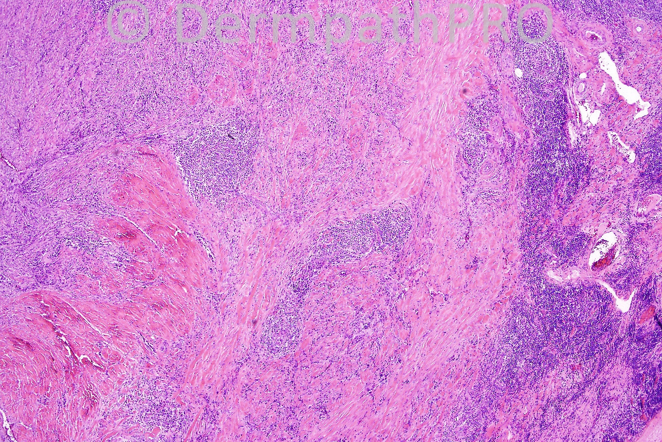
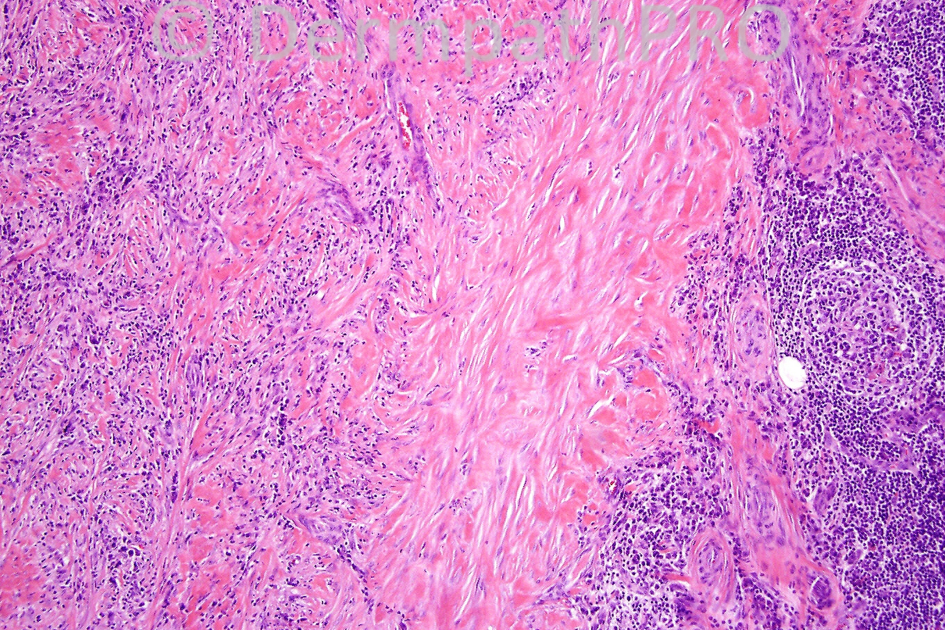
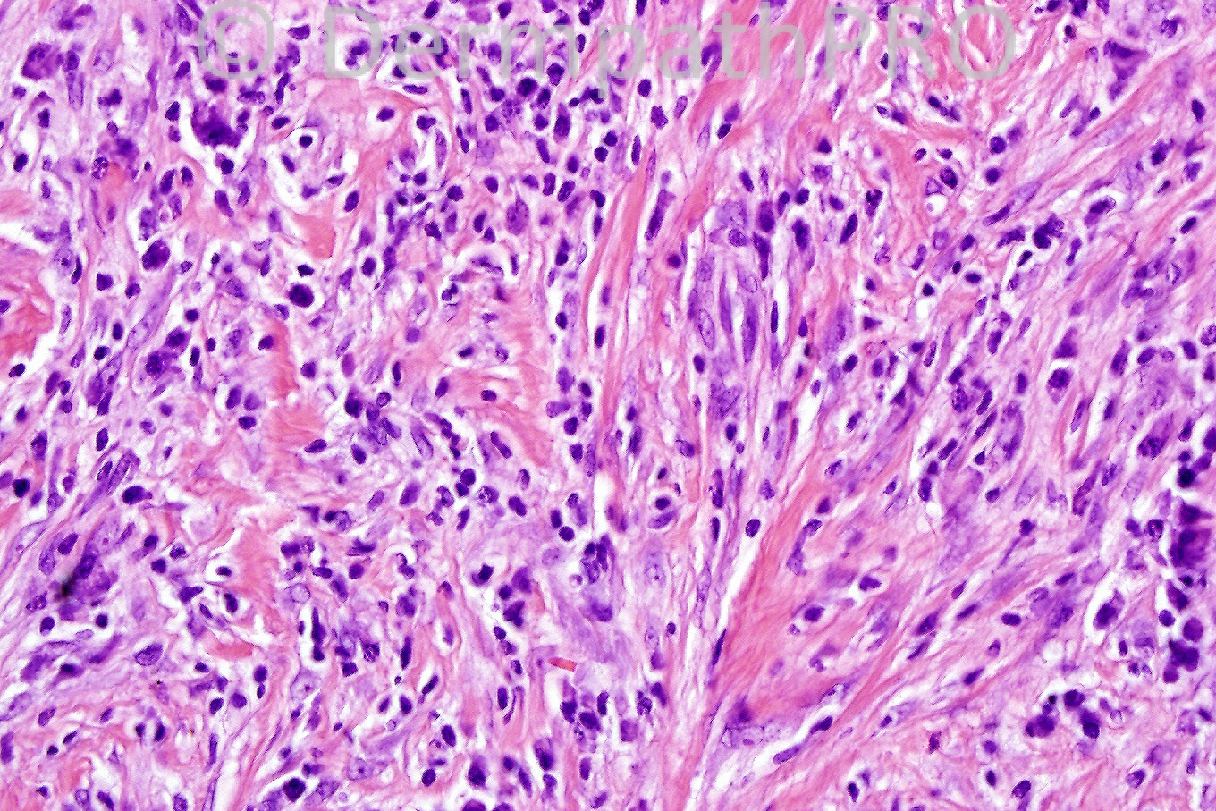
Female 8 years, subcutaneous nodule in lower leg.


  Report Record

User Feedback


Admin_Dermpath

Posted

Mona Abdel Halim - () Wrote:

I share the same request to MSMD!!!!!'!!!!!!!!!!!!!!!!!

Submitted on 28/06/2011 16:19
________________________________________
Phillip McKee - Overseas consultations (Cave Creek, Arizona, USA) Wrote:

It would be great if MSMC-() would come out of the dark void and let us know who he/she is and where he/she comes from. There is no need to be shy!

Submitted on 28/06/2011 13:26
________________________________________
Phillip McKee - Overseas consultations (Cave Creek, Arizona, USA) Wrote:

It is very clear that you all like soft tissue tumors. Again a difficult case but you all are confident of the diagnosis of inflammatory myofibroblastic tumor which is correct. In addition to the features that you mention, lymphoid follicles are present in the first picture and are a diagnostic pointer

Submitted on 28/06/2011 13:23
________________________________________
Mona Abdel Halim - Dermatology Dept. Faculty of Medicine, Cairo University (Egypt) Wrote:

Now this should be inflammatory myofibroblastic tumor, short fascicles of spindle cells, collection of lymphocytes and plasma cells forming clusters. Together with collagenous stroma.

Submitted on 28/06/2011 12:06
________________________________________
Eman El-Nabarawy - Cairo University (Egypt) Wrote:

Inflammatory myofibroblastic tumor.

Submitted on 28/06/2011 11:34
________________________________________
MSMD - () Wrote:

Hybrid of fibroblastic and myofibroblastic features: I believe this is inflammatory myofibroblastic tumor.

Submitted on 28/06/2011 10:22
________________________________________
Rodrigo Restrepo - UPB/CES (Colombia) Wrote:

This slide at low magnification looks like a sclerotic lymph node. D: Inflammatory myofibroblastic tumor

Submitted on 28/06/2011 08:27
________________________________________
Amira Tawdy - Cairo University (Egypt) Wrote:

Again a very confusing case. I see some dilated vessels at the right edge of the first photo , two populations of cells, rounded and spindle shaped I think there is pleomorphism and some large hperchromatic cells in the last 2 photos, besides some areas of sclerosis intervening I suggest the following D.D:plexiformhistocytic tumor, infantile myofibromatosis, inflammatory pseudotumor

Submitted on 28/06/2011 05:41
________________________________________
ANDREW DANCKWERTS - NHLS, WITS UNIVERSITY (JOHANNESBURG, RSA) Wrote:

INFLAMMATORY MYOFIBROBLASTIC TUMOUR (IMT) OF SOFT TISSUE - DIFFICULT CASE!

Submitted on 28/06/2011 05:13
________________________________________

Share this comment


Link to comment
share_externally


×
×
  • Create New...