Jump to content
In this section we have spot diagnoses posted on a daily basis since June 2010, now over 4000! You can review the archived cases and read the suggested diagnoses by users and the final comment by the contributors.
Case are uploaded each week day by 10 am UK time with the correct diagnosis will generally be posted at 8 pm UK time. Why not view the most recent spot diagnosis and proffer a diagnosis?

Case Number : Case 480 Posted By: Guest

Please read the clinical history and view the images by clicking on them before you proffer your diagnosis.
Submitted Date :
   (0 reviews)

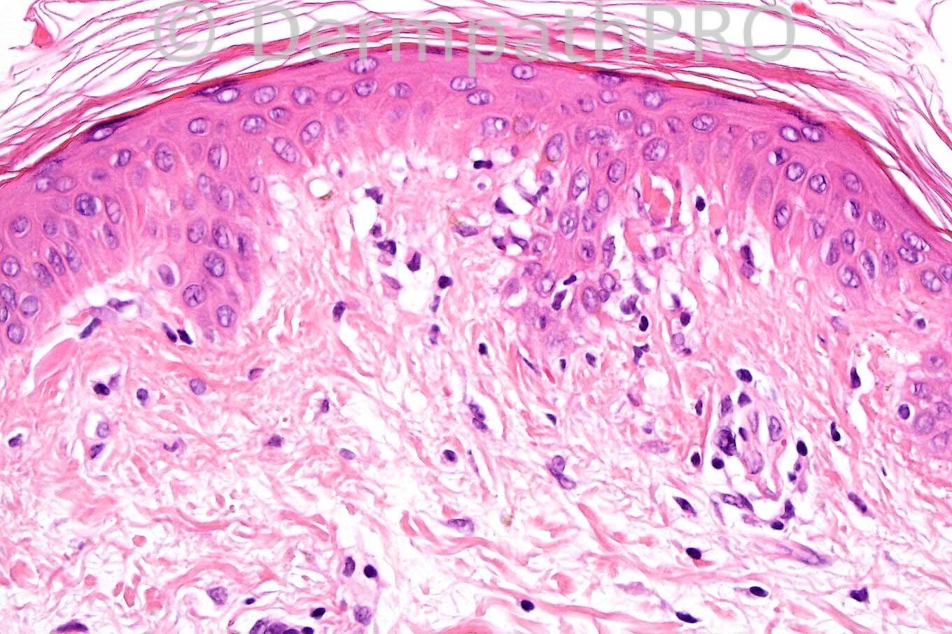
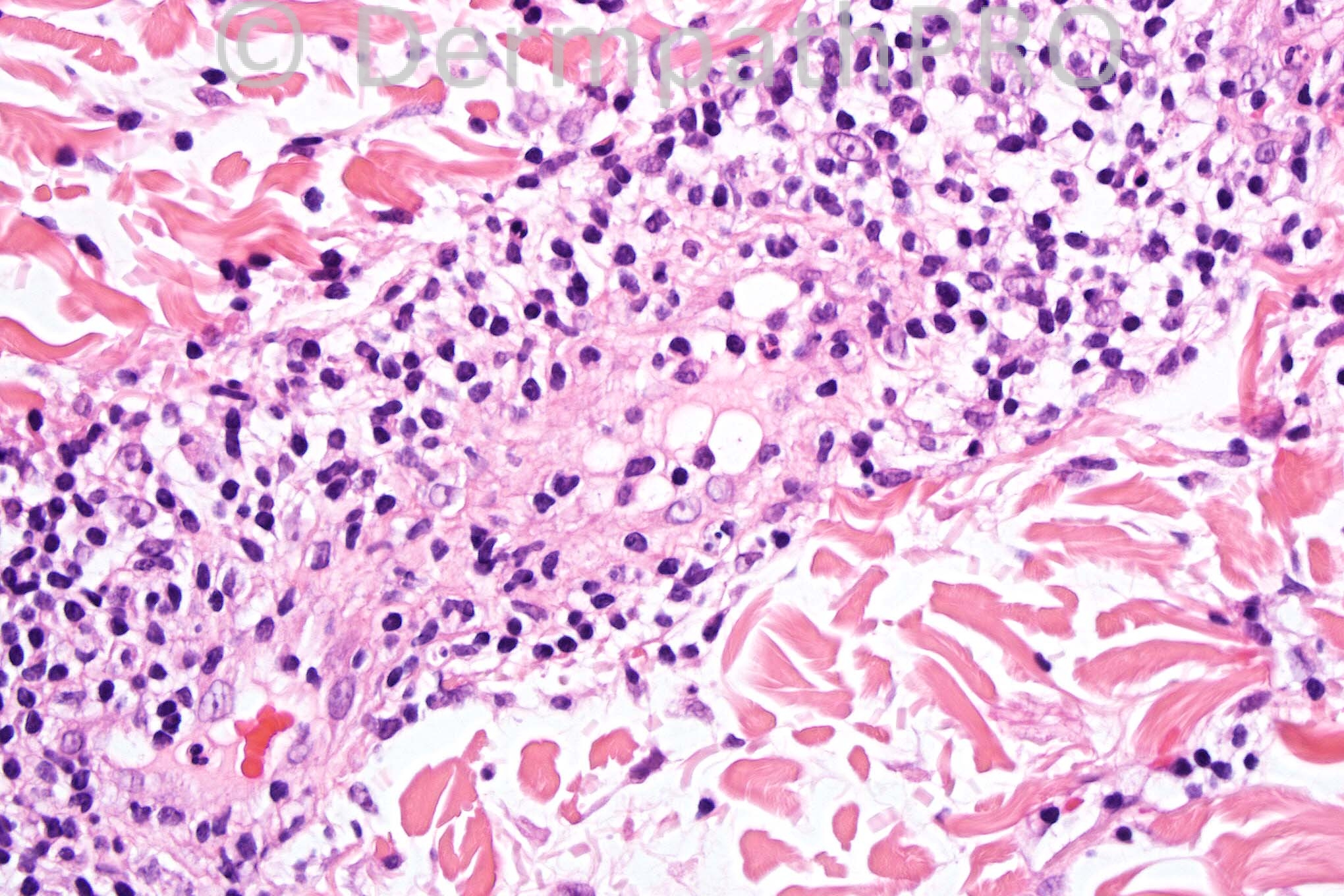
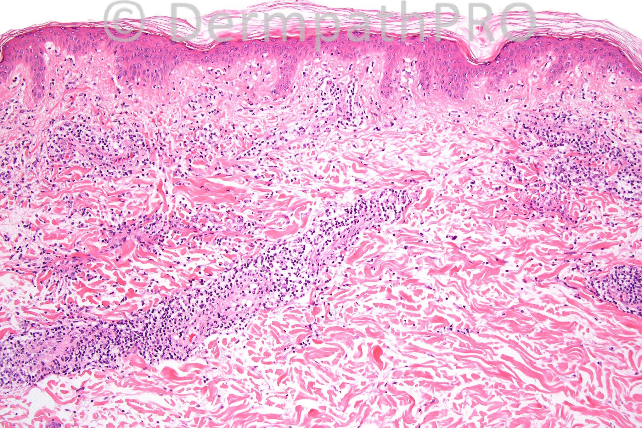
6 month history of erythematous monomorphic papules clinically thought to represent lupus erythematosus.


  Report Record

User Feedback


Admin_Dermpath

Posted

[b]Phillip H McKee - Overseas Consultations (Cave Creek, Arizona, United States) Wrote:[/b]

I agree that the clinical presentation is a little misleading but the clinical diagnosis was lupus erythematosus. The presence of basal cell hydropic degeneration with a superfical and deep lymphocytic infiltrate is entirely consistent with lupus. The patient in fact suffered from systemic lupus erythematosus.

[size=2]Submitted on 11/04/2012 19:06[/size]
[b]Sasi Attili - (United Kingdom) Wrote:[/b]

The superficial and deep infiltrate + vacuolar change makes me favour lupus. However a tough one and can't rule out the other differentials!! Clinical picture of papules is against PLC or PLEVA. Drugs is another possibility.

[size=2]Submitted on 11/04/2012 19:05[/size]
[b]Graham Reilly - PEH (Guernsey) Wrote:[/b]

perivascular lymphocytic infiltrate some endothelial swelling.Interface dermatitis with keratinocyte apoptosis.In view of papular eruption I favour PLC.

[size=2]Submitted on 11/04/2012 18:53[/size]
[b]Marie MD - () Wrote:[/b]

interface derm with neuts and endo swelling... acute LE (SLE) vs. dermatomyositis

[size=2]Submitted on 11/04/2012 18:38[/size]
[b]Richard Carr - Warwick (UK) Wrote:[/b]

If we go with a dense perivascular lymphocytic infiltrate as the main pattern L diseases i.e. Lupus, Lues, Light, Jesner's Lymphocytic infiltrate, Lyme (borreliosis), Lepidotera (insect reaction); chiLL blains (cold injury), Lymphoma, pseudoLymphomatous (e.g. drugs), erythema annuLare. Given that there is some keratinocyte necrosis I would not rule out some "acute" lupus variant here but slightey favoured pityriasis Lichenoides (the clinical does not always match the histology in terms of acuta versus chronica). Probably all wrong!

[size=2]Submitted on 11/04/2012 18:28[/size]
[b]Richard Carr - Warwick (UK) Wrote:[/b]

Difficult today. I was thinking slighly wedge shaped. Perivascular lymphocytes dominating. Rather acute (in view of endothelial swelling) and focal keratinocyte apoptosis. I wondered about pitriasis lichenoides too but won't be surprised to be wrong.

[size=2]Submitted on 11/04/2012 18:21[/size]
[b]Marcela Saeb Lima - INCMNSZ (Mexico City ) Wrote:[/b]

non bullous neutrophilic dermatosis associated to SLE

[size=2]Submitted on 11/04/2012 15:32[/size]
[b]omar erdoÄŸan - () Wrote:[/b]

leukocytoclastic vasculitis

[size=2]Submitted on 11/04/2012 12:55[/size]
[b]Izzat Abdul-kadir - ST2 - York Hospital (UK) Wrote:[/b]

Pityriasis lichenoides chronica.

[size=2]Submitted on 11/04/2012 12:20[/size]
[b]ANDREW DANCKWERTS - (LONDON, UNITED KINGDOM) Wrote:[/b]

MYCOSIS FUNGOIDES.

[size=2]Submitted on 11/04/2012 11:10[/size]

Share this comment


Link to comment
share_externally


×
×
  • Create New...