Jump to content
In this section we have spot diagnoses posted on a daily basis since June 2010, now over 4000! You can review the archived cases and read the suggested diagnoses by users and the final comment by the contributors.
Case are uploaded each week day by 10 am UK time with the correct diagnosis will generally be posted at 8 pm UK time. Why not view the most recent spot diagnosis and proffer a diagnosis?

Case Number : Case 560 - 1 Aug Posted By: Guest

Please read the clinical history and view the images by clicking on them before you proffer your diagnosis.
Submitted Date :
   (0 reviews)

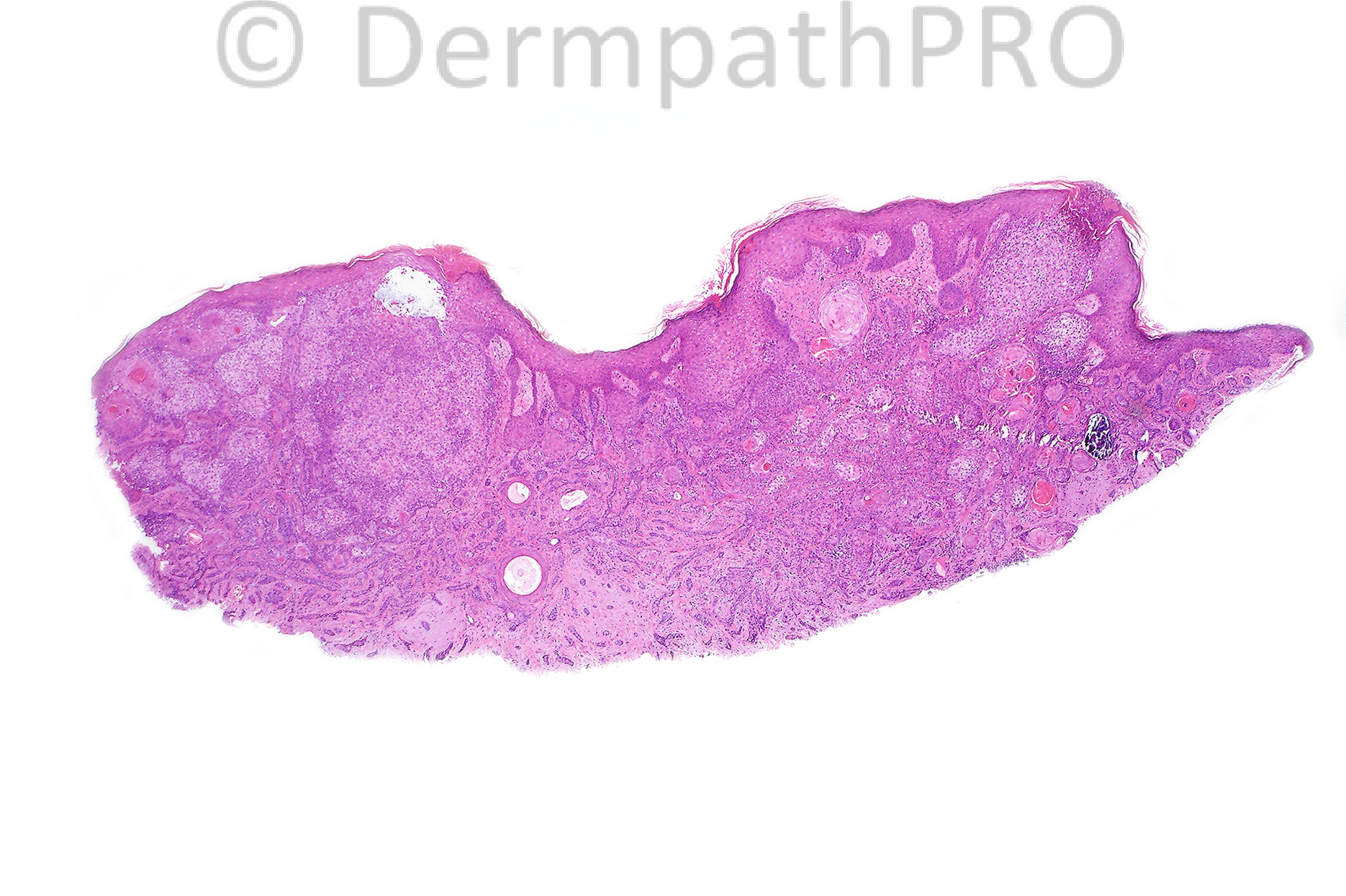
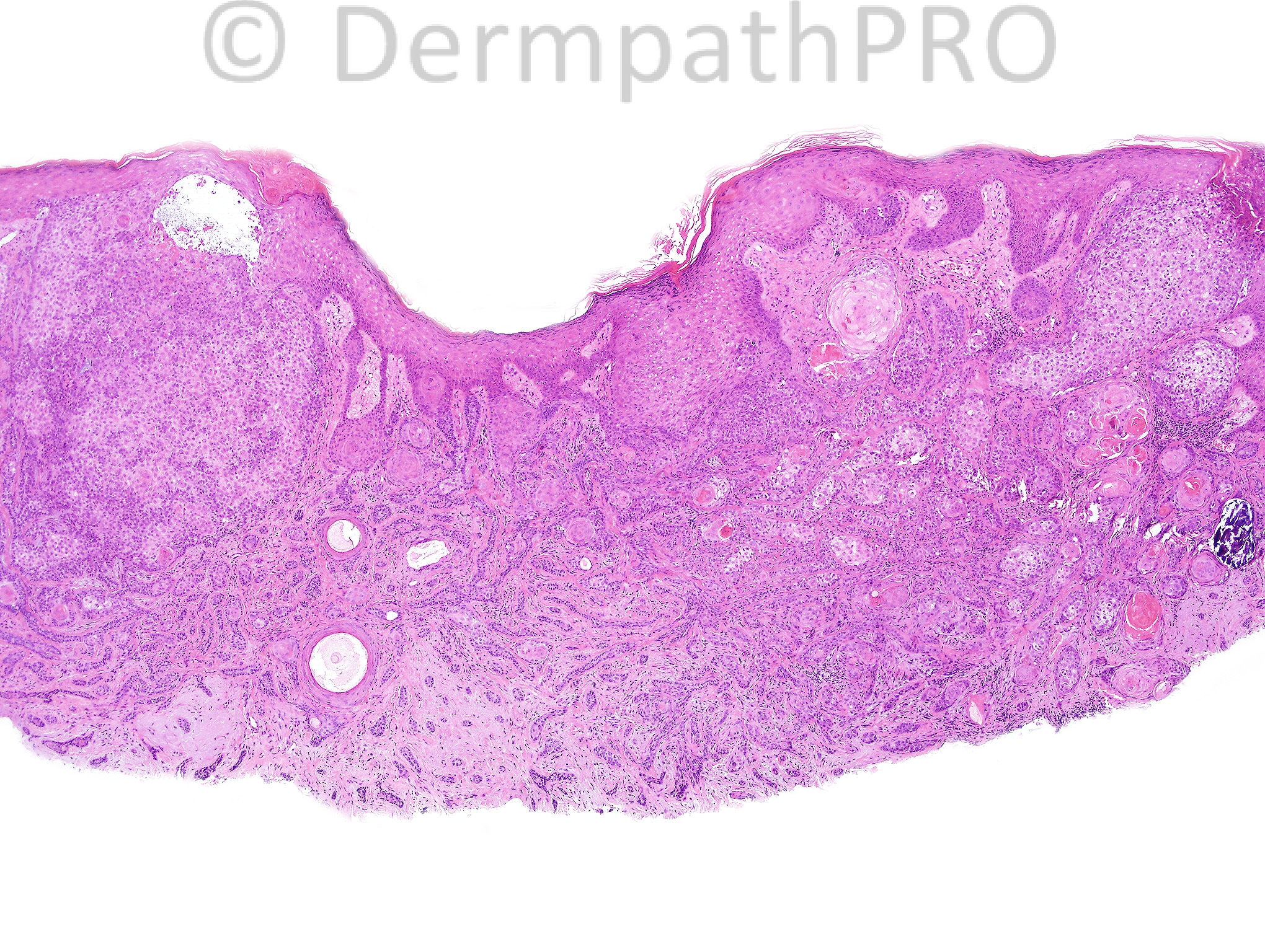
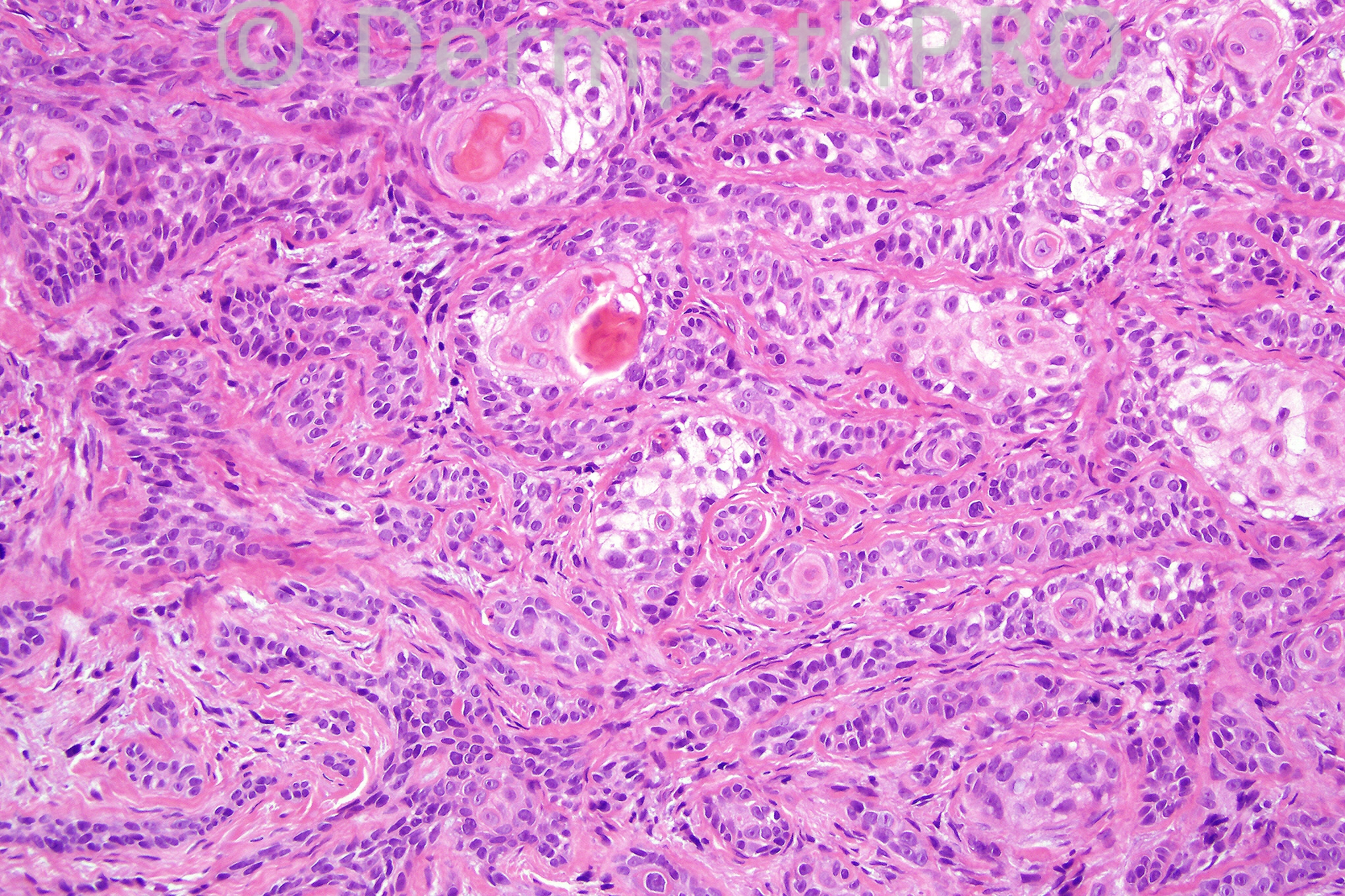
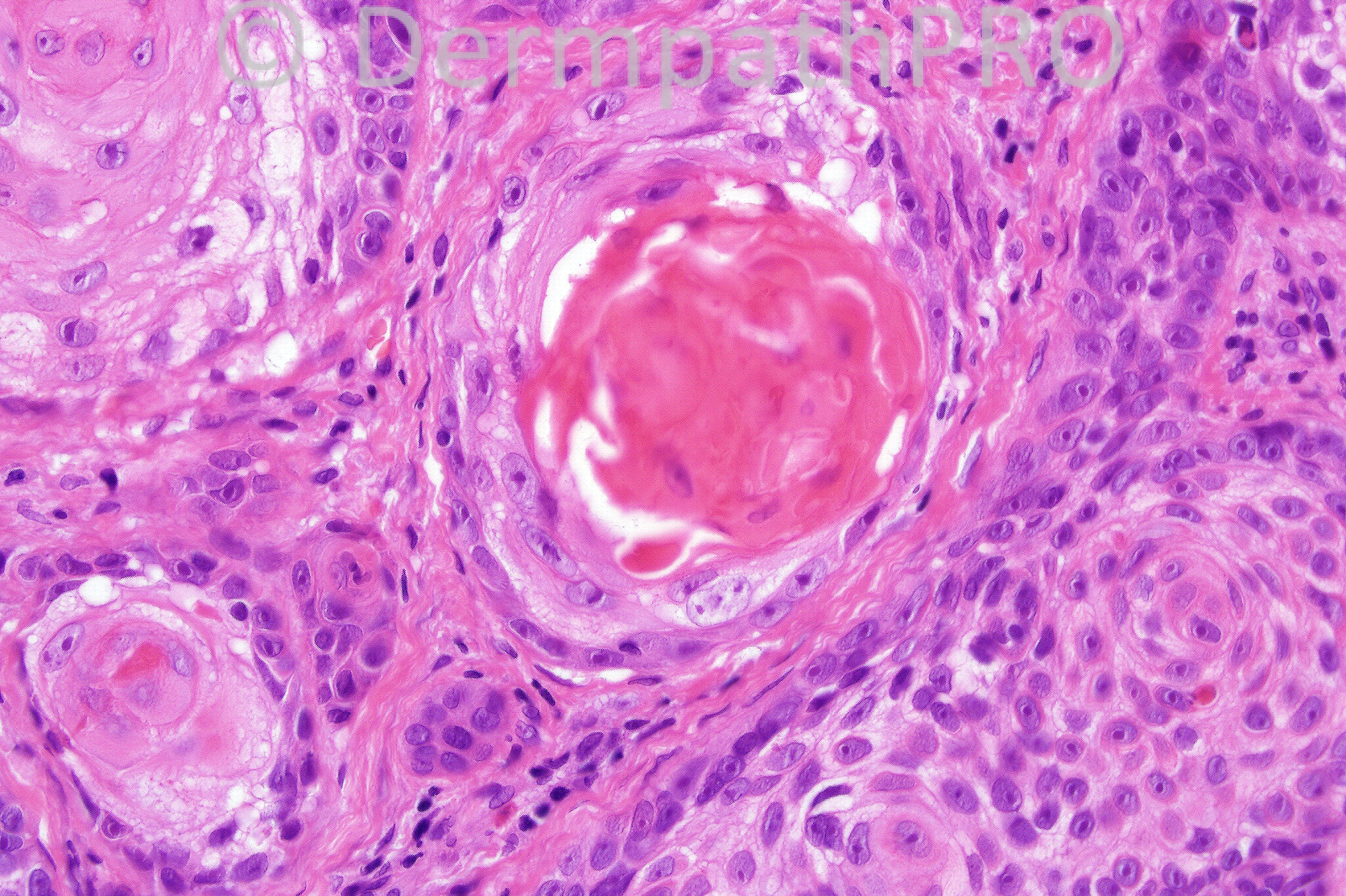
Male 72 years, with a crusted nodule on his forehead.

We are grateful to Dr. Richard Carr who has provided this case.


  Report Record

User Feedback


Guest Hazem Hamed

Posted

Desmoplastic trichoepithelioma. The lesion, however, is incompletely excised, so microcystic adenxal carcinoma can not be entirely excluded. Wider excision would be recommended.

Share this comment


Link to comment
share_externally

Hanan Vaknine MD

Posted

desmoplastic trichoepithelioma is my diagnosis
no ductal differentiation or perineural invasion were observed in the slides provided therfore the possibility of a coexsisting adnexal carcinoma appears less like
nontheless the lesion is large and involves the margins therefore i would also recommend conservative wider excision in this case

Share this comment


Link to comment
share_externally

Hanan Vaknine MD

Posted

[color=#222222][font=Arial][size=1]Desmoplastic trichoepithelioma is also my diagnosis[/size][/font][/color]
[color=#222222][font=Arial][size=1]No ductal differentiation or perineural invasion were observed in the slides provided therefore the possibility of a coexisting adnexal carcinoma appears less like [/size][/font][/color]
[color=#222222][font=Arial][size=1]Nonetheless the lesion is large and involves the margins therefore I would also recommend conservative wider excision in this case [/size][/font][/color]

Share this comment


Link to comment
share_externally

Guest Dr. Francisco Vílchez

Posted

Desmoplastic trichoepithelioma.

Share this comment


Link to comment
share_externally

Desmoplastic trichoepithelioma vs MAC, it is a shave biopsy

Share this comment


Link to comment
share_externally

[size=4][color=#000000][b]Incomplete biopsy[/b]. An overlying central depression, cords and islands of cells set in a fibrous stroma ([b]cords, the [/b][b]predominant morphologic pattern[/b]), horn cysts are also present. D[color=#333333]esmoplastic [sclerosing] trichoepithelioma is in the differential diagnosis[/color].

[color=black]Need to search for [b]Merkel cells[/b] by [/color][color=#231F20]immunohistochemical markers ([/color][color=black]Merkel cells are present in desmoplastic[/color][color=#333333] trichoepithelioma[/color][color=black], [/color]and absent in microcystic adnexal carcinoma and in most cases of morphoeic basal cell carcinoma[color=black]) and to test for [/color][color=#231F20]the immunohistochemical expression of [b]androgen receptor [/b](usually negative in [/color][color=#333333]trichoepithelioma[/color][color=#231F20] and positive in basal cell carcinomas) and to test the more or less [/color]spindle-shaped cells surrounding the cellular islands in desmoplastic trichoepithelioma for [b]CD34[/b] (focally positive in desmoplastic trichoepithelioma, whereas the stromal cells around basal cell carcinomas and microcystic adnexal carcinomas are usually negative).

Certainly, small incomplete biopsies may cause uncertainty about excluding sclerosing BCC or microcystic adnexal carcinoma or others, and [b]re-excision or resampling may be necessary for a definitive diagnosis or complete eradication in uncertain cases[/b].[/color][/size]

Share this comment


Link to comment
share_externally

Guest Dr Gonzalo de Toro

Posted

Desmoplastic trichoephitelioma

Share this comment


Link to comment
share_externally

Dr. Mona Abdel-Halim

Posted

Well, I might be out of the track, but:
1. I am not comfortable with the braod connections with the epidermis in the first and second images.
2. The appearence of the islands in images 3 and 4, foci of trichilemmal (pilar) keratinization, cells appear pleomorphic with ? acantholysis (image 3).
Could this be trichilemmal carcinaoma (with unsuaual infiltrative growth pattern) or ? Follicular SCC??

Share this comment


Link to comment
share_externally

[size=4][font=arial,helvetica,sans-serif][color=black]In [/color][b][color=#231F20]trichilemmal (tricholemmal) carcinoma[/color][/b][color=black], the lesion[b] invades downwards in a multilobular fashion[/b]. [b]There should be [/b][/color][color=#231F20][b]a high mitotic rate; with abnormal mitoses[/b]. I [b]also need to see the atypical clear cells resembling those of the outer root sheath[/b].[/color][color=black] Once again [/color][color=#231F20]Immunohistochemical studies[/color][color=black] are helpful in showing the [/color][color=#231F20]trichilemmal differentiation details if present. [color=#000000]Trichoepitheliomas are dermal tumours with focal continuity with the epidermis at times though the continuity shown here is certainly not a typical example. [/color] [/color][/font][/size]

Share this comment


Link to comment
share_externally

Guest MarieMD

Posted

trichilemmal carcinoma vs. infundibular SCC

Share this comment


Link to comment
share_externally

Guest Hazem Hamed

Posted

Cup-shaped depression of the epidermis is a good clue to Desmoplastic trichoephitelioma. I do not believe in doing immuno for most of skin tumours. I learnt that from Dr Eduardo and completely agree with his statement "Morphology remains the gold standard in most of skin tumours".

Share this comment


Link to comment
share_externally

[color=black][b][font=Revival565BT-Italic]Follicular [/font][/b][font=Revival565BT-Roman][b]squamous cell carcinoma[/b]. [b]Considered, but I need to search for any degree of anaplasia if present[/b]. [/font][/color]

Share this comment


Link to comment
share_externally

Guest Eman El-Nabarawy

Posted

My first impression was trichilemmal carcinoma.

Share this comment


Link to comment
share_externally

[color=#231F20]Immunohistochemical studies are for research purposes on many occasions but are certainly useful in reaching the correct diagnosis at times. [/color]

Share this comment


Link to comment
share_externally

Guest A Bansal

Posted

? Trichilemmal carcinoma

Share this comment


Link to comment
share_externally

Dr. Phillip McKee

Posted

A difficult case. Well done those of you who diagnosed follicular squamous cell carcinoma. This is a case of Richard Carr's and he would be best to comment on it since he has a huge collection of cases which he will soon publish. I think that there is overlap with trichilemmal carcinoma but I reserve that diagnosis for a timor which is obviously malignant and composed of clear cells often showing an association with a hair follicle in addition to the epidermis and showing pilar keratinization as is seen in this tumor. The other two tumors mentioned are good differential diagnoses. Perhaps I made this spot diagnosis too dificult by not including the more marked pleomorphism present at high power examination and the mitotic activity but I wanted to see where you would go with just these 4 figures.

Share this comment


Link to comment
share_externally

Dr. Mona Abdel-Halim

Posted

Thank you Dr McKee, I loved this challenge :-)

Share this comment


Link to comment
share_externally

Sasi Attili

Posted

I am a bit late but I had gone for desmoplastic trichoepithelioma as well, based on the pictures here.

Share this comment


Link to comment
share_externally

Dr. Richard Carr

Posted

Hello all I'm very very pleased that you were all in the right ball park on this one.
Mona was outstanding with her comments. You really don't see such good circumscript
connections with the follicular infundibular units as illustrated in this case in
desmoplastic trichoepithelioma. Also Merkel cells are present in every DTE I have
ever diagnosed and they were absent in this case. The proper history by the way was a
lesion in an even older man and on the nose (it took me a while to work out which
case it was and review the original H&E). Many follicular SCC have rather subtle atypia
as in this case and I have to agree most of the deep cord-like growth in this case
was rather bland and mitotically inactive. The lesion had focal strong CD34 epithelial
staining (negative for BerEP4 and EMA with no glands). The former is more typical of
benign (tricholemmoma) but in this case I was not happy to call it benign with
such deep infiltrative cords (permeating through deep solar elastosis as well!!).
I think this is a low-grade, infiltrative follicular
(tricholemmaal) squamous cell carcinoma with MAC less likely (no glands seen).
I advised margin
control surgery but I think this was done at St Elsewhere so have been unable to
review the excision. Some authorities think MAC is a follicular tumour with glandular
differentiation! i.e. follicular-apocrine(duct)-sebaceous unit tumour.
I like the discussion about immunos - we use it often but usually to support our
H&E diagnosis or help refute differentials. I am not sure these markers are helpful to
generalists because there are just so many pitfalls!
Regards to all

Share this comment


Link to comment
share_externally

Guest Hazem Hamed

Posted

Thank you Dr Richard for the useful comment. With focal strong staining for CD34, did you consider Desmplastic tricholemmoma as a Differential?. Thank you

Share this comment


Link to comment
share_externally

Dr. Richard Carr

Posted

Yes definitely. It does not have the typical stranded infiltrative SCC-like
pattern of desmoplastic tricholemmoma and as indicated by Phillip and me
above I was not happy with the degree of atypia and the way the cords were infiltrating
through solar elastosis without much stromal reaction (at the base of image 1 and 2).
I should point out this was an exceedingly difficult case and as so often is the case lesions
often don't read the textbooks so we have to make a judgement call.

Share this comment


Link to comment
share_externally


×
×
  • Create New...