Jump to content
In this section we have spot diagnoses posted on a daily basis since June 2010, now over 4000! You can review the archived cases and read the suggested diagnoses by users and the final comment by the contributors.
Case are uploaded each week day by 10 am UK time with the correct diagnosis will generally be posted at 8 pm UK time. Why not view the most recent spot diagnosis and proffer a diagnosis?

Case Number : Case 562 - 3 Aug Posted By: Guest

Please read the clinical history and view the images by clicking on them before you proffer your diagnosis.
Submitted Date :
   (0 reviews)

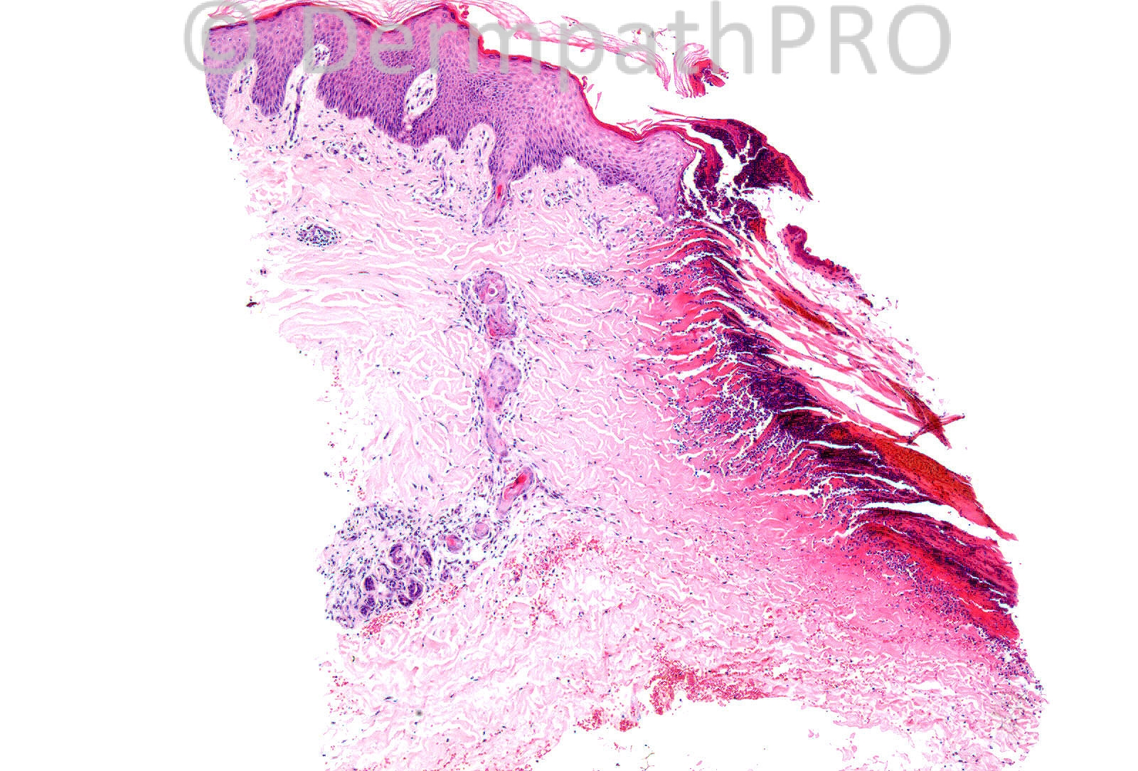
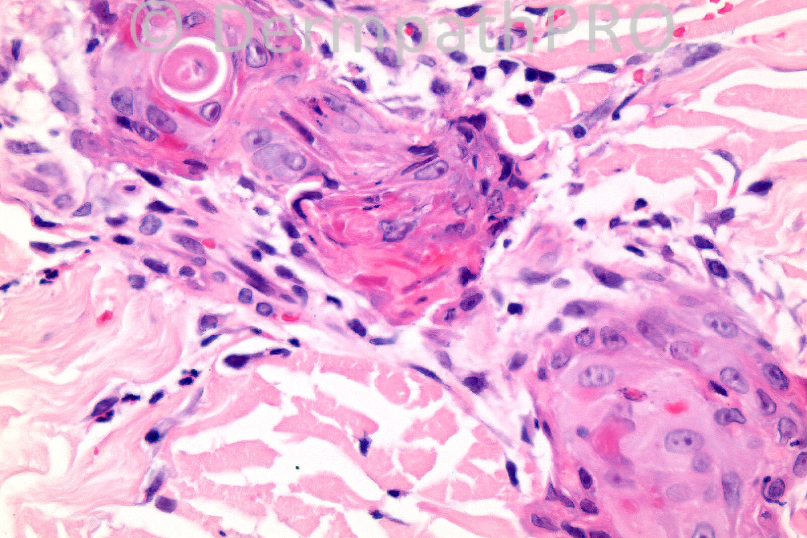
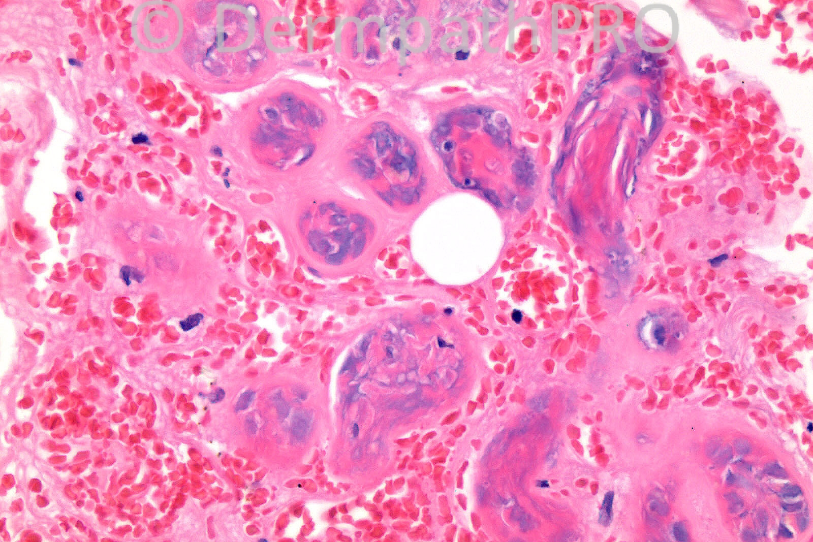
Male 74 years, dorsum of foot. Odd red lesion with central necrotic eshcar. Patient currently investigated for progressive neurological problem and uveitis. Is the skin lesion related? DD: Infective e.g. ecthyma, tick

Case posted by Dr. Richard Carr


  Report Record

User Feedback


Guest Hazem Hamed

Posted

Epidermal ulceration (adjacent epidermis shows acanthosis), neutrophils and apoptotic debris, syringometaplasia and in the 3[sup]rd[/sup] image there is necrosis of the sweat gland ducts (most likely due to ischemia) and haemorrhage. PAS, Gram and Groccott are required to exclude an infectious aetiology.
Syringometaplasia can be seen in areas of ischemia, adjacent to ulcers or healing surgical wounds. It may follow burn, radiation, cryotherapy or curettage. It also can occur in patients receiving chemotherapy for malignant tumours.

[b]The changes are most likely due to ischemia[/b].

DD: Dermatitis artefacta.

Share this comment


Link to comment
share_externally

Sasi Attili

Posted

This might just be a decubitus ulcer. However would like to rule out an infective etiology with PAS, Gram and ZN.

Share this comment


Link to comment
share_externally

Guest MarieMD

Posted

Overall the histologic changes appear to be those of ischemia. However, given the site (not typical for pressure ulcers) and history of progressive neuropathy and uveitis, I'm wondering if this patient might have CMV infection.

Share this comment


Link to comment
share_externally

Dr. Mona Abdel-Halim

Posted

Histopathological manifestations are of Ischemia. Would think of vasculopathy caused by infective agent (ecthyma gangrenosusm due to pseudomonas infection for example or mengiococcemia etc...) or other causes such as: antiphospholipid syndrome, cryoglobulins, Ptn C and S deffeciency. In either cases, the associated neurological and occular manifestations are definitly related..

Share this comment


Link to comment
share_externally

Dr. Richard Carr

Posted

Thank you all for your contributions. I agree with Hazem's comments entirely.
This is a quite old case and I reported it as follows:
The biopsy shows an area of apparent wedge shaped dermal infarction
including necrosis of eccrine sweat coils and ecrine squamous
syringometaplasia in the adjacent dermis. There is a moderate
accompanying neutrophilic infiltrate in the vicinity of dermal
infarction, which extends on to the surface of the biopsy. No
specific organisms seen on gram stain.

Histologically my thoughts would include a local "pressure"
necrosis, possibly related to footwear in somebody with a
neurological problem. I cannot rule out a vascular problem in this
punch biopsy.

Share this comment


Link to comment
share_externally


×
×
  • Create New...